- EMDB-34200: Complex Structure of Arginine Kinase McsB and McsA from Staphyloc... -
+
Open data
ID or keywords:
Loading...
-
Basic information
Entry
Database: EMDB / ID: EMD-34200
Title
Complex Structure of Arginine Kinase McsB and McsA from Staphylococcus aureus
Map data
Complex Structure of Arginine Kinase McsB and McsA from Staphylococcus aureus
Sample
Complex: McsA-McsB complex
Protein or peptide: Protein-arginine kinase
Protein or peptide: Protein-arginine kinase activator protein
Ligand: ZINC ION
Keywords
Arginine kinase / Protein quality control / Zinc finger / Enzyme activation / TRANSFERASE
Function / homology
Function and homology information
protein arginine kinase / protein arginine kinase activity / stress response to cadmium ion / phosphocreatine biosynthetic process / stress response to copper ion / creatine kinase activity / cobalt ion binding / cadmium ion binding / kinase activity / copper ion binding ...protein arginine kinase / protein arginine kinase activity / stress response to cadmium ion / phosphocreatine biosynthetic process / stress response to copper ion / creatine kinase activity / cobalt ion binding / cadmium ion binding / kinase activity / copper ion binding / : / DNA binding / zinc ion binding / ATP binding Similarity search - Function
YacH protein / Protein arginine kinase / UvrB/uvrC motif / ATP:guanido phosphotransferase active site / Phosphagen kinase active site signature. / ATP:guanido phosphotransferase / ATP:guanido phosphotransferase, catalytic domain / ATP:guanido phosphotransferase, C-terminal catalytic domain / Phosphagen kinase C-terminal domain profile. / UVR domain ...YacH protein / Protein arginine kinase / UvrB/uvrC motif / ATP:guanido phosphotransferase active site / Phosphagen kinase active site signature. / ATP:guanido phosphotransferase / ATP:guanido phosphotransferase, catalytic domain / ATP:guanido phosphotransferase, C-terminal catalytic domain / Phosphagen kinase C-terminal domain profile. / UVR domain / UVR domain profile. / Zinc finger, PARP-type / Glutamine synthetase/guanido kinase, catalytic domain Similarity search - Domain/homology
National Natural Science Foundation of China (NSFC)
22077142
China
National Natural Science Foundation of China (NSFC)
22022706
China
National Natural Science Foundation of China (NSFC)
21837006
China
National Natural Science Foundation of China (NSFC)
91953117
China
National Natural Science Foundation of China (NSFC)
21877131
China
National Natural Science Foundation of China (NSFC)
32070049
China
National Natural Science Foundation of China (NSFC)
32222040
China
Ministry of Science and Technology (MoST, China)
2021YFA1301900
China
Citation
Journal: Nat Chem Biol / Year: 2025 Title: Complex structure and activation mechanism of arginine kinase McsB by McsA. Authors: Kai Lu / Bingnan Luo / Xuan Tao / Yongbo Luo / Mingjun Ao / Bin Zheng / Xiang Xu / Xiaoyan Ma / Jingling Niu / Huinan Li / Yanxuan Xie / Zhennan Zhao / Peng Zheng / Guanbo Wang / Song Gao / ...Authors: Kai Lu / Bingnan Luo / Xuan Tao / Yongbo Luo / Mingjun Ao / Bin Zheng / Xiang Xu / Xiaoyan Ma / Jingling Niu / Huinan Li / Yanxuan Xie / Zhennan Zhao / Peng Zheng / Guanbo Wang / Song Gao / Chao Wang / Wei Xia / Zhaoming Su / Zong-Wan Mao / Abstract: Protein phosphorylation is a pivotal post-translational modification modulating various cellular processes. In Gram-positive bacteria, the protein arginine kinase McsB, along with its activator McsA, ...Protein phosphorylation is a pivotal post-translational modification modulating various cellular processes. In Gram-positive bacteria, the protein arginine kinase McsB, along with its activator McsA, has a key role in labeling misfolded and damaged proteins during stress. However, the activation mechanism of McsB by McsA remains elusive. Here we report the cryo-electron microscopy structure of a tetrameric McsA-McsB complex at 3.41 Å resolution. Biochemical analysis indicates that the homotetrameric assembly is essential for McsB's kinase activity. The conserved C-terminal zinc finger of McsA interacts with an extended loop in McsB, optimally orienting a critical catalytic cysteine residue. In addition, McsA binding decreases the CtsR's affinity for McsB, enhancing McsB's kinase activity and accelerating the turnover rate of CtsR phosphorylation. Furthermore, McsA binding also increases McsB's thermostability, ensuring its activity under heat stress. These findings elucidate the structural basis and activation mechanism of McsB in stress response.
In the structure databanks used in Yorodumi, some data are registered as the other names, "COVID-19 virus" and "2019-nCoV". Here are the details of the virus and the list of structure data.
Jan 31, 2019. EMDB accession codes are about to change! (news from PDBe EMDB page)
EMDB accession codes are about to change! (news from PDBe EMDB page)
The allocation of 4 digits for EMDB accession codes will soon come to an end. Whilst these codes will remain in use, new EMDB accession codes will include an additional digit and will expand incrementally as the available range of codes is exhausted. The current 4-digit format prefixed with “EMD-” (i.e. EMD-XXXX) will advance to a 5-digit format (i.e. EMD-XXXXX), and so on. It is currently estimated that the 4-digit codes will be depleted around Spring 2019, at which point the 5-digit format will come into force.
The EM Navigator/Yorodumi systems omit the EMD- prefix.
Related info.:Q: What is EMD? / ID/Accession-code notation in Yorodumi/EM Navigator
Yorodumi is a browser for structure data from EMDB, PDB, SASBDB, etc.
This page is also the successor to EM Navigator detail page, and also detail information page/front-end page for Omokage search.
The word "yorodu" (or yorozu) is an old Japanese word meaning "ten thousand". "mi" (miru) is to see.
Related info.:EMDB / PDB / SASBDB / Comparison of 3 databanks / Yorodumi Search / Aug 31, 2016. New EM Navigator & Yorodumi / Yorodumi Papers / Jmol/JSmol / Function and homology information / Changes in new EM Navigator and Yorodumi
 Movie
Movie Controller

 Yorodumi
 Open data
 Basic information
 Map data
 Sample
 Keywords
 Function and homology information
 Staphylococcus aureus (bacteria)
 Authors
 China, 8 items
  Citation
 Structure visualization
 Downloads & links
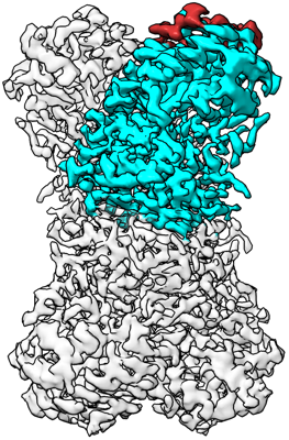 emd_34200.png
 http://ftp.pdbj.org/pub/emdb/structures/EMD-34200

 Z (Sec.)
 Y (Row.)
 X (Col.)


 Sample components
 Processing
 Electron microscopy
 FIELD EMISSION GUN
