 Movie
Movie Controller


+ Open data

- Basic information
| Entry |  | |||||||||
|---|---|---|---|---|---|---|---|---|---|---|
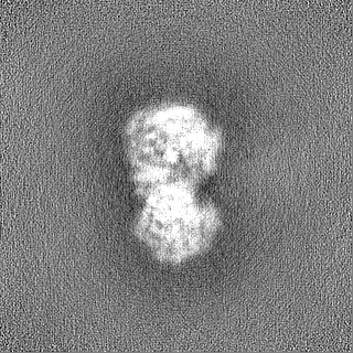
| Title | Cryo-EM map of a dimeric form of Ecoli Malate Synthase G (MSG) | |||||||||
 Map data Map data | ||||||||||
 Sample Sample |
| |||||||||
 Keywords Keywords | glyoxylate / malate / MSG / citric acid cycel / malate synthase G / BIOSYNTHETIC PROTEIN | |||||||||
| Biological species |  | |||||||||
| Method | single particle reconstruction / cryo EM / Resolution: 4.14 Å | |||||||||
 Authors Authors | Wu K-P / Wu Y-M / Lu Y-C | |||||||||
| Funding support |  Taiwan, 1 items Taiwan, 1 items
| |||||||||
 Citation Citation | Journal: J Struct Biol / Year: 2023 Title: Cryo-EM reveals the structure and dynamics of a 723-residue malate synthase G. Authors: Meng-Ru Ho / Yi-Ming Wu / Yen-Chen Lu / Tzu-Ping Ko / Kuen-Phon Wu / Abstract: Determination of sub-100 kDa (kDa) structures by cryo-electron microscopy (EM) is a longstanding but not straightforward goal. Here, we present a 2.9-Å cryo-EM structure of a 723-amino acid apo- ...Determination of sub-100 kDa (kDa) structures by cryo-electron microscopy (EM) is a longstanding but not straightforward goal. Here, we present a 2.9-Å cryo-EM structure of a 723-amino acid apo-form malate synthase G (MSG) from Escherichia coli. The cryo-EM structure of the 82-kDa MSG exhibits the same global folding as structures resolved by crystallography and nuclear magnetic resonance (NMR) spectroscopy, and the crystal and cryo-EM structures are indistinguishable. Analyses of MSG dynamics reveal consistent conformational flexibilities among the three experimental approaches, most notably that the α/β domain exhibits structural heterogeneity. We observed that sidechains of F453, L454, M629, and E630 residues involved in hosting the cofactor acetyl-CoA and substrate rotate differently between the cryo-EM apo-form and complex crystal structures. Our work demonstrates that the cryo-EM technique can be used to determine structures and conformational heterogeneity of sub-100 kDa biomolecules to a quality as high as that obtained from X-ray crystallography and NMR spectroscopy. | |||||||||
| History |
|
- Structure visualization
| Supplemental images |
|---|

- Downloads & links
-EMDB archive
| Map data | emd_34030.map.gz | 118.2 MB |  EMDB map data format EMDB map data format | |
|---|---|---|---|---|
| Header (meta data) | emd-34030-v30.xmlemd-34030.xml | 12.9 KB 12.9 KB | Display Display | EMDB header |
| FSC (resolution estimation) | emd_34030_fsc.xml | 10.5 KB | Display | FSC data file |
| Images | 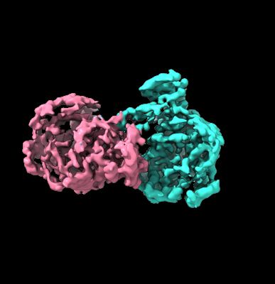 emd_34030.png emd_34030.png | 76.2 KB | ||
| Others | emd_34030_half_map_1.map.gzemd_34030_half_map_2.map.gz | 115.9 MB 115.9 MB | ||
| Archive directory |  http://ftp.pdbj.org/pub/emdb/structures/EMD-34030ftp://ftp.pdbj.org/pub/emdb/structures/EMD-34030 http://ftp.pdbj.org/pub/emdb/structures/EMD-34030ftp://ftp.pdbj.org/pub/emdb/structures/EMD-34030 | HTTPS FTP |
-Validation report
| Summary document | emd_34030_validation.pdf.gz | 1.2 MB | Display | EMDB validaton report |
|---|---|---|---|---|
| Full document | emd_34030_full_validation.pdf.gz | 1.2 MB | Display | |
| Data in XML | emd_34030_validation.xml.gz | 19.1 KB | Display | |
| Data in CIF | emd_34030_validation.cif.gz | 24.4 KB | Display | |
| Arichive directory | https://ftp.pdbj.org/pub/emdb/validation_reports/EMD-34030ftp://ftp.pdbj.org/pub/emdb/validation_reports/EMD-34030 | HTTPS FTP |
-Related structure data
-Links
| EMDB pages | EMDB (EBI/PDBe) / EMDataResource |
|---|
-Map
| File | Download / File: emd_34030.map.gz / Format: CCP4 / Size: 125 MB / Type: IMAGE STORED AS FLOATING POINT NUMBER (4 BYTES) | ||||||||||||||||||||||||||||||||||||
|---|---|---|---|---|---|---|---|---|---|---|---|---|---|---|---|---|---|---|---|---|---|---|---|---|---|---|---|---|---|---|---|---|---|---|---|---|---|
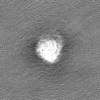
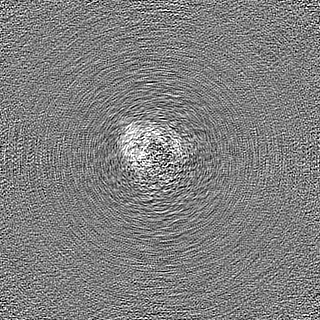
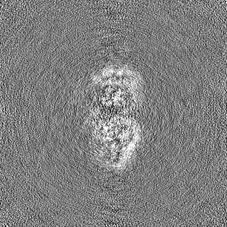
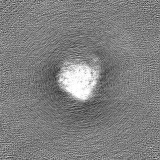
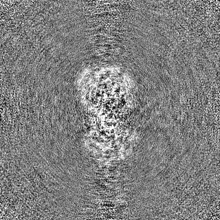
| Projections & slices | Image control
Images are generated by Spider. | ||||||||||||||||||||||||||||||||||||
| Voxel size | X=Y=Z: 0.822 Å | ||||||||||||||||||||||||||||||||||||
| Density |
| ||||||||||||||||||||||||||||||||||||
| Symmetry | Space group: 1 | ||||||||||||||||||||||||||||||||||||
| Details | EMDB XML:
|
 Z (Sec.)
 Y (Row.)
 X (Col.)


-Supplemental data
-Half map: #1
| File | emd_34030_half_map_1.map | ||||||||||||
|---|---|---|---|---|---|---|---|---|---|---|---|---|---|
| Projections & Slices |
| ||||||||||||
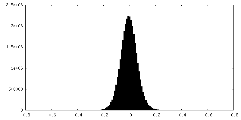
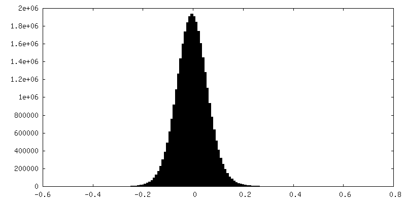
| Density Histograms |


-Half map: #2
| File | emd_34030_half_map_2.map | ||||||||||||
|---|---|---|---|---|---|---|---|---|---|---|---|---|---|
| Projections & Slices |
| ||||||||||||
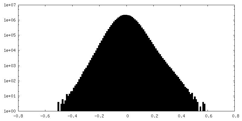
| Density Histograms |


- Sample components
-Entire : 2.9-angstrom cryo-EM structure of 723-aa malate synthase G
| Entire | Name: 2.9-angstrom cryo-EM structure of 723-aa malate synthase G |
|---|---|
| Components |
|
-Supramolecule #1: 2.9-angstrom cryo-EM structure of 723-aa malate synthase G
| Supramolecule | Name: 2.9-angstrom cryo-EM structure of 723-aa malate synthase G type: cell / ID: 1 / Parent: 0 / Macromolecule list: #1 |
|---|---|
| Source (natural) | Organism: |
-Experimental details
-Structure determination
| Method | cryo EM |
|---|---|
 Processing Processing | single particle reconstruction |
| Aggregation state | particle |
-Sample preparation
| Concentration | 0.3 mg/mL |
|---|---|
| Buffer | pH: 7.6 |
| Vitrification | Cryogen name: ETHANE |
- Electron microscopy
| Microscope | FEI TITAN KRIOS |
|---|---|
| Image recording | Film or detector model: GATAN K2 QUANTUM (4k x 4k) / Detector mode: COUNTING / Average electron dose: 51.2 e/Å2 |
| Electron beam | Acceleration voltage: 300 kV / Electron source:  FIELD EMISSION GUN FIELD EMISSION GUN |
| Electron optics | Illumination mode: FLOOD BEAM / Imaging mode: BRIGHT FIELD / Nominal defocus max: 1.8 µm / Nominal defocus min: 1.0 µm |
| Experimental equipment |  Model: Titan Krios / Image courtesy: FEI Company |
+Image processing

-Atomic model buiding 1
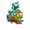
| Initial model | PDB ID: Chain - Chain ID: A |
|---|

