 Movie
Movie Controller


+ Open data

- Basic information
| Entry | Database: PDB / ID: 7yqm | |||||||||||||||||||||||||||||||||||||||||||||||||||||||||||||||||||||||||||||||||
|---|---|---|---|---|---|---|---|---|---|---|---|---|---|---|---|---|---|---|---|---|---|---|---|---|---|---|---|---|---|---|---|---|---|---|---|---|---|---|---|---|---|---|---|---|---|---|---|---|---|---|---|---|---|---|---|---|---|---|---|---|---|---|---|---|---|---|---|---|---|---|---|---|---|---|---|---|---|---|---|---|---|---|
| Title | 2.9-angstrom cryo-EM structure of Ecoli malate synthase G | |||||||||||||||||||||||||||||||||||||||||||||||||||||||||||||||||||||||||||||||||
 Components Components | Malate synthase G | |||||||||||||||||||||||||||||||||||||||||||||||||||||||||||||||||||||||||||||||||
 Keywords Keywords | BIOSYNTHETIC PROTEIN / glyoxylate / malate / MSG / citric acid cycel / malate synthase G | |||||||||||||||||||||||||||||||||||||||||||||||||||||||||||||||||||||||||||||||||
| Function / homology |  Function and homology information Function and homology informationmalate synthase / malate synthase activity / glyoxylate catabolic process / glyoxylate cycle / tricarboxylic acid cycle / magnesium ion binding / cytosol Similarity search - Function | |||||||||||||||||||||||||||||||||||||||||||||||||||||||||||||||||||||||||||||||||
| Biological species |  | |||||||||||||||||||||||||||||||||||||||||||||||||||||||||||||||||||||||||||||||||
| Method | ELECTRON MICROSCOPY / single particle reconstruction / cryo EM / Resolution: 2.89 Å | |||||||||||||||||||||||||||||||||||||||||||||||||||||||||||||||||||||||||||||||||
 Authors Authors | Wu, K.-P. / Wu, Y.-M. / Lu, Y.-C. | |||||||||||||||||||||||||||||||||||||||||||||||||||||||||||||||||||||||||||||||||
| Funding support |  Taiwan, 1items Taiwan, 1items
| |||||||||||||||||||||||||||||||||||||||||||||||||||||||||||||||||||||||||||||||||
 Citation Citation | Journal: J Struct Biol / Year: 2023 Title: Cryo-EM reveals the structure and dynamics of a 723-residue malate synthase G. Authors: Meng-Ru Ho / Yi-Ming Wu / Yen-Chen Lu / Tzu-Ping Ko / Kuen-Phon Wu / Abstract: Determination of sub-100 kDa (kDa) structures by cryo-electron microscopy (EM) is a longstanding but not straightforward goal. Here, we present a 2.9-Å cryo-EM structure of a 723-amino acid apo- ...Determination of sub-100 kDa (kDa) structures by cryo-electron microscopy (EM) is a longstanding but not straightforward goal. Here, we present a 2.9-Å cryo-EM structure of a 723-amino acid apo-form malate synthase G (MSG) from Escherichia coli. The cryo-EM structure of the 82-kDa MSG exhibits the same global folding as structures resolved by crystallography and nuclear magnetic resonance (NMR) spectroscopy, and the crystal and cryo-EM structures are indistinguishable. Analyses of MSG dynamics reveal consistent conformational flexibilities among the three experimental approaches, most notably that the α/β domain exhibits structural heterogeneity. We observed that sidechains of F453, L454, M629, and E630 residues involved in hosting the cofactor acetyl-CoA and substrate rotate differently between the cryo-EM apo-form and complex crystal structures. Our work demonstrates that the cryo-EM technique can be used to determine structures and conformational heterogeneity of sub-100 kDa biomolecules to a quality as high as that obtained from X-ray crystallography and NMR spectroscopy. | |||||||||||||||||||||||||||||||||||||||||||||||||||||||||||||||||||||||||||||||||
| History |
|
- Structure visualization
| Structure viewer | Molecule: MolmilJmol/JSmol |
|---|
- Downloads & links
-Download
| PDBx/mmCIF format | 7yqm.cif.gz | 132.2 KB | Display | PDBx/mmCIF format |
|---|---|---|---|---|
| PDB format | pdb7yqm.ent.gz | 100.7 KB | Display | PDB format |
| PDBx/mmJSON format | 7yqm.json.gz | Tree view | PDBx/mmJSON format | |
| Others |  Other downloads Other downloads |
-Validation report
| Arichive directory | https://data.pdbj.org/pub/pdb/validation_reports/yq/7yqmftp://data.pdbj.org/pub/pdb/validation_reports/yq/7yqm | HTTPS FTP |
|---|
-Related structure data
| Related structure data | 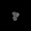 34029MC 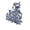 7yqnC M: map data used to model this data C: citing same article ( |
|---|---|
| Similar structure data |

-Links
 PDBj
PDBj- Assembly
| Deposited unit | 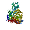
|
|---|---|
| 1 |
|
-Components
| #1: Protein | Mass: 80581.344 Da / Num. of mol.: 1 Source method: isolated from a genetically manipulated source Source: (gene. exp.) |
|---|---|
| Has protein modification | N |
-Experimental details
-Experiment
| Experiment | Method: ELECTRON MICROSCOPY |
|---|---|
| EM experiment | Aggregation state: PARTICLE / 3D reconstruction method: single particle reconstruction |
- Sample preparation
| Component | Name: 2.9-angstrom cryo-EM structure of 723-aa malate synthase G Type: CELL / Entity ID: all / Source: RECOMBINANT |
|---|---|
| Source (natural) | Organism: |
| Source (recombinant) | Organism: |
| Buffer solution | pH: 7.6 |
| Specimen | Conc.: 0.3 mg/ml / Embedding applied: NO / Shadowing applied: NO / Staining applied: NO / Vitrification applied: YES |
| Specimen support | Grid material: COPPER / Grid mesh size: 300 divisions/in. |
| Vitrification | Cryogen name: ETHANE |
- Electron microscopy imaging
| Experimental equipment |  Model: Titan Krios / Image courtesy: FEI Company |
|---|---|
| Microscopy | Model: FEI TITAN KRIOS |
| Electron gun | Electron source:  FIELD EMISSION GUN / Accelerating voltage: 300 kV / Illumination mode: FLOOD BEAM FIELD EMISSION GUN / Accelerating voltage: 300 kV / Illumination mode: FLOOD BEAM |
| Electron lens | Mode: BRIGHT FIELD / Nominal defocus max: 1800 nm / Nominal defocus min: 1000 nm |
| Image recording | Electron dose: 51.2 e/Å2 / Detector mode: COUNTING / Film or detector model: GATAN K2 QUANTUM (4k x 4k) |
- Processing
| Software | Name: PHENIX / Version: 1.19.2_4158: / Classification: refinement | |||||||||||||||||||||||||||
|---|---|---|---|---|---|---|---|---|---|---|---|---|---|---|---|---|---|---|---|---|---|---|---|---|---|---|---|---|
| EM software |
| |||||||||||||||||||||||||||
| CTF correction | Type: NONE | |||||||||||||||||||||||||||
| Symmetry | Point symmetry: C1 (asymmetric) | |||||||||||||||||||||||||||
| 3D reconstruction | Resolution: 2.89 Å / Resolution method: FSC 0.143 CUT-OFF / Num. of particles: 211582 / Algorithm: FOURIER SPACE / Symmetry type: POINT | |||||||||||||||||||||||||||
| Atomic model building | PDB-ID: 1P7T Pdb chain-ID: A / Accession code: 1P7T / Source name: PDB / Type: experimental model | |||||||||||||||||||||||||||
| Refine LS restraints |
|

