[日本語] English
- EMDB-33540: Cryo-EM structure of the Mycobacterium smegmatis DNA integrity sc... -

+
データを開く


IDまたはキーワード:

読み込み中...

-
基本情報

登録情報
データベース: EMDB / ID: EMD-33540
タイトルCryo-EM structure of the Mycobacterium smegmatis DNA integrity scanning protein (MsDisA).
マップデータCombined map and sharpened with Bfactor of -50 in Relion
試料
  • 複合体: Cryo-EM structure of the Mycobacterium smegmatis DNA integrity scanning protein (MsDisA).
    • タンパク質・ペプチド: DNA integrity scanning protein DisA
キーワードsecond messengers / stress response / c-di-AMP / cryo-EM / CELL CYCLE
機能・相同性
機能・相同性情報


: / diadenylate cyclase activity / diadenylate cyclase / DNA repair / DNA binding / ATP binding
類似検索 - 分子機能
DNA integrity scanning, DisA, linker region / DNA integrity scanning protein, DisA / DisA, linker domain superfamily / DisA bacterial checkpoint controller linker region / DNA integrity scanning protein, DisA, N-terminal / DNA integrity scanning protein, DisA, N-terminal domain superfamily / DisA bacterial checkpoint controller nucleotide-binding / Diadenylate cyclase (DAC) domain profile. / DisA/LigA, helix-hairpin-helix motif / Helix-hairpin-helix motif / RuvA domain 2-like
類似検索 - ドメイン・相同性
DNA integrity scanning protein DisA
類似検索 - 構成要素
生物種Mycolicibacterium smegmatis MC2 155 (バクテリア)
手法単粒子再構成法 / クライオ電子顕微鏡法 / 解像度: 3.1 Å
データ登録者Gautam S / Vinothkumar KR / Chatterji D
資金援助 インド, 2件
OrganizationGrant number
Science and Engineering Research Board (SERB)RJN-094/2017 インド
Department of Biotechnology (DBT, India)DBT/PR12422/MED/31/287/2014 インド
引用ジャーナル: Protein Sci / : 2023
タイトル: Regulatory mechanisms of c-di-AMP synthase from Mycobacterium smegmatis revealed by a structure: Function analysis.
著者: Sudhanshu Gautam / Avisek Mahapa / Lahari Yeramala / Apoorv Gandhi / Sushma Krishnan / Vinothkumar Kutti R / Dipankar Chatterji /
要旨: Cyclic-di-nucleotide-based secondary messengers regulate various physiological functions, including stress responses in bacteria. Cyclic diadenosine monophosphate (c-di-AMP) has recently emerged as a ...Cyclic-di-nucleotide-based secondary messengers regulate various physiological functions, including stress responses in bacteria. Cyclic diadenosine monophosphate (c-di-AMP) has recently emerged as a crucial second messenger with implications in processes including osmoregulation, antibiotic resistance, biofilm formation, virulence, DNA repair, ion homeostasis, and sporulation, and has potential therapeutic applications. The contrasting activities of the enzymes diadenylate cyclase (DAC) and phosphodiesterase (PDE) determine the equilibrium levels of c-di-AMP. Although c-di-AMP is suspected of playing an essential role in the pathophysiology of bacterial infections and in regulating host-pathogen interactions, the mechanisms of its regulation remain relatively unexplored in mycobacteria. In this report, we biochemically and structurally characterize the c-di-AMP synthase (MsDisA) from Mycobacterium smegmatis. The enzyme activity is regulated by pH and substrate concentration; conditions of significance in the homoeostasis of c-di-AMP levels. Substrate binding stimulates conformational changes in the protein, and pApA and ppApA are synthetic intermediates detectable when enzyme efficiency is low. Unlike the orthologous Bacillus subtilis enzyme, MsDisA does not bind to, and its activity is not influenced in the presence of DNA. Furthermore, we have determined the cryo-EM structure of MsDisA, revealing asymmetry in its structure in contrast to the symmetric crystal structure of Thermotoga maritima DisA. We also demonstrate that the N-terminal minimal region alone is sufficient and essential for oligomerization and catalytic activity. Our data shed light on the regulation of mycobacterial DisA and possible future directions to pursue.
履歴
登録2022年6月4日-
ヘッダ(付随情報) 公開2023年2月8日-
マップ公開2023年2月8日-
更新2024年7月3日-
現状2024年7月3日処理サイト: PDBj / 状態: 公開

-
構造の表示

添付画像

ダウンロードとリンク

-
マップ

ファイルダウンロード / ファイル: emd_33540.map.gz / 形式: CCP4 / 大きさ: 325 MB / タイプ: IMAGE STORED AS FLOATING POINT NUMBER (4 BYTES)
注釈Combined map and sharpened with Bfactor of -50 in Relion
ボクセルのサイズX=Y=Z: 1.07 Å
密度
表面レベル登録者による: 1.4
最小 - 最大-5.6700954 - 8.803793000000001
平均 (標準偏差)0.0020104963 (±0.12417459)
対称性空間群: 1
詳細

EMDB XML:

マップ形状
Axis orderXYZ
Origin000
サイズ440440440
Spacing440440440
セルA=B=C: 470.80002 Å
α=β=γ: 90.0 °

-
添付データ

-
ハーフマップ: One of the half maps from Cryosparc

ファイルemd_33540_half_map_1.map
注釈One of the half maps from Cryosparc
投影像・断面図
ZYX

投影像

断面 (1/2)
密度ヒストグラム

-
ハーフマップ: One of the half maps from Cryosparc

ファイルemd_33540_half_map_2.map
注釈One of the half maps from Cryosparc
投影像・断面図
ZYX

投影像

断面 (1/2)
密度ヒストグラム

-
試料の構成要素

-
全体 : Cryo-EM structure of the Mycobacterium smegmatis DNA integrity sc...

全体名称: Cryo-EM structure of the Mycobacterium smegmatis DNA integrity scanning protein (MsDisA).
要素
  • 複合体: Cryo-EM structure of the Mycobacterium smegmatis DNA integrity scanning protein (MsDisA).
    • タンパク質・ペプチド: DNA integrity scanning protein DisA

-
超分子 #1: Cryo-EM structure of the Mycobacterium smegmatis DNA integrity sc...

超分子名称: Cryo-EM structure of the Mycobacterium smegmatis DNA integrity scanning protein (MsDisA).
タイプ: complex / ID: 1 / 親要素: 0 / 含まれる分子: all
由来(天然)生物種: Mycolicibacterium smegmatis MC2 155 (バクテリア)
分子量理論値: 330 KDa

-
分子 #1: DNA integrity scanning protein DisA

分子名称: DNA integrity scanning protein DisA / タイプ: protein_or_peptide / ID: 1 / コピー数: 8 / 光学異性体: LEVO / EC番号: diadenylate cyclase
由来(天然)生物種: Mycolicibacterium smegmatis MC2 155 (バクテリア)
分子量理論値: 41.844199 KDa
組換発現生物種: Escherichia coli BL21(DE3) (大腸菌)
配列文字列: MAVKSGARSG RNVVHLARPT LRETLGRLAP GTPLRDGLER ILRGRTGALI VLGYDDSVEA ICDGGFVLDV RYAPTRLREL SKMDGAVVL SSDGSRILRA NVQLVPDPSI PTDESGTRHR SAERTAIQTG YPVISVSHSM SIVTVYVAGE RHVVPDSATI L SRANQTIA ...文字列:
MAVKSGARSG RNVVHLARPT LRETLGRLAP GTPLRDGLER ILRGRTGALI VLGYDDSVEA ICDGGFVLDV RYAPTRLREL SKMDGAVVL SSDGSRILRA NVQLVPDPSI PTDESGTRHR SAERTAIQTG YPVISVSHSM SIVTVYVAGE RHVVPDSATI L SRANQTIA TLERYKGRLD EVSRQLSTAE IEDFVTLRDV MTVVQRLEMV RRISLEIDAD VVELGTDGRQ LKLQLDELVG DN ETARELI VRDYHANPDP PTAAQVAATL EELDSLSDSE LLDFTVLARV FGYPSTAEAQ DSAMSSRGYR AMAAIPRLQF AHV DLLVRS FGSLQNLLAA SADDLQSVDG IGSMWARHIR EGLSLLAEST IADRLAAAAL EHHHHHH

UniProtKB: DNA integrity scanning protein DisA

-
実験情報

-
構造解析

手法クライオ電子顕微鏡法
解析単粒子再構成法
試料の集合状態particle

-
試料調製

濃度0.1 mg/mL
緩衝液pH: 7.5 / 構成要素 - 濃度: 75.0 mM / 構成要素 - 式: NaCl / 構成要素 - 名称: Sodium Chloride
詳細: Buffer were made freshly with 75mM NaCl and 50mM Tris (pH 7.5)
グリッドモデル: Quantifoil R2/2 / 材質: COPPER / メッシュ: 300 / 支持フィルム - 材質: CARBON / 支持フィルム - トポロジー: HOLEY / 前処理 - タイプ: GLOW DISCHARGE / 前処理 - 雰囲気: AIR
凍結凍結剤: ETHANE / チャンバー内湿度: 100 % / 装置: FEI VITROBOT MARK IV / 詳細: blotted for 3.5s.
詳細The protein sample was made on a holey carbon grid with an additional layer of thin carbon.

-
電子顕微鏡法

顕微鏡FEI TITAN KRIOS
撮影フィルム・検出器のモデル: FEI FALCON III (4k x 4k)
検出モード: COUNTING / デジタル化 - サイズ - 横: 4096 pixel / デジタル化 - サイズ - 縦: 4096 pixel / 撮影したグリッド数: 1 / 実像数: 1587 / 平均露光時間: 60.0 sec. / 平均電子線量: 27.75 e/Å2
電子線加速電圧: 300 kV / 電子線源: FIELD EMISSION GUN
電子光学系C2レンズ絞り径: 50.0 µm / 倍率(補正後): 130841 / 照射モード: FLOOD BEAM / 撮影モード: BRIGHT FIELD / Cs: 2.7 mm
最大 デフォーカス(公称値): 3.3000000000000003 µm
最小 デフォーカス(公称値): 2.1 µm / 倍率(公称値): 75000
試料ステージ試料ホルダーモデル: FEI TITAN KRIOS AUTOGRID HOLDER
ホルダー冷却材: NITROGEN
実験機器
モデル: Titan Krios / 画像提供: FEI Company

+
画像解析

粒子像選択
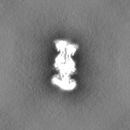
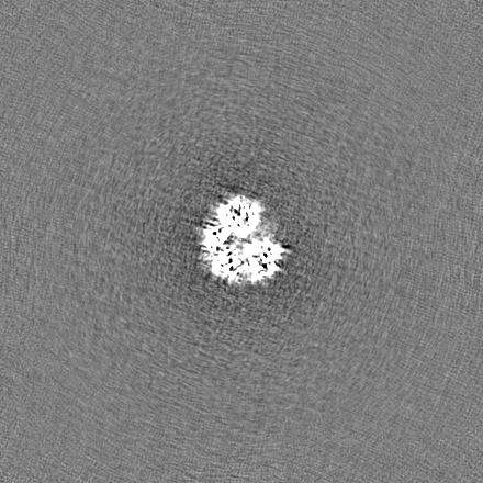
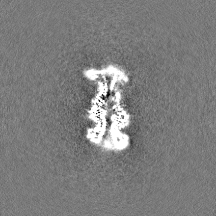
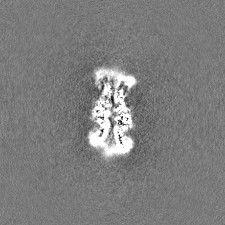
選択した数: 973755
詳細: Template Picker from cryoSparc 3.0 were used to pick total particles in the first extraction. Three rounds of reference-free 2d classification were carried to remove bad particles followed by NU-refinement.
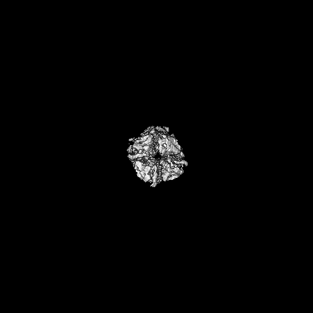
初期モデルモデルのタイプ: OTHER / 詳細: Initial model was obtained with cryosparc 3.0.
最終 再構成使用したクラス数: 4 / 想定した対称性 - 点群: C1 (非対称) / アルゴリズム: BACK PROJECTION / 解像度のタイプ: BY AUTHOR / 解像度: 3.1 Å / 解像度の算出法: FSC 0.143 CUT-OFF / ソフトウェア - 名称: cryoSPARC (ver. 3.0) / 使用した粒子像数: 204932
初期 角度割当タイプ: MAXIMUM LIKELIHOOD / ソフトウェア - 名称: cryoSPARC (ver. 3.0)
最終 角度割当タイプ: MAXIMUM LIKELIHOOD / ソフトウェア - 名称: cryoSPARC (ver. 3.0)
FSC曲線 (解像度の算出)

-
原子モデル構築 1

詳細Model from AlphaFold was used as the starting point (AF-A0R564-F1-model_v2.pdb).
精密化空間: REAL / プロトコル: OTHER / 温度因子: 138.7
得られたモデル

PDB-7y0d:
Cryo-EM structure of the Mycobacterium smegmatis DNA integrity scanning protein (MsDisA).

+
万見について

-
お知らせ

-
2022年2月9日: EMDBエントリの付随情報ファイルのフォーマットが新しくなりました

EMDBエントリの付随情報ファイルのフォーマットが新しくなりました

  • EMDBのヘッダファイルのバージョン3が、公式のフォーマットとなりました。
  • これまでは公式だったバージョン1.9は、アーカイブから削除されます。

関連情報:EMDBヘッダ

外部リンク:wwPDBはEMDBデータモデルのバージョン3へ移行します

-
2020年8月12日: 新型コロナ情報

新型コロナ情報

URL: https://pdbjlvh1.pdbj.org/emnavi/covid19.php

新ページ: EM Navigatorに新型コロナウイルスの特設ページを開設しました。

関連情報:Covid-19情報 / 2020年3月5日: 新型コロナウイルスの構造データ

+
2020年3月5日: 新型コロナウイルスの構造データ

新型コロナウイルスの構造データ

関連情報:万見生物種 / 2020年8月12日: 新型コロナ情報

外部リンク:COVID-19特集ページ - PDBj / 今月の分子2020年2月:コロナウイルスプロテーアーゼ

+
2019年1月31日: EMDBのIDの桁数の変更

EMDBのIDの桁数の変更

  • EMDBエントリに付与されているアクセスコード(EMDB-ID)は4桁の数字(例、EMD-1234)でしたが、間もなく枯渇します。これまでの4桁のID番号は4桁のまま変更されませんが、4桁の数字を使い切った後に発行されるIDは5桁以上の数字(例、EMD-12345)になります。5桁のIDは2019年の春頃から発行される見通しです。
  • EM Navigator/万見では、接頭語「EMD-」は省略されています。

関連情報:Q: 「EMD」とは何ですか? / 万見/EM NavigatorにおけるID/アクセスコードの表記

外部リンク:EMDB Accession Codes are Changing Soon! / PDBjへお問い合わせ

+
2017年7月12日: PDB大規模アップデート

PDB大規模アップデート

  • 新バージョンのPDBx/mmCIF辞書形式に基づくデータがリリースされました。
  • 今回の更新はバージョン番号が4から5になる大規模なもので、全エントリデータの書き換えが行われる「Remediation」というアップデートに該当します。
  • このバージョンアップで、電子顕微鏡の実験手法に関する多くの項目の書式が改定されました(例:em_softwareなど)。
  • EM NavigatorとYorodumiでも、この改定に基づいた表示内容になります。

外部リンク:wwPDB Remediation / OneDepデータ基準に準拠した、より強化された内容のモデル構造ファイルが、PDBアーカイブで公開されました。

-
万見 (Yorodumi)

幾万の構造データを、幾万の視点から

  • 万見(Yorodumi)は、EMDB/PDB/SASBDBなどの構造データを閲覧するためのページです。
  • EM Navigatorの詳細ページの後継、Omokage検索のフロントエンドも兼ねています。

関連情報:EMDB / PDB / SASBDB / 3つのデータバンクの比較 / 万見検索 / 2016年8月31日: 新しいEM Navigatorと万見 / 万見文献 / Jmol/JSmol / 機能・相同性情報 / 新しいEM Navigatorと万見の変更点

他の情報も見る