+ データを開く
- 基本情報
基本情報
| 登録情報 | データベース: EMDB / ID: EMD-3347 | |||||||||
|---|---|---|---|---|---|---|---|---|---|---|
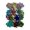
| タイトル | Volta phase plate 3.2A resolution reconstruction of the T20S proteasome | |||||||||
 マップデータ マップデータ | Volta phase plate cryo-EM reconstruction of T20S proteasome | |||||||||
 試料 試料 |
| |||||||||
 キーワード キーワード | proteasome / 20S | |||||||||
| 生物種 |   Thermoplasma acidophilum (好酸性) Thermoplasma acidophilum (好酸性) | |||||||||
| 手法 | 単粒子再構成法 / クライオ電子顕微鏡法 / 解像度: 3.2 Å | |||||||||
 データ登録者 データ登録者 | Danev R / Baumeister W | |||||||||
 引用 引用 |  ジャーナル: Elife / 年: 2016 ジャーナル: Elife / 年: 2016タイトル: Cryo-EM single particle analysis with the Volta phase plate. 著者: Radostin Danev / Wolfgang Baumeister /  要旨: We present a method for in-focus data acquisition with a phase plate that enables near-atomic resolution single particle reconstructions. Accurate focusing is the determining factor for obtaining ...We present a method for in-focus data acquisition with a phase plate that enables near-atomic resolution single particle reconstructions. Accurate focusing is the determining factor for obtaining high quality data. A double-area focusing strategy was implemented in order to achieve the required precision. With this approach we obtained a 3.2 Å resolution reconstruction of the Thermoplasma acidophilum 20S proteasome. The phase plate matches or slightly exceeds the performance of the conventional defocus approach. Spherical aberration becomes a limiting factor for achieving resolutions below 3 Å with in-focus phase plate images. The phase plate could enable single particle analysis of challenging samples in terms of small size, heterogeneity and flexibility that are difficult to solve by the conventional defocus approach. | |||||||||
| 履歴 |
|
- 構造の表示
構造の表示
| ムービー |
 ムービービューア ムービービューア |
|---|---|
| 構造ビューア | EMマップ:  SurfView SurfView Molmil Molmil Jmol/JSmol Jmol/JSmol |
| 添付画像 |
- ダウンロードとリンク
-EMDBアーカイブ
| マップデータ |  emd_3347.map.gz emd_3347.map.gz | 19.4 MB |  EMDBマップデータ形式 EMDBマップデータ形式 | |
|---|---|---|---|---|
| ヘッダ (付随情報) |  emd-3347-v30.xml emd-3347-v30.xml emd-3347.xml emd-3347.xml | 11.1 KB 11.1 KB | 表示 表示 |  EMDBヘッダ EMDBヘッダ |
| FSC (解像度算出) |  emd_3347_fsc.xml emd_3347_fsc.xml | 7.3 KB | 表示 |  FSCデータファイル FSCデータファイル |
| 画像 |  emd_3347.png emd_3347.png | 268.5 KB | ||
| マスクデータ |  emd_3347_msk_1.map emd_3347_msk_1.map | 20.8 MB |  マスクマップ マスクマップ | |
| アーカイブディレクトリ |  http://ftp.pdbj.org/pub/emdb/structures/EMD-3347 http://ftp.pdbj.org/pub/emdb/structures/EMD-3347 ftp://ftp.pdbj.org/pub/emdb/structures/EMD-3347 ftp://ftp.pdbj.org/pub/emdb/structures/EMD-3347 | HTTPS FTP |
-検証レポート
| 文書・要旨 |  emd_3347_validation.pdf.gz emd_3347_validation.pdf.gz | 381.6 KB | 表示 |  EMDB検証レポート EMDB検証レポート |
|---|---|---|---|---|
| 文書・詳細版 |  emd_3347_full_validation.pdf.gz emd_3347_full_validation.pdf.gz | 380.7 KB | 表示 | |
| XML形式データ |  emd_3347_validation.xml.gz emd_3347_validation.xml.gz | 9.2 KB | 表示 | |
| アーカイブディレクトリ |  https://ftp.pdbj.org/pub/emdb/validation_reports/EMD-3347 https://ftp.pdbj.org/pub/emdb/validation_reports/EMD-3347 ftp://ftp.pdbj.org/pub/emdb/validation_reports/EMD-3347 ftp://ftp.pdbj.org/pub/emdb/validation_reports/EMD-3347 | HTTPS FTP |
-関連構造データ
| 関連構造データ |  3348C C: 同じ文献を引用 ( |
|---|---|
| 類似構造データ | |
| 電子顕微鏡画像生データ |  EMPIAR-10057 (タイトル: Volta phase plate in-focus dataset of T20S proteasome EMPIAR-10057 (タイトル: Volta phase plate in-focus dataset of T20S proteasomeData size: 50.3 Data #1: Volta phase plate in-focus unaligned multi-frame micrographs of T20S proteasome [micrographs - multiframe]) |
- リンク
リンク
| EMDBのページ |  EMDB (EBI/PDBe) / EMDB (EBI/PDBe) /  EMDataResource EMDataResource |
|---|
- マップ
マップ
| ファイル |  ダウンロード / ファイル: emd_3347.map.gz / 形式: CCP4 / 大きさ: 20.3 MB / タイプ: IMAGE STORED AS FLOATING POINT NUMBER (4 BYTES) ダウンロード / ファイル: emd_3347.map.gz / 形式: CCP4 / 大きさ: 20.3 MB / タイプ: IMAGE STORED AS FLOATING POINT NUMBER (4 BYTES) | ||||||||||||||||||||||||||||||||||||||||||||||||||||||||||||||||||||
|---|---|---|---|---|---|---|---|---|---|---|---|---|---|---|---|---|---|---|---|---|---|---|---|---|---|---|---|---|---|---|---|---|---|---|---|---|---|---|---|---|---|---|---|---|---|---|---|---|---|---|---|---|---|---|---|---|---|---|---|---|---|---|---|---|---|---|---|---|---|
| 注釈 | Volta phase plate cryo-EM reconstruction of T20S proteasome | ||||||||||||||||||||||||||||||||||||||||||||||||||||||||||||||||||||
| 投影像・断面図 | 画像のコントロール
画像は Spider により作成 | ||||||||||||||||||||||||||||||||||||||||||||||||||||||||||||||||||||
| ボクセルのサイズ | X=Y=Z: 1.35 Å | ||||||||||||||||||||||||||||||||||||||||||||||||||||||||||||||||||||
| 密度 |
| ||||||||||||||||||||||||||||||||||||||||||||||||||||||||||||||||||||
| 対称性 | 空間群: 1 | ||||||||||||||||||||||||||||||||||||||||||||||||||||||||||||||||||||
| 詳細 | EMDB XML:
CCP4マップ ヘッダ情報:
| ||||||||||||||||||||||||||||||||||||||||||||||||||||||||||||||||||||
-添付データ
-セグメンテーションマップ: Automatically generated mask during postprocessing in Relion.
| 注釈 | Automatically generated mask during postprocessing in Relion. | ||||||||||||
|---|---|---|---|---|---|---|---|---|---|---|---|---|---|
| ファイル |  emd_3347_msk_1.map emd_3347_msk_1.map | ||||||||||||
| 投影像・断面図 |
| ||||||||||||
| 密度ヒストグラム |
- 試料の構成要素
-全体 : Thermoplasma acidophilum 20S proteasome
| 全体 | 名称: Thermoplasma acidophilum 20S proteasome |
|---|---|
| 要素 |
|
-超分子 #1000: Thermoplasma acidophilum 20S proteasome
| 超分子 | 名称: Thermoplasma acidophilum 20S proteasome / タイプ: sample / ID: 1000 / 集合状態: 28-mer / Number unique components: 1 |
|---|---|
| 分子量 | 理論値: 700 KDa |
-分子 #1: 20S proteasome
| 分子 | 名称: 20S proteasome / タイプ: protein_or_peptide / ID: 1 / コピー数: 1 / 集合状態: 28-mer / 組換発現: Yes |
|---|---|
| 由来(天然) | 生物種:   Thermoplasma acidophilum (好酸性) / 別称: Thermoplasma Thermoplasma acidophilum (好酸性) / 別称: Thermoplasma |
| 分子量 | 理論値: 700 KDa |
| 組換発現 | 生物種:  |
-実験情報
-構造解析
| 手法 | クライオ電子顕微鏡法 |
|---|---|
 解析 解析 | 単粒子再構成法 |
| 試料の集合状態 | particle |
- 試料調製
試料調製
| 濃度 | 0.5 mg/mL |
|---|---|
| 緩衝液 | pH: 7 / 詳細: 25 mM Tris-HCL |
| グリッド | 詳細: Quantifoil R 1.2/1.3 |
| 凍結 | 凍結剤: ETHANE-PROPANE MIXTURE / チャンバー内湿度: 95 % / チャンバー内温度: 80 K / 装置: FEI VITROBOT MARK III 手法: 3 ul sample on Quantifoil R1.2/1.3, Vitrobot chamber temperature 20 degC, blot time 5s. |
- 電子顕微鏡法
| 顕微鏡 | FEI TITAN KRIOS |
|---|---|
| 温度 | 平均: 80 K |
| アライメント法 | Legacy - 非点収差: Objective lens astigmatis was corrected manually at the same mag. as the data acquisition. |
| 特殊光学系 | エネルギーフィルター - 名称: Gatan Quantum エネルギーフィルター - エネルギー下限: 0.0 eV エネルギーフィルター - エネルギー上限: 10.0 eV |
| 日付 | 2015年7月21日 |
| 撮影 | カテゴリ: CCD フィルム・検出器のモデル: GATAN K2 SUMMIT (4k x 4k) デジタル化 - サンプリング間隔: 5 µm / 実像数: 158 / 平均電子線量: 30 e/Å2 / 詳細: every image consisted of 12 frames 0.5 s each. |
| 電子線 | 加速電圧: 300 kV / 電子線源:  FIELD EMISSION GUN FIELD EMISSION GUN |
| 電子光学系 | 倍率(補正後): 37037 / 照射モード: FLOOD BEAM / 撮影モード: BRIGHT FIELD / Cs: 2.7 mm / 最大 デフォーカス(公称値): 0.02 µm / 最小 デフォーカス(公称値): 0.02 µm / 倍率(公称値): 37000 |
| 試料ステージ | 試料ホルダーモデル: FEI TITAN KRIOS AUTOGRID HOLDER |
| 実験機器 |  モデル: Titan Krios / 画像提供: FEI Company |
 ムービー
ムービー コントローラー


 Z (Sec.)
 Y (Row.)
 X (Col.)


