 Movie
Movie Controller

[English] 日本語
 Yorodumi
- EMDB-33334: Minor polymorph in alpha-synuclein fibril seeded by cerebrospinal... -
+ Open data

- Basic information
| Entry | 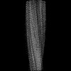 | ||||||||||||||||||||||||
|---|---|---|---|---|---|---|---|---|---|---|---|---|---|---|---|---|---|---|---|---|---|---|---|---|---|
| Title | Minor polymorph in alpha-synuclein fibril seeded by cerebrospinal fluid from a mid-to-late stage (mid-PD-4) Parkinson's disease patient | ||||||||||||||||||||||||
 Map data Map data | |||||||||||||||||||||||||
 Sample Sample |
| ||||||||||||||||||||||||
 Keywords Keywords | amyloid fibril / PROTEIN FIBRIL | ||||||||||||||||||||||||
| Function / homology |  Function and homology information Function and homology informationnegative regulation of mitochondrial electron transport, NADH to ubiquinone / negative regulation of dopamine uptake involved in synaptic transmission / negative regulation of norepinephrine uptake / response to desipramine / positive regulation of SNARE complex assembly / positive regulation of hydrogen peroxide catabolic process / supramolecular fiber / regulation of synaptic vesicle recycling / negative regulation of chaperone-mediated autophagy / regulation of reactive oxygen species biosynthetic process ...negative regulation of mitochondrial electron transport, NADH to ubiquinone / negative regulation of dopamine uptake involved in synaptic transmission / negative regulation of norepinephrine uptake / response to desipramine / positive regulation of SNARE complex assembly / positive regulation of hydrogen peroxide catabolic process / supramolecular fiber / regulation of synaptic vesicle recycling / negative regulation of chaperone-mediated autophagy / regulation of reactive oxygen species biosynthetic process / positive regulation of protein localization to cell periphery / negative regulation of exocytosis / dopamine biosynthetic process / dopamine uptake involved in synaptic transmission / response to iron(II) ion / negative regulation of dopamine metabolic process / negative regulation of platelet-derived growth factor receptor signaling pathway / SNARE complex assembly / negative regulation of thrombin-activated receptor signaling pathway / Lewy body / negative regulation of microtubule polymerization / synaptic vesicle priming / synaptic vesicle transport / regulation of norepinephrine uptake / transporter regulator activity / protein kinase inhibitor activity / positive regulation of inositol phosphate biosynthetic process / regulation of dopamine secretion / positive regulation of receptor recycling / cuprous ion binding / positive regulation of exocytosis / nuclear outer membrane / dynein complex binding / synaptic transmission, dopaminergic / synaptic vesicle exocytosis / response to magnesium ion / positive regulation of endocytosis / negative regulation of serotonin uptake / cysteine-type endopeptidase inhibitor activity / kinesin binding / regulation of presynapse assembly / synaptic vesicle endocytosis / alpha-tubulin binding / beta-tubulin binding / phospholipase binding / behavioral response to cocaine / supramolecular fiber organization / cellular response to fibroblast growth factor stimulus / response to type II interferon / cellular response to epinephrine stimulus / inclusion body / Hsp70 protein binding / response to interleukin-1 / cellular response to copper ion / axon terminus / positive regulation of release of sequestered calcium ion into cytosol / enzyme inhibitor activity / glutathione metabolic process / regulation of microtubule cytoskeleton organization / SNARE binding / protein tetramerization / protein sequestering activity / microglial cell activation / phosphoprotein binding / receptor internalization / tubulin binding / protein destabilization / ferrous iron binding / synapse organization / phospholipid binding / PKR-mediated signaling / tau protein binding / enzyme activator activity / positive regulation of inflammatory response / terminal bouton / actin cytoskeleton / synaptic vesicle membrane / growth cone / actin binding / cellular response to oxidative stress / response to lipopolysaccharide / histone binding / cell cortex / microtubule binding / negative regulation of neuron apoptotic process / amyloid fibril formation / mitochondrial outer membrane / lysosome / oxidoreductase activity / mitochondrial inner membrane / transcription cis-regulatory region binding / positive regulation of apoptotic process / ribosome / mitochondrial matrix / Amyloid fiber formation / copper ion binding / protein domain specific binding / axon / neuronal cell body / calcium ion binding Similarity search - Function | ||||||||||||||||||||||||
| Biological species |  Homo sapiens (human) Homo sapiens (human) | ||||||||||||||||||||||||
| Method | helical reconstruction / cryo EM / Resolution: 3.0 Å | ||||||||||||||||||||||||
 Authors Authors | Fan Y / Sun YP / Wang J / Liu C | ||||||||||||||||||||||||
| Funding support |  China, 7 items China, 7 items
| ||||||||||||||||||||||||
 Citation Citation | Journal: Structure / Year: 2023 Title: Conformational change of α-synuclein fibrils in cerebrospinal fluid from different clinical phases of Parkinson's disease. Authors: Yun Fan / Yunpeng Sun / Wenbo Yu / Youqi Tao / Wencheng Xia / Yiqi Liu / Qinyue Zhao / Yilin Tang / Yimin Sun / Fengtao Liu / Qin Cao / Jianjun Wu / Cong Liu / Jian Wang / Dan Li / Abstract: α-Synuclein (α-syn) has been shown to form various conformational fibrils associated with different synucleinopathies. But whether the conformation of α-syn fibrils changes during disease ...α-Synuclein (α-syn) has been shown to form various conformational fibrils associated with different synucleinopathies. But whether the conformation of α-syn fibrils changes during disease progression is unclear. Here, we amplified α-syn aggregates from the cerebrospinal fluid (CSF) of patients with Parkinson's disease (PD) staged in preclinical PD (pre-PD), middle- to late-stage PD (mid-PD), and late-stage PD (late-PD). Our results show that α-syn fibrils derived from the late-PD patient are most potent in inducing endogenous α-syn aggregation in primary neurons, followed by the mid-PD and pre-PD fibrils. By using cryo-electron microscopy, we further determined the high-resolution structures of the CSF-amplified fibrils. The structures exhibit remarkable differences in a minor but significant population of conformational species in different staged samples. Our work demonstrates structural and pathological differences between α-syn fibrils derived from PD patients at a spectrum of clinical stages, which suggests potential conformational transition of α-syn fibrils during the progression of PD. | ||||||||||||||||||||||||
| History |
|
- Structure visualization
| Supplemental images |
|---|

- Downloads & links
-EMDB archive
| Map data | emd_33334.map.gz | 13.5 MB | EMDB map data format | |
|---|---|---|---|---|
| Header (meta data) | emd-33334-v30.xmlemd-33334.xml | 15.4 KB 15.4 KB | Display Display | EMDB header |
| FSC (resolution estimation) | emd_33334_fsc.xml | 10.2 KB | Display | FSC data file |
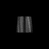
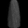
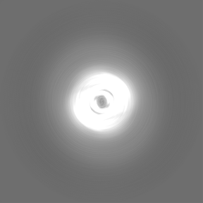
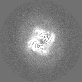
| Images |  emd_33334.png emd_33334.png | 38.3 KB | ||
| Filedesc metadata | emd-33334.cif.gz | 5.2 KB | ||
| Others | emd_33334_half_map_1.map.gzemd_33334_half_map_2.map.gz | 70.8 MB 70.8 MB | ||
| Archive directory |  http://ftp.pdbj.org/pub/emdb/structures/EMD-33334ftp://ftp.pdbj.org/pub/emdb/structures/EMD-33334 http://ftp.pdbj.org/pub/emdb/structures/EMD-33334ftp://ftp.pdbj.org/pub/emdb/structures/EMD-33334 | HTTPS FTP |
-Related structure data
| Related structure data |  7xo2MC  7v47C  7v48C  7v49C  7xo0C  7xo1C  7xo3C  8h03C  8h04C  8h05C M: atomic model generated by this map C: citing same article ( |
|---|---|
| Similar structure data |

-Links
| EMDB pages | EMDB (EBI/PDBe) / EMDataResource |
|---|
-Map
| File | Download / File: emd_33334.map.gz / Format: CCP4 / Size: 91.1 MB / Type: IMAGE STORED AS FLOATING POINT NUMBER (4 BYTES) | ||||||||||||||||||||||||||||||||||||
|---|---|---|---|---|---|---|---|---|---|---|---|---|---|---|---|---|---|---|---|---|---|---|---|---|---|---|---|---|---|---|---|---|---|---|---|---|---|
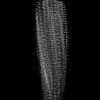
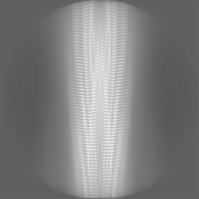
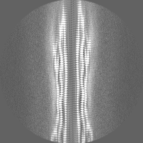
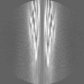
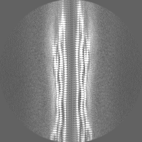
| Projections & slices | Image control
Images are generated by Spider. | ||||||||||||||||||||||||||||||||||||
| Voxel size | X=Y=Z: 1.06 Å | ||||||||||||||||||||||||||||||||||||
| Density |
| ||||||||||||||||||||||||||||||||||||
| Symmetry | Space group: 1 | ||||||||||||||||||||||||||||||||||||
| Details | EMDB XML:
|
 Z (Sec.)
 Y (Row.)
 X (Col.)


-Supplemental data
-Half map: #2
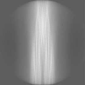
| File | emd_33334_half_map_1.map | ||||||||||||
|---|---|---|---|---|---|---|---|---|---|---|---|---|---|
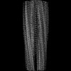
| Projections & Slices |
| ||||||||||||
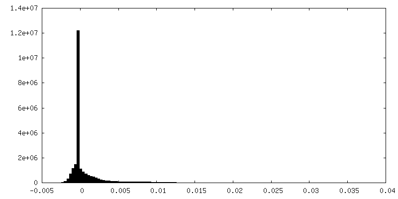
| Density Histograms |

-Half map: #1
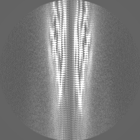
| File | emd_33334_half_map_2.map | ||||||||||||
|---|---|---|---|---|---|---|---|---|---|---|---|---|---|
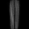
| Projections & Slices |
| ||||||||||||
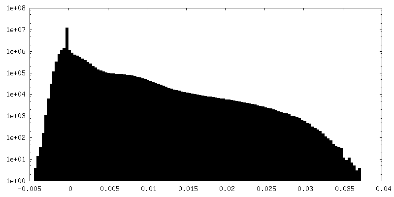
| Density Histograms |


- Sample components
-Entire : Minor polymorph in alpha-synuclein fibril seeded by cerebrospinal...
| Entire | Name: Minor polymorph in alpha-synuclein fibril seeded by cerebrospinal fluid from a mid-to-late stage (mid-PD-4) Parkinson's disease patient |
|---|---|
| Components |
|
-Supramolecule #1: Minor polymorph in alpha-synuclein fibril seeded by cerebrospinal...
| Supramolecule | Name: Minor polymorph in alpha-synuclein fibril seeded by cerebrospinal fluid from a mid-to-late stage (mid-PD-4) Parkinson's disease patient type: organelle_or_cellular_component / ID: 1 / Parent: 0 / Macromolecule list: all |
|---|---|
| Source (natural) | Organism: Homo sapiens (human) |
-Macromolecule #1: Alpha-synuclein
| Macromolecule | Name: Alpha-synuclein / type: protein_or_peptide / ID: 1 / Number of copies: 6 / Enantiomer: LEVO |
|---|---|
| Source (natural) | Organism: Homo sapiens (human) |
| Molecular weight | Theoretical: 14.476108 KDa |
| Recombinant expression | Organism:  |
| Sequence | String: MDVFMKGLSK AKEGVVAAAE KTKQGVAEAA GKTKEGVLYV GSKTKEGVVH GVATVAEKTK EQVTNVGGAV VTGVTAVAQK TVEGAGSIA AATGFVKKDQ LGKNEEGAPQ EGILEDMPVD PDNEAYEMPS EEGYQDYEPE A UniProtKB: Alpha-synuclein |
-Experimental details
-Structure determination
| Method | cryo EM |
|---|---|
 Processing Processing | helical reconstruction |
| Aggregation state | filament |
-Sample preparation
| Buffer | pH: 6.5 |
|---|---|
| Vitrification | Cryogen name: ETHANE |
- Electron microscopy
| Microscope | FEI TITAN KRIOS |
|---|---|
| Image recording | Film or detector model: GATAN K3 (6k x 4k) / Average electron dose: 55.0 e/Å2 |
| Electron beam | Acceleration voltage: 300 kV / Electron source:  FIELD EMISSION GUN FIELD EMISSION GUN |
| Electron optics | Illumination mode: FLOOD BEAM / Imaging mode: BRIGHT FIELD / Nominal defocus max: 2.0 µm / Nominal defocus min: 1.0 µm |
| Experimental equipment |  Model: Titan Krios / Image courtesy: FEI Company |
-Image processing
| Final reconstruction | Applied symmetry - Helical parameters - Δz: 2.42 Å Applied symmetry - Helical parameters - Δ&Phi: 179.34 ° Applied symmetry - Helical parameters - Axial symmetry: C1 (asymmetric) Resolution.type: BY AUTHOR / Resolution: 3.0 Å / Resolution method: FSC 0.143 CUT-OFF / Number images used: 14416 |
|---|---|
| Startup model | Type of model: PDB ENTRY PDB model - PDB ID: |
| Final angle assignment | Type: NOT APPLICABLE |
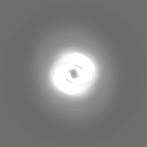
| FSC plot (resolution estimation) | 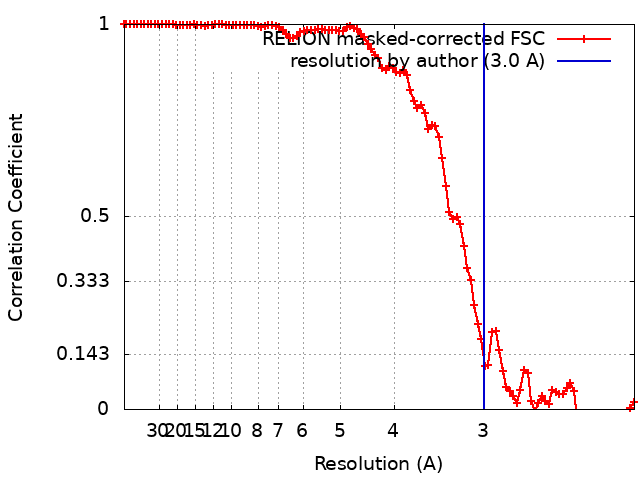 |

