 Movie
Movie Controller


+ Open data

- Basic information
| Entry |  | |||||||||
|---|---|---|---|---|---|---|---|---|---|---|
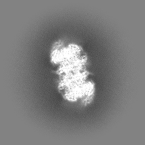
| Title | Cryo-EM map of hMCM-DH R195A/L209G mutant | |||||||||
 Map data Map data | ||||||||||
 Sample Sample |
| |||||||||
| Biological species |  Homo sapiens (human) Homo sapiens (human) | |||||||||
| Method | single particle reconstruction / cryo EM / Resolution: 2.95 Å | |||||||||
 Authors Authors | Li J / Dong JQ / Dang SY / Zhai YL | |||||||||
| Funding support |  Hong Kong, 1 items Hong Kong, 1 items
| |||||||||
 Citation Citation | Journal: Cell / Year: 2023 Title: The human pre-replication complex is an open complex. Authors: Jian Li / Jiangqing Dong / Weitao Wang / Daqi Yu / Xinyu Fan / Yan Chit Hui / Clare S K Lee / Wai Hei Lam / Nathan Alary / Yang Yang / Yingyi Zhang / Qian Zhao / Chun-Long Chen / Bik-Kwoon ...Authors: Jian Li / Jiangqing Dong / Weitao Wang / Daqi Yu / Xinyu Fan / Yan Chit Hui / Clare S K Lee / Wai Hei Lam / Nathan Alary / Yang Yang / Yingyi Zhang / Qian Zhao / Chun-Long Chen / Bik-Kwoon Tye / Shangyu Dang / Yuanliang Zhai /   Abstract: In eukaryotes, DNA replication initiation requires assembly and activation of the minichromosome maintenance (MCM) 2-7 double hexamer (DH) to melt origin DNA strands. However, the mechanism for this ...In eukaryotes, DNA replication initiation requires assembly and activation of the minichromosome maintenance (MCM) 2-7 double hexamer (DH) to melt origin DNA strands. However, the mechanism for this initial melting is unknown. Here, we report a 2.59-Å cryo-electron microscopy structure of the human MCM-DH (hMCM-DH), also known as the pre-replication complex. In this structure, the hMCM-DH with a constricted central channel untwists and stretches the DNA strands such that almost a half turn of the bound duplex DNA is distorted with 1 base pair completely separated, generating an initial open structure (IOS) at the hexamer junction. Disturbing the IOS inhibits DH formation and replication initiation. Mapping of hMCM-DH footprints indicates that IOSs are distributed across the genome in large clusters aligning well with initiation zones designed for stochastic origin firing. This work unravels an intrinsic mechanism that couples DH formation with initial DNA melting to license replication initiation in human cells. | |||||||||
| History |
|
- Structure visualization
| Supplemental images |
|---|

- Downloads & links
-EMDB archive
| Map data | emd_33320.map.gz | 398.9 MB |  EMDB map data format EMDB map data format | |
|---|---|---|---|---|
| Header (meta data) | emd-33320-v30.xmlemd-33320.xml | 25 KB 25 KB | Display Display | EMDB header |
| Images |  emd_33320.png emd_33320.png | 104.5 KB | ||
| Others | emd_33320_additional_1.map.gzemd_33320_additional_2.map.gzemd_33320_additional_3.map.gzemd_33320_additional_4.map.gzemd_33320_half_map_1.map.gzemd_33320_half_map_2.map.gz | 399 MB 398.6 MB 398.5 MB 398.6 MB 392 MB 392 MB | ||
| Archive directory |  http://ftp.pdbj.org/pub/emdb/structures/EMD-33320ftp://ftp.pdbj.org/pub/emdb/structures/EMD-33320 http://ftp.pdbj.org/pub/emdb/structures/EMD-33320ftp://ftp.pdbj.org/pub/emdb/structures/EMD-33320 | HTTPS FTP |
-Related structure data


-Links
| EMDB pages | EMDB (EBI/PDBe) / EMDataResource |
|---|
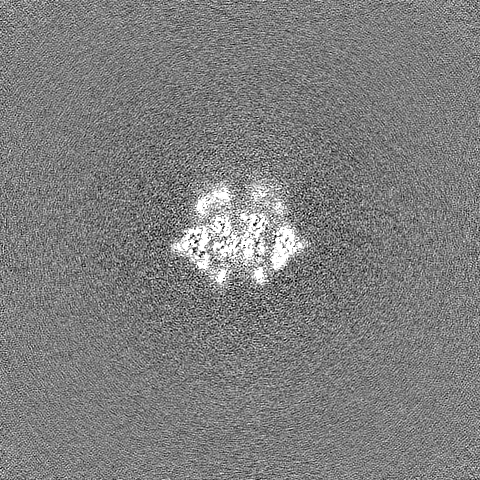
-Map
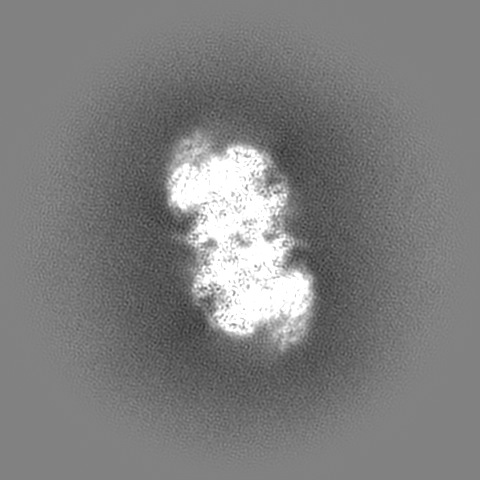
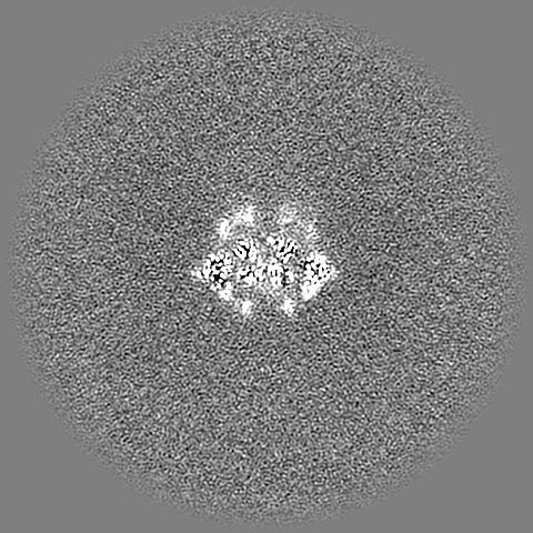
| File | Download / File: emd_33320.map.gz / Format: CCP4 / Size: 421.9 MB / Type: IMAGE STORED AS FLOATING POINT NUMBER (4 BYTES) | ||||||||||||||||||||||||||||||||||||
|---|---|---|---|---|---|---|---|---|---|---|---|---|---|---|---|---|---|---|---|---|---|---|---|---|---|---|---|---|---|---|---|---|---|---|---|---|---|
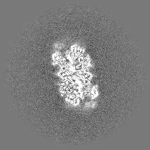
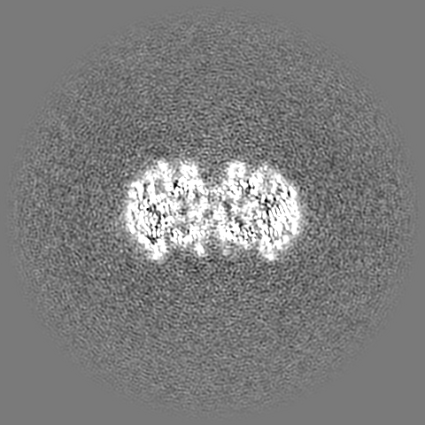
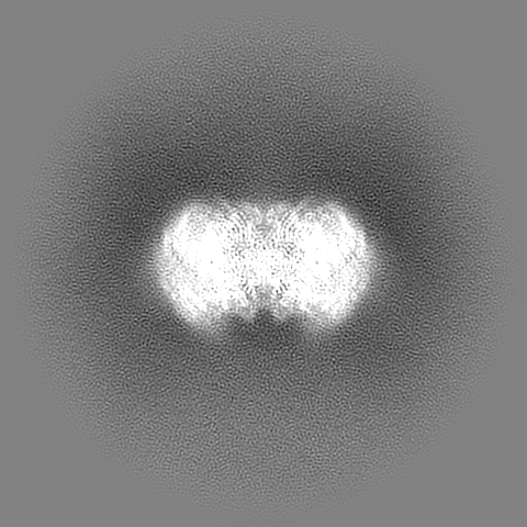
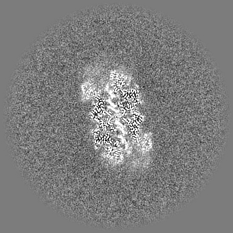
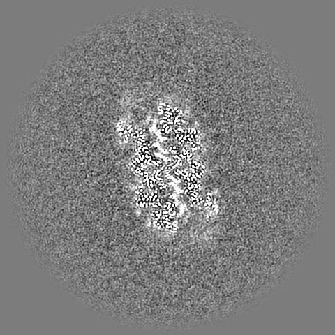
| Projections & slices | Image control
Images are generated by Spider. | ||||||||||||||||||||||||||||||||||||
| Voxel size | X=Y=Z: 1.06 Å | ||||||||||||||||||||||||||||||||||||
| Density |
| ||||||||||||||||||||||||||||||||||||
| Symmetry | Space group: 1 | ||||||||||||||||||||||||||||||||||||
| Details | EMDB XML:
|
 Z (Sec.)
 Y (Row.)
 X (Col.)


-Supplemental data
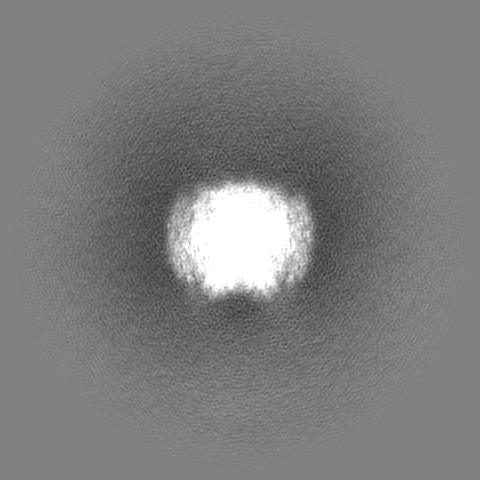
-Additional map: #3
| File | emd_33320_additional_1.map | ||||||||||||
|---|---|---|---|---|---|---|---|---|---|---|---|---|---|
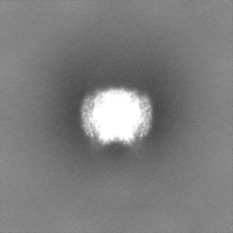
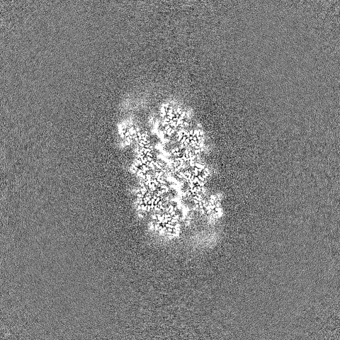
| Projections & Slices |
| ||||||||||||
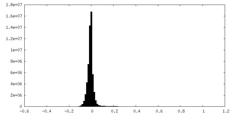
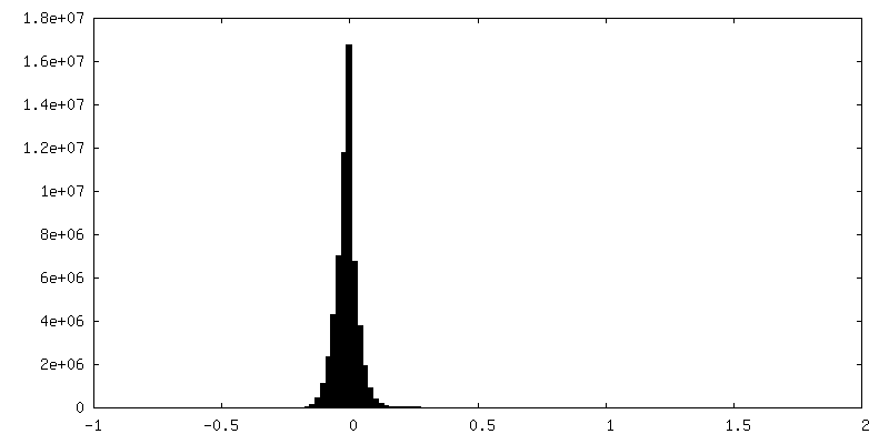
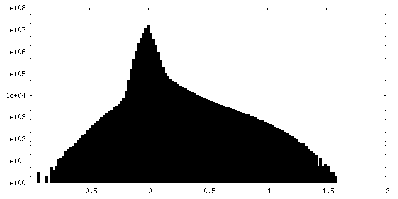
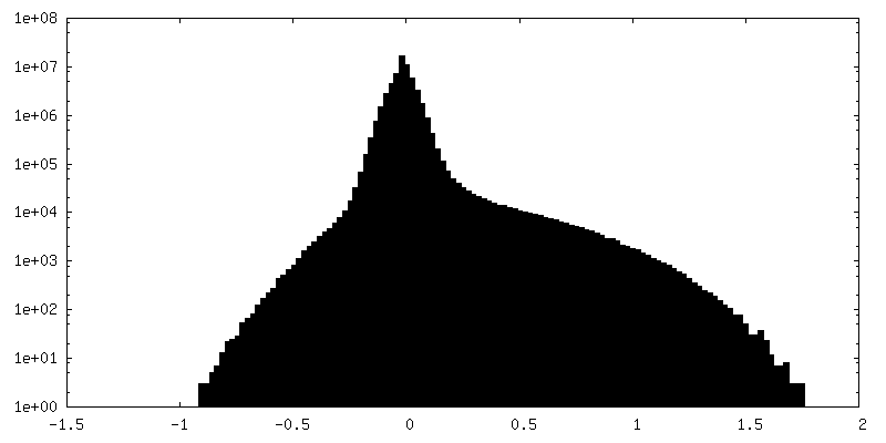
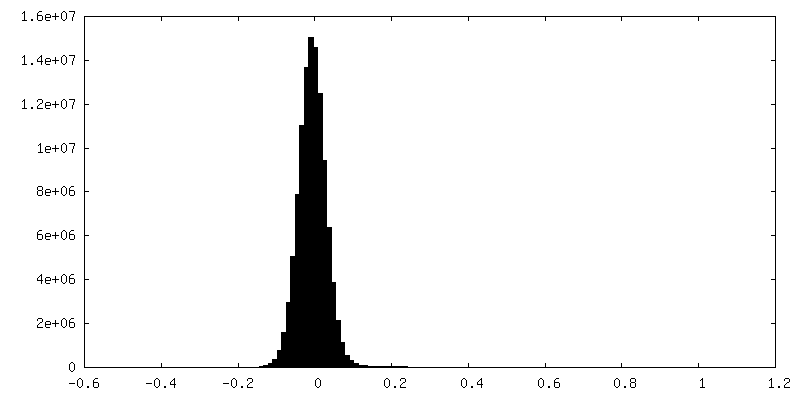
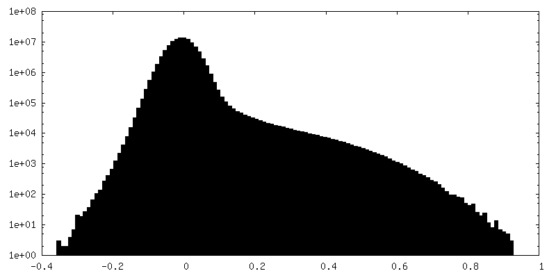
| Density Histograms |
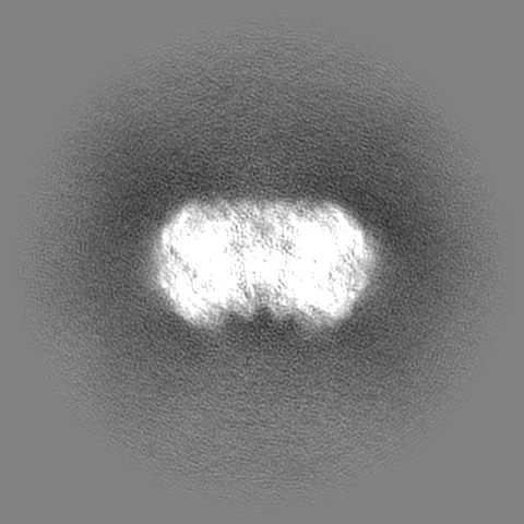


-Additional map: #2
| File | emd_33320_additional_2.map | ||||||||||||
|---|---|---|---|---|---|---|---|---|---|---|---|---|---|
| Projections & Slices |
| ||||||||||||
| Density Histograms |


-Additional map: #1
| File | emd_33320_additional_3.map | ||||||||||||
|---|---|---|---|---|---|---|---|---|---|---|---|---|---|
| Projections & Slices |
| ||||||||||||
| Density Histograms |


-Additional map: #4
| File | emd_33320_additional_4.map | ||||||||||||
|---|---|---|---|---|---|---|---|---|---|---|---|---|---|
| Projections & Slices |
| ||||||||||||
| Density Histograms |


-Half map: #2
| File | emd_33320_half_map_1.map | ||||||||||||
|---|---|---|---|---|---|---|---|---|---|---|---|---|---|
| Projections & Slices |
| ||||||||||||
| Density Histograms |


-Half map: #1
| File | emd_33320_half_map_2.map | ||||||||||||
|---|---|---|---|---|---|---|---|---|---|---|---|---|---|
| Projections & Slices |
| ||||||||||||
| Density Histograms |


- Sample components
-Entire : humman MCM-DH
| Entire | Name: humman MCM-DH |
|---|---|
| Components |
|
-Supramolecule #1: humman MCM-DH
| Supramolecule | Name: humman MCM-DH / type: complex / ID: 1 / Chimera: Yes / Parent: 0 / Macromolecule list: all |
|---|---|
| Source (natural) | Organism: Homo sapiens (human) |
-Macromolecule #1: hMCM5
| Macromolecule | Name: hMCM5 / type: protein_or_peptide / ID: 1 / Enantiomer: LEVO |
|---|---|
| Sequence | String: MSGFDDPGIF YSDSFGGDAQ ADEGQARKSQ LQRRFKEFLR QYRVGTDRTG FTFKYRDELK RHYNLGEYWI EVEMEDLASF DEDLADYLYK QPAEHLQLLE EAAKEVADEV TRPRPSGEEV LQDIQVMLKS DASPSSIRSL KSDMMSHLVK IPGIIIAASA VRAKATRISI ...String: MSGFDDPGIF YSDSFGGDAQ ADEGQARKSQ LQRRFKEFLR QYRVGTDRTG FTFKYRDELK RHYNLGEYWI EVEMEDLASF DEDLADYLYK QPAEHLQLLE EAAKEVADEV TRPRPSGEEV LQDIQVMLKS DASPSSIRSL KSDMMSHLVK IPGIIIAASA VRAKATRISI QCRSCRNTLT NIAMRPGLEG YALPAKCNTD QAGRPKCPGD PYFIMPDKCK CVDFQTLKLQ ELPDAVPHGE MPRHMQLYCD RYLCDKVVPG NRVTIMGIYS IKKFGLTTSR GRDRVGVGIR SSYIRVLGIQ VDTDGSGRSF AGAVSPQEEE EFRRLAALPN VYEVISKSIA PSIFGGTDMK KAIACLLFGG SRKRLPDGLT RRGDINLLML GDPGTAKSQL LKFVEKCSPI GVYTSGKGSS AAGLTASVMR DPSSRNFIME GGAMVLADGG VVCIDEFDKM REDDRVAIHE AMEQQTISIA KAGITTTLNS RCSVLAAANS VFGRWDETKG EDNIDFMPTI LSRFDMIFIV KDEHNEERDV MLAKHVITLH VSALTQTQAV EGEIDLAKLK KFIAYCRVKC GPRLSAEAAE KLKNRYIIMR SGARQHERDS DRRSSIPITV RQLEAIVRIA EALSKMKLQP FATEADVEEA LRLFQVSTLD AALSGTLSGV EGFTSQEDQE MLSRIEKQLK RRFAIGSQVS EHSIIKDFTK QKYPEHAIHK VLQLMLRRGE IQHRMQRKVL YRLK |
-Experimental details
-Structure determination
| Method | cryo EM |
|---|---|
 Processing Processing | single particle reconstruction |
| Aggregation state | particle |
-Sample preparation
| Buffer | pH: 7.5 Component:
| ||||||||||||||||||||||||
|---|---|---|---|---|---|---|---|---|---|---|---|---|---|---|---|---|---|---|---|---|---|---|---|---|---|
| Grid | Model: Quantifoil R1.2/1.3 / Material: COPPER / Mesh: 400 / Support film - Material: CARBON / Support film - topology: CONTINUOUS / Support film - Film thickness: 2.0 nm | ||||||||||||||||||||||||
| Vitrification | Cryogen name: ETHANE |
- Electron microscopy
| Microscope | FEI TITAN KRIOS |
|---|---|
| Specialist optics | Energy filter - Name: GIF Bioquantum / Energy filter - Slit width: 20 eV |
| Image recording | Film or detector model: GATAN K3 BIOQUANTUM (6k x 4k) / Digitization - Dimensions - Width: 5760 pixel / Digitization - Dimensions - Height: 4092 pixel / Number real images: 4748 / Average exposure time: 5.0 sec. / Average electron dose: 50.04 e/Å2 |
| Electron beam | Acceleration voltage: 300 kV / Electron source:  FIELD EMISSION GUN FIELD EMISSION GUN |
| Electron optics | Illumination mode: FLOOD BEAM / Imaging mode: BRIGHT FIELD / Cs: 2.7 mm / Nominal defocus max: 2.5 µm / Nominal defocus min: 1.2 µm / Nominal magnification: 81000 |
| Sample stage | Specimen holder model: FEI TITAN KRIOS AUTOGRID HOLDER / Cooling holder cryogen: NITROGEN |
| Experimental equipment |  Model: Titan Krios / Image courtesy: FEI Company |
-Image processing
| Final reconstruction | Applied symmetry - Point group: C1 (asymmetric) / Resolution.type: BY AUTHOR / Resolution: 2.95 Å / Resolution method: FSC 0.143 CUT-OFF / Software - Name: cryoSPARC (ver. v3.0.1) / Number images used: 349893 |
|---|---|
| Initial angle assignment | Type: MAXIMUM LIKELIHOOD / Software - Name: cryoSPARC (ver. v3.0.1) |
| Final angle assignment | Type: MAXIMUM LIKELIHOOD / Software - Name: cryoSPARC (ver. v3.0.1) |
