[English] 日本語
Yorodumi
- EMDB-33103: Cryo-EM structure of human galanin receptor 2 -

+
Open data


ID or keywords:

Loading...

-
Basic information

Entry
Database: EMDB / ID: EMD-33103
TitleCryo-EM structure of human galanin receptor 2
Map datasharp map
Sample
  • Complex: Human galanin receptor 2 complex with Gq heterotrimer
    • Complex: Human galanin receptor 2
      • Protein or peptide: Galanin receptor type 2
      • Protein or peptide: Guanine nucleotide-binding protein G(q) subunit alpha
      • Protein or peptide: Guanine nucleotide-binding protein G(I)/G(S)/G(T) subunit beta-1
      • Protein or peptide: Guanine nucleotide-binding protein G(I)/G(S)/G(O) subunit gamma-2
    • Complex: scFv16
      • Protein or peptide: scFv16
    • Complex: Galanin
      • Protein or peptide: Galanin
Function / homology
Function and homology information


galanin-activated signaling pathway / galanin receptor binding / type 1 galanin receptor binding / type 2 galanin receptor binding / type 3 galanin receptor binding / galanin receptor activity / positive regulation of large conductance calcium-activated potassium channel activity / positive regulation of timing of catagen / positive regulation of cortisol secretion / regulation of glucocorticoid metabolic process ...galanin-activated signaling pathway / galanin receptor binding / type 1 galanin receptor binding / type 2 galanin receptor binding / type 3 galanin receptor binding / galanin receptor activity / positive regulation of large conductance calcium-activated potassium channel activity / positive regulation of timing of catagen / positive regulation of cortisol secretion / regulation of glucocorticoid metabolic process / inositol phosphate metabolic process / negative regulation of lymphocyte proliferation / phosphatidylinositol metabolic process / neuropeptide hormone activity / neuropeptide binding / feeding behavior / insulin secretion / peptide hormone binding / cAMP-mediated signaling / response to immobilization stress / neuropeptide signaling pathway / protein kinase A signaling / Peptide ligand-binding receptors / muscle contraction / secretory granule / Olfactory Signaling Pathway / Activation of the phototransduction cascade / response to insulin / G beta:gamma signalling through PLC beta / Presynaptic function of Kainate receptors / Thromboxane signalling through TP receptor / adenylate cyclase-modulating G protein-coupled receptor signaling pathway / G-protein activation / G protein-coupled acetylcholine receptor signaling pathway / Activation of G protein gated Potassium channels / Inhibition of voltage gated Ca2+ channels via Gbeta/gamma subunits / cilium / Prostacyclin signalling through prostacyclin receptor / Glucagon signaling in metabolic regulation / G beta:gamma signalling through CDC42 / adenylate cyclase-activating G protein-coupled receptor signaling pathway / ADP signalling through P2Y purinoceptor 12 / G beta:gamma signalling through BTK / Sensory perception of sweet, bitter, and umami (glutamate) taste / Synthesis, secretion, and inactivation of Glucagon-like Peptide-1 (GLP-1) / Adrenaline,noradrenaline inhibits insulin secretion / photoreceptor disc membrane / Glucagon-type ligand receptors / Vasopressin regulates renal water homeostasis via Aquaporins / G alpha (z) signalling events / cellular response to catecholamine stimulus / Glucagon-like Peptide-1 (GLP1) regulates insulin secretion / ADORA2B mediated anti-inflammatory cytokines production / adenylate cyclase-activating dopamine receptor signaling pathway / ADP signalling through P2Y purinoceptor 1 / G beta:gamma signalling through PI3Kgamma / cellular response to prostaglandin E stimulus / Cooperation of PDCL (PhLP1) and TRiC/CCT in G-protein beta folding / sensory perception of taste / response to estrogen / GPER1 signaling / neuron projection development / G-protein beta-subunit binding / heterotrimeric G-protein complex / Inactivation, recovery and regulation of the phototransduction cascade / extracellular vesicle / G alpha (12/13) signalling events / signaling receptor complex adaptor activity / Thrombin signalling through proteinase activated receptors (PARs) / retina development in camera-type eye / GTPase binding / Ca2+ pathway / phospholipase C-activating G protein-coupled receptor signaling pathway / positive regulation of cytosolic calcium ion concentration / G alpha (i) signalling events / fibroblast proliferation / G alpha (s) signalling events / G alpha (q) signalling events / cell population proliferation / Ras protein signal transduction / Extra-nuclear estrogen signaling / learning or memory / cell surface receptor signaling pathway / response to xenobiotic stimulus / positive regulation of apoptotic process / G protein-coupled receptor signaling pathway / lysosomal membrane / GTPase activity / neuronal cell body / synapse / protein-containing complex binding / signal transduction / positive regulation of transcription by RNA polymerase II / extracellular space / extracellular exosome / extracellular region / membrane / plasma membrane / cytosol / cytoplasm
Similarity search - Function
Galanin receptor 2 / Galanin receptor family / Galanin / Galanin precursor / Galanin message associated peptide (GMAP) / Galanin / Galanin message associated peptide (GMAP) / Galanin signature. / Galanin / Serpentine type 7TM GPCR chemoreceptor Srsx ...Galanin receptor 2 / Galanin receptor family / Galanin / Galanin precursor / Galanin message associated peptide (GMAP) / Galanin / Galanin message associated peptide (GMAP) / Galanin signature. / Galanin / Serpentine type 7TM GPCR chemoreceptor Srsx / G-protein, gamma subunit / G-protein gamma subunit domain profile. / GGL domain / G-protein gamma-like domain superfamily / G-protein gamma-like domain / GGL domain / G protein gamma subunit-like motifs / Guanine nucleotide-binding protein, beta subunit / G-protein, beta subunit / G-protein coupled receptors family 1 signature. / G protein-coupled receptor, rhodopsin-like / GPCR, rhodopsin-like, 7TM / G-protein coupled receptors family 1 profile. / 7 transmembrane receptor (rhodopsin family) / G-protein beta WD-40 repeat / WD40 repeat, conserved site / Trp-Asp (WD) repeats signature. / WD domain, G-beta repeat / WD40 repeats / WD40 repeat / Trp-Asp (WD) repeats profile. / Trp-Asp (WD) repeats circular profile. / WD40-repeat-containing domain superfamily / WD40/YVTN repeat-like-containing domain superfamily
Similarity search - Domain/homology
Galanin receptor type 2 / Galanin peptides / Guanine nucleotide-binding protein G(I)/G(S)/G(O) subunit gamma-2 / Guanine nucleotide-binding protein G(I)/G(S)/G(T) subunit beta-1
Similarity search - Component
Biological speciesHomo sapiens (human) / Mus musculus (house mouse)
Methodsingle particle reconstruction / cryo EM / Resolution: 3.11 Å
AuthorsIshimoto N / Kita S / Park SY
Funding support1 items
OrganizationGrant numberCountry
Not funded
Citation
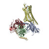
Journal: PLoS Biol / Year: 2022
Title: Structure of the human galanin receptor 2 bound to galanin and Gq reveals the basis of ligand specificity and how binding affects the G-protein interface.
Authors: Yunseok Heo / Naito Ishimoto / Ye-Eun Jeon / Ji-Hye Yun / Mio Ohki / Yuki Anraku / Mina Sasaki / Shunsuke Kita / Hideo Fukuhara / Tatsuya Ikuta / Kouki Kawakami / Asuka Inoue / Katsumi ...Authors: Yunseok Heo / Naito Ishimoto / Ye-Eun Jeon / Ji-Hye Yun / Mio Ohki / Yuki Anraku / Mina Sasaki / Shunsuke Kita / Hideo Fukuhara / Tatsuya Ikuta / Kouki Kawakami / Asuka Inoue / Katsumi Maenaka / Jeremy R H Tame / Weontae Lee / Sam-Yong Park /
Abstract: Galanin is a neuropeptide expressed in the central and peripheral nervous systems, where it regulates various processes including neuroendocrine release, cognition, and nerve regeneration. Three G- ...Galanin is a neuropeptide expressed in the central and peripheral nervous systems, where it regulates various processes including neuroendocrine release, cognition, and nerve regeneration. Three G-protein coupled receptors (GPCRs) for galanin have been discovered, which is the focus of efforts to treat diseases including Alzheimer's disease, anxiety, and addiction. To understand the basis of the ligand preferences of the receptors and to assist structure-based drug design, we used cryo-electron microscopy (cryo-EM) to solve the molecular structure of GALR2 bound to galanin and a cognate heterotrimeric G-protein, providing a molecular view of the neuropeptide binding site. Mutant proteins were assayed to help reveal the basis of ligand specificity, and structural comparison between the activated GALR2 and inactive hβ2AR was used to relate galanin binding to the movements of transmembrane (TM) helices and the G-protein interface.
History
DepositionMar 21, 2022-
Header (metadata) releaseJul 13, 2022-
Map releaseJul 13, 2022-
UpdateAug 10, 2022-
Current statusAug 10, 2022Processing site: PDBj / Status: Released

-
Structure visualization

Supplemental images

Downloads & links

-
Map

FileDownload / File: emd_33103.map.gz / Format: CCP4 / Size: 52.7 MB / Type: IMAGE STORED AS FLOATING POINT NUMBER (4 BYTES)
Annotationsharp map
Voxel sizeX=Y=Z: 1.11667 Å
Density
Contour LevelBy AUTHOR: 0.26
Minimum - Maximum-1.8493583 - 2.6497397
Average (Standard dev.)-0.00073055975 (±0.047740012)
SymmetrySpace group: 1
Details

EMDB XML:

Map geometry
Axis orderXYZ
Origin000
Dimensions240240240
Spacing240240240
CellA=B=C: 268.0 Å
α=β=γ: 90.0 °

-
Supplemental data

-
Mask #1

Fileemd_33103_msk_1.map
Projections & Slices
AxesZYX

Projections

Slices (1/2)
Density Histograms

-
Half map: half map 1

Fileemd_33103_half_map_1.map
Annotationhalf map 1
Projections & Slices
AxesZYX

Projections

Slices (1/2)
Density Histograms

-
Half map: half map 2

Fileemd_33103_half_map_2.map
Annotationhalf map 2
Projections & Slices
AxesZYX

Projections

Slices (1/2)
Density Histograms

-
Sample components

+
Entire : Human galanin receptor 2 complex with Gq heterotrimer

EntireName: Human galanin receptor 2 complex with Gq heterotrimer
Components
  • Complex: Human galanin receptor 2 complex with Gq heterotrimer
    • Complex: Human galanin receptor 2
      • Protein or peptide: Galanin receptor type 2
      • Protein or peptide: Guanine nucleotide-binding protein G(q) subunit alpha
      • Protein or peptide: Guanine nucleotide-binding protein G(I)/G(S)/G(T) subunit beta-1
      • Protein or peptide: Guanine nucleotide-binding protein G(I)/G(S)/G(O) subunit gamma-2
    • Complex: scFv16
      • Protein or peptide: scFv16
    • Complex: Galanin
      • Protein or peptide: Galanin

+
Supramolecule #1: Human galanin receptor 2 complex with Gq heterotrimer

SupramoleculeName: Human galanin receptor 2 complex with Gq heterotrimer / type: complex / Chimera: Yes / ID: 1 / Parent: 0 / Macromolecule list: all
Source (natural)Organism: Homo sapiens (human)
Molecular weightTheoretical: 150 KDa

+
Supramolecule #2: Human galanin receptor 2

SupramoleculeName: Human galanin receptor 2 / type: complex / ID: 2 / Parent: 1 / Macromolecule list: #1-#4
Source (natural)Organism: Mus musculus (house mouse)
Recombinant expressionOrganism: Spodoptera frugiperda (fall armyworm)

+
Supramolecule #3: scFv16

SupramoleculeName: scFv16 / type: complex / ID: 3 / Parent: 1 / Macromolecule list: #5

+
Supramolecule #4: Galanin

SupramoleculeName: Galanin / type: complex / ID: 4 / Parent: 1 / Macromolecule list: #6

+
Macromolecule #1: Galanin receptor type 2

MacromoleculeName: Galanin receptor type 2 / type: protein_or_peptide / ID: 1 / Number of copies: 1 / Enantiomer: LEVO
Source (natural)Organism: Homo sapiens (human)
Molecular weightTheoretical: 41.744082 KDa
Recombinant expressionOrganism: Spodoptera frugiperda (fall armyworm)
SequenceString: MNVSGCPGAG NASQAGGGGG WHPEAVIVPL LFALIFLVGT VGNTLVLAVL LRGGQAVSTT NLFILNLGVA DLCFILCCVP FQATIYTLD GWVFGSLLCK AVHFLIFLTM HASSFTLAAV SLDRYLAIRY PLHSRELRTP RNALAAIGLI WGLSLLFSGP Y LSYYRQSQ ...String:
MNVSGCPGAG NASQAGGGGG WHPEAVIVPL LFALIFLVGT VGNTLVLAVL LRGGQAVSTT NLFILNLGVA DLCFILCCVP FQATIYTLD GWVFGSLLCK AVHFLIFLTM HASSFTLAAV SLDRYLAIRY PLHSRELRTP RNALAAIGLI WGLSLLFSGP Y LSYYRQSQ LANLTVCHPA WSAPRRRAMD ICTFVFSYLL PVLVLGLTYA RTLRYLWRAV DPVAAGSGAR RAKRKVTRMI LI VAALFCL CWMPHHALIL CVWFGQFPLT RATYALRILS HLVSYANSCV NPIVYALVSK HFRKGFRTIC AGLLGRAPGR ASG RVCAAA RGTHSGSVLE RESSDLLHMS EAAGALRPCP GASQPCILEP CPGPSWQGPK AGDSILTVDV A

+
Macromolecule #2: Guanine nucleotide-binding protein G(q) subunit alpha

MacromoleculeName: Guanine nucleotide-binding protein G(q) subunit alpha / type: protein_or_peptide / ID: 2 / Number of copies: 1 / Enantiomer: LEVO
Source (natural)Organism: Homo sapiens (human)
Molecular weightTheoretical: 28.084832 KDa
Recombinant expressionOrganism: Spodoptera frugiperda (fall armyworm)
SequenceString: MGSTVSAEDK AAAERSKMID KNLREDGEKA RRTLRLLLLG ADNSGKSTIV KQMRILHGGS GGSGGTSGIF ETKFQVDKVN FHMFDVGGQ RDERRKWIQC FNDVTAIIFV VDSSDYNRLQ EALNDFKSIW NNRWLRTISV ILFLNKQDLL AEKVLAGKSK I EDYFPEFA ...String:
MGSTVSAEDK AAAERSKMID KNLREDGEKA RRTLRLLLLG ADNSGKSTIV KQMRILHGGS GGSGGTSGIF ETKFQVDKVN FHMFDVGGQ RDERRKWIQC FNDVTAIIFV VDSSDYNRLQ EALNDFKSIW NNRWLRTISV ILFLNKQDLL AEKVLAGKSK I EDYFPEFA RYTTPEDATP EPGEDPRVTR AKYFIRKEFV DISTASGDGR HICYPHFTCA VDTENARRIF NDCKDIILQM NL REYNLV

+
Macromolecule #3: Guanine nucleotide-binding protein G(I)/G(S)/G(T) subunit beta-1

MacromoleculeName: Guanine nucleotide-binding protein G(I)/G(S)/G(T) subunit beta-1
type: protein_or_peptide / ID: 3 / Number of copies: 1 / Enantiomer: LEVO
Source (natural)Organism: Homo sapiens (human)
Molecular weightTheoretical: 37.41693 KDa
Recombinant expressionOrganism: Spodoptera frugiperda (fall armyworm)
SequenceString: MSELDQLRQE AEQLKNQIRD ARKACADATL SQITNNIDPV GRIQMRTRRT LRGHLAKIYA MHWGTDSRLL VSASQDGKLI IWDSYTTNK VHAIPLRSSW VMTCAYAPSG NYVACGGLDN ICSIYNLKTR EGNVRVSREL AGHTGYLSCC RFLDDNQIVT S SGDTTCAL ...String:
MSELDQLRQE AEQLKNQIRD ARKACADATL SQITNNIDPV GRIQMRTRRT LRGHLAKIYA MHWGTDSRLL VSASQDGKLI IWDSYTTNK VHAIPLRSSW VMTCAYAPSG NYVACGGLDN ICSIYNLKTR EGNVRVSREL AGHTGYLSCC RFLDDNQIVT S SGDTTCAL WDIETGQQTT TFTGHTGDVM SLSLAPDTRL FVSGACDASA KLWDVREGMC RQTFTGHESD INAICFFPNG NA FATGSDD ATCRLFDLRA DQELMTYSHD NIICGITSVS FSKSGRLLLA GYDDFNCNVW DALKADRAGV LAGHDNRVSC LGV TDDGMA VATGSWDSFL KIWN

+
Macromolecule #4: Guanine nucleotide-binding protein G(I)/G(S)/G(O) subunit gamma-2

MacromoleculeName: Guanine nucleotide-binding protein G(I)/G(S)/G(O) subunit gamma-2
type: protein_or_peptide / ID: 4 / Number of copies: 1 / Enantiomer: LEVO
Source (natural)Organism: Homo sapiens (human)
Molecular weightTheoretical: 7.861143 KDa
Recombinant expressionOrganism: Spodoptera frugiperda (fall armyworm)
SequenceString:
MASNNTASIA QARKLVEQLK MEANIDRIKV SKAAADLMAY CEAHAKEDPL LTPVPASENP FREKKFFCAI L

+
Macromolecule #5: scFv16

MacromoleculeName: scFv16 / type: protein_or_peptide / ID: 5 / Number of copies: 1 / Enantiomer: LEVO
Source (natural)Organism: Mus musculus (house mouse)
Molecular weightTheoretical: 27.474574 KDa
Recombinant expressionOrganism: Spodoptera frugiperda (fall armyworm)
SequenceString: DVQLVESGGG LVQPGGSRKL SCSASGFAFS SFGMHWVRQA PEKGLEWVAY ISSGSGTIYY ADTVKGRFTI SRDDPKNTLF LQMTSLRSE DTAMYYCVRS IYYYGSSPFD FWGQGTTLTV SSGGGGSGGG GSGGGGSDIV MTQATSSVPV TPGESVSISC R SSKSLLHS ...String:
DVQLVESGGG LVQPGGSRKL SCSASGFAFS SFGMHWVRQA PEKGLEWVAY ISSGSGTIYY ADTVKGRFTI SRDDPKNTLF LQMTSLRSE DTAMYYCVRS IYYYGSSPFD FWGQGTTLTV SSGGGGSGGG GSGGGGSDIV MTQATSSVPV TPGESVSISC R SSKSLLHS NGNTYLYWFL QRPGQSPQLL IYRMSNLASG VPDRFSGSGS GTAFTLTISR LEAEDVGVYY CMQHLEYPLT FG AGTKLEL KAAAENLYFQ

+
Macromolecule #6: Galanin

MacromoleculeName: Galanin / type: protein_or_peptide / ID: 6 / Number of copies: 1 / Enantiomer: LEVO
Source (natural)Organism: Homo sapiens (human)
Molecular weightTheoretical: 3.161446 KDa
SequenceString:
GWTLNSAGYL LGPHAVGNHR SFSDKNGLTS

-
Experimental details

-
Structure determination

Methodcryo EM
Processingsingle particle reconstruction
Aggregation stateparticle

-
Sample preparation

Concentration1.5 mg/mL
BufferpH: 7.5
Component:
ConcentrationFormulaName
20.0 mMHEPES2-[4-(2-Hydroxyethyl)-1-piperazinyl]ethanesulfonic acid
100.0 mMNaClSodium chloridesodium chloride
1.0 mMMgCl2magnesium chloride
0.5 mMTCEPtris(2-carboxyethyl)phosphine
0.001 %(w/v)LMNGLauryl Maltose Neopentyl Glycol
0.0001 %(w/v)CHScholesteryl homosuccinate
GridModel: Quantifoil R1.2/1.3 / Material: COPPER / Mesh: 300 / Support film - Material: CARBON
VitrificationCryogen name: ETHANE

-
Electron microscopy

MicroscopeFEI TITAN KRIOS
Electron beamAcceleration voltage: 300 kV / Electron source: FIELD EMISSION GUN
Electron opticsIllumination mode: FLOOD BEAM / Imaging mode: BRIGHT FIELDBright-field microscopy / Cs: 2.7 mm / Nominal defocus max: 2.0 µm / Nominal defocus min: 0.8 µm / Nominal magnification: 130000
Sample stageSpecimen holder model: FEI TITAN KRIOS AUTOGRID HOLDER / Cooling holder cryogen: NITROGEN
Image recordingFilm or detector model: GATAN K3 (6k x 4k) / Number grids imaged: 1 / Number real images: 7216 / Average exposure time: 4.7 sec. / Average electron dose: 53.17 e/Å2
Experimental equipment
Model: Titan Krios / Image courtesy: FEI Company

-
Image processing

Particle selectionNumber selected: 2666499
CTF correctionSoftware - Name: cryoSPARC (ver. 3.3.1)
Startup modelType of model: PDB ENTRY
PDB model - PDB ID:
Initial angle assignmentType: MAXIMUM LIKELIHOOD
Final angle assignmentType: MAXIMUM LIKELIHOOD
Final reconstructionResolution.type: BY AUTHOR / Resolution: 3.11 Å / Resolution method: FSC 0.143 CUT-OFF / Number images used: 479312
FSC plot (resolution estimation)

-
Atomic model buiding 1

Initial modelPDB ID:
RefinementSpace: REAL
Output model

PDB-7xbd:
Cryo-EM structure of human galanin receptor 2

+
About Yorodumi

-
News

-
Feb 9, 2022. New format data for meta-information of EMDB entries

New format data for meta-information of EMDB entries

  • Version 3 of the EMDB header file is now the official format.
  • The previous official version 1.9 will be removed from the archive.

Related info.:EMDB header

External links:wwPDB to switch to version 3 of the EMDB data model

-
Aug 12, 2020. Covid-19 info

Covid-19 info

URL: https://pdbj.org/emnavi/covid19.php

New page: Covid-19 featured information page in EM Navigator.

Related info.:Covid-19 info / Mar 5, 2020. Novel coronavirus structure data

+
Mar 5, 2020. Novel coronavirus structure data

Novel coronavirus structure data

Related info.:Yorodumi Speices / Aug 12, 2020. Covid-19 info

External links:COVID-19 featured content - PDBj / Molecule of the Month (242):Coronavirus Proteases

+
Jan 31, 2019. EMDB accession codes are about to change! (news from PDBe EMDB page)

EMDB accession codes are about to change! (news from PDBe EMDB page)

  • The allocation of 4 digits for EMDB accession codes will soon come to an end. Whilst these codes will remain in use, new EMDB accession codes will include an additional digit and will expand incrementally as the available range of codes is exhausted. The current 4-digit format prefixed with “EMD-” (i.e. EMD-XXXX) will advance to a 5-digit format (i.e. EMD-XXXXX), and so on. It is currently estimated that the 4-digit codes will be depleted around Spring 2019, at which point the 5-digit format will come into force.
  • The EM Navigator/Yorodumi systems omit the EMD- prefix.

Related info.:Q: What is EMD? / ID/Accession-code notation in Yorodumi/EM Navigator

External links:EMDB Accession Codes are Changing Soon! / Contact to PDBj

+
Jul 12, 2017. Major update of PDB

Major update of PDB

  • wwPDB released updated PDB data conforming to the new PDBx/mmCIF dictionary.
  • This is a major update changing the version number from 4 to 5, and with Remediation, in which all the entries are updated.
  • In this update, many items about electron microscopy experimental information are reorganized (e.g. em_software).
  • Now, EM Navigator and Yorodumi are based on the updated data.

External links:wwPDB Remediation / Enriched Model Files Conforming to OneDep Data Standards Now Available in the PDB FTP Archive

-
Yorodumi

Thousand views of thousand structures

  • Yorodumi is a browser for structure data from EMDB, PDB, SASBDB, etc.
  • This page is also the successor to EM Navigator detail page, and also detail information page/front-end page for Omokage search.
  • The word "yorodu" (or yorozu) is an old Japanese word meaning "ten thousand". "mi" (miru) is to see.

Related info.:EMDB / PDB / SASBDB / Comparison of 3 databanks / Yorodumi Search / Aug 31, 2016. New EM Navigator & Yorodumi / Yorodumi Papers / Jmol/JSmol / Function and homology information / Changes in new EM Navigator and Yorodumi

Read more