+ Open data
- Basic information
| Entry |  | |||||||||
|---|---|---|---|---|---|---|---|---|---|---|
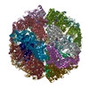
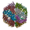
| Title | cryo-EM structure of human TRiC-ADP | |||||||||
 Map data Map data | ||||||||||
 Sample Sample |
| |||||||||
 Keywords Keywords | STRUCTURAL PROTEIN | |||||||||
| Function / homology |  Function and homology information Function and homology informationzona pellucida receptor complex / positive regulation of establishment of protein localization to telomere / positive regulation of protein localization to Cajal body / scaRNA localization to Cajal body / positive regulation of telomerase RNA localization to Cajal body / tubulin complex assembly / chaperonin-containing T-complex / : / BBSome-mediated cargo-targeting to cilium / Formation of tubulin folding intermediates by CCT/TriC ...zona pellucida receptor complex / positive regulation of establishment of protein localization to telomere / positive regulation of protein localization to Cajal body / scaRNA localization to Cajal body / positive regulation of telomerase RNA localization to Cajal body / tubulin complex assembly / chaperonin-containing T-complex / : / BBSome-mediated cargo-targeting to cilium / Formation of tubulin folding intermediates by CCT/TriC / binding of sperm to zona pellucida / Folding of actin by CCT/TriC / Prefoldin mediated transfer of substrate to CCT/TriC / RHOBTB1 GTPase cycle / WD40-repeat domain binding / pericentriolar material / Association of TriC/CCT with target proteins during biosynthesis / chaperone-mediated protein complex assembly / beta-tubulin binding / Hydrolases; Acting on acid anhydrides; In phosphorus-containing anhydrides / RHOBTB2 GTPase cycle / heterochromatin / : / positive regulation of telomere maintenance via telomerase / protein folding chaperone / acrosomal vesicle / Gene and protein expression by JAK-STAT signaling after Interleukin-12 stimulation / mRNA 3'-UTR binding / cell projection / ATP-dependent protein folding chaperone / mRNA 5'-UTR binding / response to virus / azurophil granule lumen / Cooperation of PDCL (PhLP1) and TRiC/CCT in G-protein beta folding / unfolded protein binding / melanosome / protein folding / G-protein beta-subunit binding / cell body / secretory granule lumen / ficolin-1-rich granule lumen / microtubule / cytoskeleton / protein stabilization / cilium / cadherin binding / ubiquitin protein ligase binding / centrosome / Neutrophil degranulation / Golgi apparatus / ATP hydrolysis activity / RNA binding / extracellular exosome / extracellular region / nucleoplasm / ATP binding / identical protein binding / cytoplasm / cytosol Similarity search - Function | |||||||||
| Biological species |  Homo sapiens (human) Homo sapiens (human) | |||||||||
| Method | single particle reconstruction / cryo EM / Resolution: 3.3 Å | |||||||||
 Authors Authors | Cong Y / Liu CX | |||||||||
| Funding support |  China, 1 items China, 1 items
| |||||||||
 Citation Citation |  Journal: Commun Biol / Year: 2023 Journal: Commun Biol / Year: 2023Title: Pathway and mechanism of tubulin folding mediated by TRiC/CCT along its ATPase cycle revealed using cryo-EM. Authors: Caixuan Liu / Mingliang Jin / Shutian Wang / Wenyu Han / Qiaoyu Zhao / Yifan Wang / Cong Xu / Lei Diao / Yue Yin / Chao Peng / Lan Bao / Yanxing Wang / Yao Cong /  Abstract: The eukaryotic chaperonin TRiC/CCT assists the folding of about 10% of cytosolic proteins through an ATP-driven conformational cycle, and the essential cytoskeleton protein tubulin is the obligate ...The eukaryotic chaperonin TRiC/CCT assists the folding of about 10% of cytosolic proteins through an ATP-driven conformational cycle, and the essential cytoskeleton protein tubulin is the obligate substrate of TRiC. Here, we present an ensemble of cryo-EM structures of endogenous human TRiC throughout its ATPase cycle, with three of them revealing endogenously engaged tubulin in different folding stages. The open-state TRiC-tubulin-S1 and -S2 maps show extra density corresponding to tubulin in the cis-ring chamber of TRiC. Our structural and XL-MS analyses suggest a gradual upward translocation and stabilization of tubulin within the TRiC chamber accompanying TRiC ring closure. In the closed TRiC-tubulin-S3 map, we capture a near-natively folded tubulin-with the tubulin engaging through its N and C domains mainly with the A and I domains of the CCT3/6/8 subunits through electrostatic and hydrophilic interactions. Moreover, we also show the potential role of TRiC C-terminal tails in substrate stabilization and folding. Our study delineates the pathway and molecular mechanism of TRiC-mediated folding of tubulin along the ATPase cycle of TRiC, and may also inform the design of therapeutic agents targeting TRiC-tubulin interactions. | |||||||||
| History |
|
- Structure visualization
| Supplemental images |
|---|
- Downloads & links
-EMDB archive
| Map data |  emd_32993.map.gz emd_32993.map.gz | 8.3 MB |  EMDB map data format EMDB map data format | |
|---|---|---|---|---|
| Header (meta data) |  emd-32993-v30.xml emd-32993-v30.xml emd-32993.xml emd-32993.xml | 28.4 KB 28.4 KB | Display Display |  EMDB header EMDB header |
| Images |  emd_32993.png emd_32993.png | 112.1 KB | ||
| Filedesc metadata |  emd-32993.cif.gz emd-32993.cif.gz | 9.2 KB | ||
| Others |  emd_32993_half_map_1.map.gz emd_32993_half_map_1.map.gz emd_32993_half_map_2.map.gz emd_32993_half_map_2.map.gz | 49.7 MB 49.7 MB | ||
| Archive directory |  http://ftp.pdbj.org/pub/emdb/structures/EMD-32993 http://ftp.pdbj.org/pub/emdb/structures/EMD-32993 ftp://ftp.pdbj.org/pub/emdb/structures/EMD-32993 ftp://ftp.pdbj.org/pub/emdb/structures/EMD-32993 | HTTPS FTP |
-Validation report
| Summary document |  emd_32993_validation.pdf.gz emd_32993_validation.pdf.gz | 857.7 KB | Display |  EMDB validaton report EMDB validaton report |
|---|---|---|---|---|
| Full document |  emd_32993_full_validation.pdf.gz emd_32993_full_validation.pdf.gz | 857.3 KB | Display | |
| Data in XML |  emd_32993_validation.xml.gz emd_32993_validation.xml.gz | 12.4 KB | Display | |
| Data in CIF |  emd_32993_validation.cif.gz emd_32993_validation.cif.gz | 14.6 KB | Display | |
| Arichive directory |  https://ftp.pdbj.org/pub/emdb/validation_reports/EMD-32993 https://ftp.pdbj.org/pub/emdb/validation_reports/EMD-32993 ftp://ftp.pdbj.org/pub/emdb/validation_reports/EMD-32993 ftp://ftp.pdbj.org/pub/emdb/validation_reports/EMD-32993 | HTTPS FTP |
-Related structure data
| Related structure data |  7x3uMC  7wz3C  7x0aC  7x0sC  7x0vC  7x3jC  7x6qC  7x7yC M: atomic model generated by this map C: citing same article ( |
|---|---|
| Similar structure data | Similarity search - Function & homology  F&H Search F&H Search |
- Links
Links
| EMDB pages |  EMDB (EBI/PDBe) / EMDB (EBI/PDBe) /  EMDataResource EMDataResource |
|---|---|
| Related items in Molecule of the Month |
- Map
Map
| File |  Download / File: emd_32993.map.gz / Format: CCP4 / Size: 64 MB / Type: IMAGE STORED AS FLOATING POINT NUMBER (4 BYTES) Download / File: emd_32993.map.gz / Format: CCP4 / Size: 64 MB / Type: IMAGE STORED AS FLOATING POINT NUMBER (4 BYTES) | ||||||||||||||||||||||||||||||||||||
|---|---|---|---|---|---|---|---|---|---|---|---|---|---|---|---|---|---|---|---|---|---|---|---|---|---|---|---|---|---|---|---|---|---|---|---|---|---|
| Projections & slices | Image control
Images are generated by Spider. | ||||||||||||||||||||||||||||||||||||
| Voxel size | X=Y=Z: 1.318 Å | ||||||||||||||||||||||||||||||||||||
| Density |
| ||||||||||||||||||||||||||||||||||||
| Symmetry | Space group: 1 | ||||||||||||||||||||||||||||||||||||
| Details | EMDB XML:
|
-Supplemental data
-Half map: #1
| File | emd_32993_half_map_1.map | ||||||||||||
|---|---|---|---|---|---|---|---|---|---|---|---|---|---|
| Projections & Slices |
| ||||||||||||
| Density Histograms |
-Half map: #2
| File | emd_32993_half_map_2.map | ||||||||||||
|---|---|---|---|---|---|---|---|---|---|---|---|---|---|
| Projections & Slices |
| ||||||||||||
| Density Histograms |
- Sample components
+Entire : Human TRiC-ADP state
+Supramolecule #1: Human TRiC-ADP state
+Macromolecule #1: T-complex protein 1 subunit alpha
+Macromolecule #2: T-complex protein 1 subunit beta
+Macromolecule #3: T-complex protein 1 subunit delta
+Macromolecule #4: T-complex protein 1 subunit epsilon
+Macromolecule #5: T-complex protein 1 subunit gamma
+Macromolecule #6: T-complex protein 1 subunit eta
+Macromolecule #7: T-complex protein 1 subunit theta
+Macromolecule #8: T-complex protein 1 subunit zeta
+Macromolecule #9: ADENOSINE-5'-DIPHOSPHATE
-Experimental details
-Structure determination
| Method | cryo EM |
|---|---|
 Processing Processing | single particle reconstruction |
| Aggregation state | particle |
- Sample preparation
| Buffer | pH: 7.5 / Component - Concentration: 50.0 mM / Component - Formula: NaCl / Component - Name: sodium Chloride |
|---|---|
| Vitrification | Cryogen name: ETHANE |
- Electron microscopy
| Microscope | FEI TITAN KRIOS |
|---|---|
| Image recording | Film or detector model: GATAN K2 SUMMIT (4k x 4k) / Detector mode: SUPER-RESOLUTION / Average electron dose: 38.0 e/Å2 |
| Electron beam | Acceleration voltage: 300 kV / Electron source:  FIELD EMISSION GUN FIELD EMISSION GUN |
| Electron optics | Illumination mode: FLOOD BEAM / Imaging mode: BRIGHT FIELD / Nominal defocus max: 2.5 µm / Nominal defocus min: 0.8 µm |
| Experimental equipment |  Model: Titan Krios / Image courtesy: FEI Company |
 Movie
Movie Controller


 Z (Sec.)
 Y (Row.)
 X (Col.)


