Ministry of Education, Culture, Sports, Science and Technology (Japan)
18H03978
Japan
Ministry of Education, Culture, Sports, Science and Technology (Japan)
21H04758
Japan
Ministry of Education, Culture, Sports, Science and Technology (Japan)
21H05247
Japan
Ministry of Education, Culture, Sports, Science and Technology (Japan)
21K15036
Japan
Citation
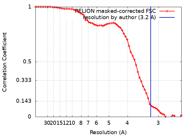
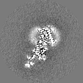
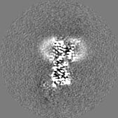
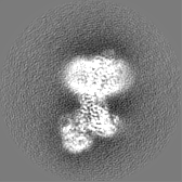
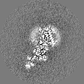
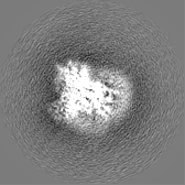
Journal: EMBO J / Year: 2021 Title: Cryo-EM analysis provides new mechanistic insight into ATP binding to Ca -ATPase SERCA2b. Authors: Yuxia Zhang / Satoshi Watanabe / Akihisa Tsutsumi / Hiroshi Kadokura / Masahide Kikkawa / Kenji Inaba / Abstract: Sarco/endoplasmic reticulum Ca -ATPase (SERCA) 2b is a ubiquitous SERCA family member that conducts Ca uptake from the cytosol to the ER. Herein, we present a 3.3 Å resolution cryo-electron ...Sarco/endoplasmic reticulum Ca -ATPase (SERCA) 2b is a ubiquitous SERCA family member that conducts Ca uptake from the cytosol to the ER. Herein, we present a 3.3 Å resolution cryo-electron microscopy (cryo-EM) structure of human SERCA2b in the E1·2Ca state, revealing a new conformation for Ca -bound SERCA2b with a much closer arrangement of cytosolic domains than in the previously reported crystal structure of Ca -bound SERCA1a. Multiple conformations generated by 3D classification of cryo-EM maps reflect the intrinsically dynamic nature of the cytosolic domains in this state. Notably, ATP binding residues of SERCA2b in the E1·2Ca state are located at similar positions to those in the E1·2Ca -ATP state; hence, the cryo-EM structure likely represents a preformed state immediately prior to ATP binding. Consistently, a SERCA2b mutant with an interdomain disulfide bridge that locks the closed cytosolic domain arrangement displayed significant autophosphorylation activity in the presence of Ca . We propose a novel mechanism of ATP binding to SERCA2b.
In the structure databanks used in Yorodumi, some data are registered as the other names, "COVID-19 virus" and "2019-nCoV". Here are the details of the virus and the list of structure data.
Jan 31, 2019. EMDB accession codes are about to change! (news from PDBe EMDB page)
EMDB accession codes are about to change! (news from PDBe EMDB page)
The allocation of 4 digits for EMDB accession codes will soon come to an end. Whilst these codes will remain in use, new EMDB accession codes will include an additional digit and will expand incrementally as the available range of codes is exhausted. The current 4-digit format prefixed with “EMD-” (i.e. EMD-XXXX) will advance to a 5-digit format (i.e. EMD-XXXXX), and so on. It is currently estimated that the 4-digit codes will be depleted around Spring 2019, at which point the 5-digit format will come into force.
The EM Navigator/Yorodumi systems omit the EMD- prefix.
Related info.:Q: What is EMD? / ID/Accession-code notation in Yorodumi/EM Navigator
Yorodumi is a browser for structure data from EMDB, PDB, SASBDB, etc.
This page is also the successor to EM Navigator detail page, and also detail information page/front-end page for Omokage search.
The word "yorodu" (or yorozu) is an old Japanese word meaning "ten thousand". "mi" (miru) is to see.
Related info.:EMDB / PDB / SASBDB / Comparison of 3 databanks / Yorodumi Search / Aug 31, 2016. New EM Navigator & Yorodumi / Yorodumi Papers / Jmol/JSmol / Function and homology information / Changes in new EM Navigator and Yorodumi
 Movie
Movie Controller


 Open data
 Basic information
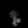
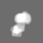
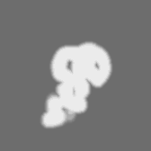
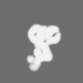
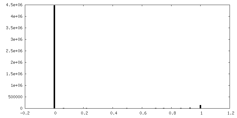
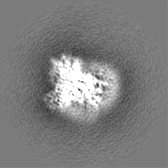
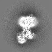
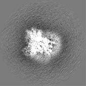
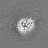
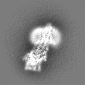
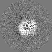
 Map data
 Sample
 Keywords
 Function and homology information
 Homo sapiens (human)
 Authors
 Japan, 4 items
  Citation
 Structure visualization
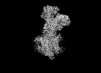
 Downloads & links
 emd_32350.png
 http://ftp.pdbj.org/pub/emdb/structures/EMD-32350
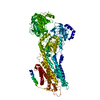


 Z (Sec.)
 Y (Row.)
 X (Col.)


 Sample components
 Processing
 Electron microscopy
 FIELD EMISSION GUN