+ Open data
- Basic information
| Entry | Database: EMDB / ID: EMD-31505 | |||||||||
|---|---|---|---|---|---|---|---|---|---|---|
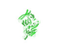
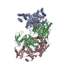
| Title | VAR2CSA 3D7 ectodomain core region | |||||||||
 Map data Map data | ||||||||||
 Sample Sample |
| |||||||||
 Keywords Keywords | Plasmodium falciparum / Complex / CSA binding / CELL ADHESION | |||||||||
| Function / homology |  Function and homology information Function and homology informationsymbiont-mediated perturbation of host erythrocyte aggregation / infected host cell surface knob / antigenic variation / adhesion of symbiont to microvasculature / cell adhesion molecule binding / cell-cell adhesion / host cell surface receptor binding / host cell plasma membrane / membrane Similarity search - Function | |||||||||
| Biological species |   | |||||||||
| Method | single particle reconstruction / cryo EM / Resolution: 3.6 Å | |||||||||
 Authors Authors | Wang L / Zhaoning W | |||||||||
| Funding support |  China, 1 items China, 1 items
| |||||||||
 Citation Citation |  Journal: Cell Discov / Year: 2021 Journal: Cell Discov / Year: 2021Title: The molecular mechanism of cytoadherence to placental or tumor cells through VAR2CSA from Plasmodium falciparum. Authors: Weiwei Wang / Zhaoning Wang / Xiuna Yang / Yan Gao / Xiang Zhang / Long Cao / Aguang Dai / Jin Sun / Lei Sun / Lubin Jiang / Zhenguo Chen / Lanfeng Wang /  | |||||||||
| History |
|
- Structure visualization
| Movie |
 Movie viewer Movie viewer |
|---|---|
| Structure viewer | EM map:  SurfView SurfView Molmil Molmil Jmol/JSmol Jmol/JSmol |
| Supplemental images |
- Downloads & links
-EMDB archive
| Map data |  emd_31505.map.gz emd_31505.map.gz | 120.1 MB |  EMDB map data format EMDB map data format | |
|---|---|---|---|---|
| Header (meta data) |  emd-31505-v30.xml emd-31505-v30.xml emd-31505.xml emd-31505.xml | 13 KB 13 KB | Display Display |  EMDB header EMDB header |
| Images |  emd_31505.png emd_31505.png | 40.5 KB | ||
| Filedesc metadata |  emd-31505.cif.gz emd-31505.cif.gz | 6.6 KB | ||
| Archive directory |  http://ftp.pdbj.org/pub/emdb/structures/EMD-31505 http://ftp.pdbj.org/pub/emdb/structures/EMD-31505 ftp://ftp.pdbj.org/pub/emdb/structures/EMD-31505 ftp://ftp.pdbj.org/pub/emdb/structures/EMD-31505 | HTTPS FTP |
-Related structure data
| Related structure data |  7fasMC  7fapC M: atomic model generated by this map C: citing same article ( |
|---|---|
| Similar structure data |
- Links
Links
| EMDB pages |  EMDB (EBI/PDBe) / EMDB (EBI/PDBe) /  EMDataResource EMDataResource |
|---|
- Map
Map
| File |  Download / File: emd_31505.map.gz / Format: CCP4 / Size: 129.7 MB / Type: IMAGE STORED AS FLOATING POINT NUMBER (4 BYTES) Download / File: emd_31505.map.gz / Format: CCP4 / Size: 129.7 MB / Type: IMAGE STORED AS FLOATING POINT NUMBER (4 BYTES) | ||||||||||||||||||||||||||||||||||||||||||||||||||||||||||||||||||||
|---|---|---|---|---|---|---|---|---|---|---|---|---|---|---|---|---|---|---|---|---|---|---|---|---|---|---|---|---|---|---|---|---|---|---|---|---|---|---|---|---|---|---|---|---|---|---|---|---|---|---|---|---|---|---|---|---|---|---|---|---|---|---|---|---|---|---|---|---|---|
| Projections & slices | Image control
Images are generated by Spider. | ||||||||||||||||||||||||||||||||||||||||||||||||||||||||||||||||||||
| Voxel size | X=Y=Z: 1.044 Å | ||||||||||||||||||||||||||||||||||||||||||||||||||||||||||||||||||||
| Density |
| ||||||||||||||||||||||||||||||||||||||||||||||||||||||||||||||||||||
| Symmetry | Space group: 1 | ||||||||||||||||||||||||||||||||||||||||||||||||||||||||||||||||||||
| Details | EMDB XML:
CCP4 map header:
| ||||||||||||||||||||||||||||||||||||||||||||||||||||||||||||||||||||
-Supplemental data
- Sample components
-Entire : VAR2CSA
| Entire | Name: VAR2CSA |
|---|---|
| Components |
|
-Supramolecule #1: VAR2CSA
| Supramolecule | Name: VAR2CSA / type: complex / ID: 1 / Parent: 0 / Macromolecule list: all |
|---|---|
| Source (natural) | Organism:  |
| Molecular weight | Theoretical: 306 kDa/nm |
-Macromolecule #1: Erythrocyte membrane protein 1, PfEMP1
| Macromolecule | Name: Erythrocyte membrane protein 1, PfEMP1 / type: protein_or_peptide / ID: 1 / Number of copies: 1 / Enantiomer: LEVO |
|---|---|
| Source (natural) | Organism:  |
| Molecular weight | Theoretical: 228.991188 KDa |
| Recombinant expression | Organism:  |
| Sequence | String: MDKSSIANKI EAYLGAKSDD SKIDQSLKAD PSEVQYYGSG GDGYYLRKNI CKITVNHSDS GTNDPCDRIP PPYGDNDQWK CAIILSKVS EKPENVFVPP RRQRMCINNL EKLNVDKIRD KHAFLADVLL TARNEGERIV QNHPDTNSSN VCNALERSFA D IADIIRGT ...String: MDKSSIANKI EAYLGAKSDD SKIDQSLKAD PSEVQYYGSG GDGYYLRKNI CKITVNHSDS GTNDPCDRIP PPYGDNDQWK CAIILSKVS EKPENVFVPP RRQRMCINNL EKLNVDKIRD KHAFLADVLL TARNEGERIV QNHPDTNSSN VCNALERSFA D IADIIRGT DLWKGTNSNL EQNLKQMFAK IRENDKVLQD KYPKDQNYRK LREDWWNANR QKVWEVITCG ARSNDLLIKR GW RTSGKSN GDNKLELCRK CGHYEEKVPT KLDYVPQFLR WLTEWIEDFY REKQNLIDDM ERHREECTSE DHKSKEGTSY CST CKDKCK KYCECVKKWK SEWENQKNKY TELYQQNKNE TSQKNTSRYD DYVKDFFKKL EANYSSLENY IKGDPYFAEY ATKL SFILN SSDANNPSEK IQKNNDEVCN CNESGIASVE QEQISDPSSN KTCITHSSIK ANKKKVCKHV KLGVRENDKD LRVCV IEHT SLSGVENCCC QDFLRILQEN CSDNKSGSSS NGSCNNKNQE ACEKNLEKVL ASLTNCYKCD KCKSEQSKKN NKNWIW KKS SGKEGGLQKE YANTIGLPPR TQSLCLVVCL DEKGKKTQEL KNIRTNSELL KEWIIAAFHE GKNLKPSHEK KNDDNGK KL CKALEYSFAD YGDLIKGTSI WDNEYTKDLE LNLQKIFGKL FRKYIKKNNT AEQDTSYSSL DELRESWWNT NKKYIWLA M KHGAGMNSTT CCGDGSVTGS GSSCDDIPTI DLIPQYLRFL QEWVEHFCKQ RQEKVKPVIE NCKSCKESGG TCNGECKTE CKNKCEVYKK FIEDCKGGDG TAGSSWVKRW DQIYKRYSKY IEDAKRNRKA GTKNCGPSST TNAAENKCVQ SDIDSFFKHL IDIGLTTPS SYLSIVLDDN ICGADKAPWT TYTTYTTTEK CNKETDKSKL QQCNTAVVVN VPSPLGNTPH GYKYACQCKI P TNEETCDD RKEYMNQWSC GSARTMKRGY KNDNYELCKY NGVDVKPTTV RSNSSKLDDK DVTFFNLFEQ WNKEIQYQIE QY MTNTKIS CNNEKNVLSR VSDEAAQPKF SDNERDRNSI THEDKNCKEK CKCYSLWIEK INDQWDKQKD NYNKFQRKQI YDA NKGSQN KKVVSLSNFL FFSCWEEYIQ KYFNGDWSKI KNIGSDTFEF LIKKCGNDSG DGETIFSEKL NNAEKKCKEN ESTN NKMKS SETSCDCSEP IYIRGCQPKI YDGKIFPGKG GEKQWICKDT IIHGDTNGAC IPPRTQNLCV GELWDKRYGG RSNIK NDTK ESLKQKIKNA IQKETELLYE YHDKGTAIIS RNPMKGQKEK EEKNNDSNGL PKGFCHAVQR SFIDYKNMIL GTSVNI YEY IGKLQEDIKK IIEKGTTKQN GKTVGSGAEN VNAWWKGIEG EMWDAVRCAI TKINKKQKKN GTFSIDECGI FPPTGND ED QSVSWFKEWS EQFCIERLQY EKNIRDACTN NGQGDKIQGD CKRKCEEYKK YISEKKQEWD KQKTKYENKY VGKSASDL L KENYPECISA NFDFIFNDNI EYKTYYPYGD YSSICSCEQV KYYEYNNAEK KNNKSLCHEK GNDRTWSKKY IKKLENGRT LEGVYVPPRR QQLCLYELFP IIIKNKNDIT NAKKELLETL QIVAEREAYY LWKQYHAHND TTYLAHKKAC CAIRGSFYDL EDIIKGNDL VHDEYTKYID SKLNEIFDSS NKNDIETKRA RTDWWENEAI AVPNITGANK SDPKTIRQLV WDAMQSGVRK A IDEEKEKK KPNENFPPCM GVQHIGIAKP QFIRWLEEWT NEFCEKYTKY FEDMKSNCNL RKGADDCDDN SNIECKKACA NY TNWLNPK RIEWNGMSNY YNKIYRKSNK ESEDGKDYSM IMEPTVIDYL NKRCNGEING NYICCSCKNI GENSTSGTVN KKL QKKETQ CEDNKGPLDL MNKVLNKMDP KYSEHKMKCT EVYLEHVEEQ LKEIDNAIKD Y UniProtKB: Erythrocyte membrane protein 1, PfEMP1 |
-Experimental details
-Structure determination
| Method | cryo EM |
|---|---|
 Processing Processing | single particle reconstruction |
| Aggregation state | particle |
- Sample preparation
| Buffer | pH: 6.5 |
|---|---|
| Vitrification | Cryogen name: ETHANE |
- Electron microscopy
| Microscope | FEI TITAN KRIOS |
|---|---|
| Image recording | Film or detector model: GATAN K2 SUMMIT (4k x 4k) / Average electron dose: 52.0 e/Å2 |
| Electron beam | Acceleration voltage: 300 kV / Electron source:  FIELD EMISSION GUN FIELD EMISSION GUN |
| Electron optics | Illumination mode: OTHER / Imaging mode: BRIGHT FIELD |
| Experimental equipment |  Model: Titan Krios / Image courtesy: FEI Company |
+ Image processing
-Atomic model buiding 1
| Refinement | Protocol: AB INITIO MODEL |
|---|---|
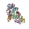
| Output model |  PDB-7fas: |
 Movie
Movie Controller






 Z (Sec.)
 Y (Row.)
 X (Col.)


