 Movie
Movie Controller

[English] 日本語
 Yorodumi
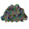
- EMDB-31393: Structure of the phycobilisome from the red alga Porphyridium pur... -
+ Open data

- Basic information
| Entry |  | |||||||||
|---|---|---|---|---|---|---|---|---|---|---|
| Title | Structure of the phycobilisome from the red alga Porphyridium purpureum in Middle Light | |||||||||
 Map data Map data | ||||||||||
 Sample Sample |
| |||||||||
 Keywords Keywords | light-harvesting complex / photosynthesis | |||||||||
| Function / homology |  Function and homology information Function and homology informationphycobilisome / chloroplast thylakoid membrane / photosynthesis / endomembrane system / lyase activity Similarity search - Function | |||||||||
| Biological species |  Porphyridium purpureum (eukaryote) Porphyridium purpureum (eukaryote) | |||||||||
| Method | single particle reconstruction / cryo EM / Resolution: 3.0 Å | |||||||||
 Authors Authors | Ma JF / Sui S-F | |||||||||
| Funding support |  China, 2 items China, 2 items
| |||||||||
 Citation Citation | Journal: Commun Biol / Year: 2023 Title: The structural basis for light acclimation in phycobilisome light harvesting systems systems in Porphyridium purpureum Authors: Dodson EJ / Ma J / Suissa Szlejf M / Maroudas-Sklare N / Paltiel Y / Adir N / Sun S / Sui SF / Keren N | |||||||||
| History |
|
- Structure visualization
| Supplemental images |
|---|

- Downloads & links
-EMDB archive
| Map data | emd_31393.map.gz | 92 MB | EMDB map data format | |
|---|---|---|---|---|
| Header (meta data) | emd-31393-v30.xmlemd-31393.xml | 60.4 KB 60.4 KB | Display Display | EMDB header |
| Images |  emd_31393.png emd_31393.png | 123.8 KB | ||
| Filedesc metadata | emd-31393.cif.gz | 13.1 KB | ||
| Archive directory |  http://ftp.pdbj.org/pub/emdb/structures/EMD-31393ftp://ftp.pdbj.org/pub/emdb/structures/EMD-31393 http://ftp.pdbj.org/pub/emdb/structures/EMD-31393ftp://ftp.pdbj.org/pub/emdb/structures/EMD-31393 | HTTPS FTP |
-Related structure data
| Related structure data |  7ezxMC C: citing same article ( M: atomic model generated by this map |
|---|---|
| Similar structure data |


-Links
| EMDB pages | EMDB (EBI/PDBe) / EMDataResource |
|---|
-Map
| File | Download / File: emd_31393.map.gz / Format: CCP4 / Size: 744.3 MB / Type: IMAGE STORED AS FLOATING POINT NUMBER (4 BYTES) | ||||||||||||||||||||||||||||||||||||
|---|---|---|---|---|---|---|---|---|---|---|---|---|---|---|---|---|---|---|---|---|---|---|---|---|---|---|---|---|---|---|---|---|---|---|---|---|---|
| Projections & slices | Image control
Images are generated by Spider. | ||||||||||||||||||||||||||||||||||||
| Voxel size | X=Y=Z: 1.066 Å | ||||||||||||||||||||||||||||||||||||
| Density |
| ||||||||||||||||||||||||||||||||||||
| Symmetry | Space group: 1 | ||||||||||||||||||||||||||||||||||||
| Details | EMDB XML:
|
 Z (Sec.)
 Y (Row.)
 X (Col.)


-Supplemental data
- Sample components
+Entire : phycobilisome
+Supramolecule #1: phycobilisome
+Macromolecule #1: Phycobilisome 31.8 kDa linker polypeptide, phycoerythrin-associat...
+Macromolecule #2: R-phycoerythrin gamma chain, chloroplastic
+Macromolecule #3: Phycobilisome rod-core linker polypeptide
+Macromolecule #4: C-phycocyanin alpha subunit
+Macromolecule #5: C-phycocyanin beta subunit
+Macromolecule #6: Phycoerythrin alpha subunit
+Macromolecule #7: B-phycoerythrin beta chain
+Macromolecule #8: Phycobilisome 31.8 kDa linker polypeptide, phycoerythrin-associat...
+Macromolecule #9: Phycobilisome 31.8 kDa linker polypeptide, phycoerythrin-associat...
+Macromolecule #10: Phycobilisome 27.9 kDa linker polypeptide, phycoerythrin-associat...
+Macromolecule #11: CaRSPs2
+Macromolecule #12: CaRSPs1
+Macromolecule #13: FAS1 domain-containing protein
+Macromolecule #14: R-phycoerythrin gamma chain, chloroplastic
+Macromolecule #15: Phycobilisome 32.1 kDa linker polypeptide, phycocyanin-associated, rod
+Macromolecule #16: R-phycoerythrin gamma chain, chloroplastic
+Macromolecule #17: R-phycoerythrin gamma chain, chloroplastic
+Macromolecule #18: Phycobilisome 27.9 kDa linker polypeptide, phycoerythrin-associat...
+Macromolecule #19: Phycobilisome 31.8 kDa linker polypeptide, phycoerythrin-associat...
+Macromolecule #20: Allophycocyanin alpha subunit
+Macromolecule #21: Allophycocyanin beta subunit
+Macromolecule #22: Allophycocyanin gamma subunit
+Macromolecule #23: Allophycocyanin beta 18 subunit
+Macromolecule #24: Phycobilisome 7.8 kDa linker polypeptide, allophycocyanin-associa...
+Macromolecule #25: Phycobilisome linker polypeptide
+Macromolecule #26: Lrc4
+Macromolecule #27: LRC5
+Macromolecule #28: FAS1 domain-containing protein
+Macromolecule #29: PHYCOERYTHROBILIN

+Macromolecule #30: PHYCOUROBILIN
+Macromolecule #31: PHYCOCYANOBILIN

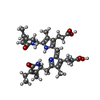
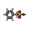
+Macromolecule #32: phenylmethanesulfonic acid
-Experimental details
-Structure determination
| Method | cryo EM |
|---|---|
 Processing Processing | single particle reconstruction |
| Aggregation state | particle |
-Sample preparation
| Buffer | pH: 7 Component:
| |||||||||
|---|---|---|---|---|---|---|---|---|---|---|
| Grid | Model: PELCO Ultrathin Carbon with Lacey Carbon / Material: COPPER / Mesh: 200 / Support film - Material: CARBON / Support film - topology: LACEY / Support film - Film thickness: 2 / Pretreatment - Type: GLOW DISCHARGE / Pretreatment - Time: 30 sec. / Pretreatment - Atmosphere: AIR / Pretreatment - Pressure: 0.1 kPa | |||||||||
| Vitrification | Cryogen name: ETHANE / Chamber humidity: 100 % / Chamber temperature: 18 K / Instrument: FEI VITROBOT MARK IV |
- Electron microscopy
| Microscope | FEI TITAN KRIOS |
|---|---|
| Temperature | Min: 70.0 K / Max: 70.0 K |
| Specialist optics | Energy filter - Name: GIF Quantum ER |
| Image recording | Film or detector model: GATAN K2 SUMMIT (4k x 4k) / Detector mode: SUPER-RESOLUTION / Digitization - Dimensions - Width: 4096 pixel / Digitization - Dimensions - Height: 4096 pixel / Digitization - Frames/image: 1-32 / Number grids imaged: 2 / Number real images: 6438 / Average exposure time: 8.2 sec. / Average electron dose: 50.0 e/Å2 |
| Electron beam | Acceleration voltage: 300 kV / Electron source:  FIELD EMISSION GUN FIELD EMISSION GUN |
| Electron optics | C2 aperture diameter: 100.0 µm / Calibrated defocus max: 2.3000000000000003 µm / Calibrated defocus min: 1.3 µm / Calibrated magnification: 130000 / Illumination mode: FLOOD BEAM / Imaging mode: BRIGHT FIELD / Cs: 2.7 mm / Nominal defocus max: 2.3000000000000003 µm / Nominal defocus min: 1.3 µm / Nominal magnification: 130000 |
| Sample stage | Specimen holder model: FEI TITAN KRIOS AUTOGRID HOLDER / Cooling holder cryogen: NITROGEN |
| Experimental equipment |  Model: Titan Krios / Image courtesy: FEI Company |
+Image processing
-Atomic model buiding 1
| Initial model | PDB ID: Chain - Source name: PDB / Chain - Initial model type: experimental model |
|---|---|
| Refinement | Space: REAL / Protocol: RIGID BODY FIT / Overall B value: 62.05 / Target criteria: 0,99 |
| Output model | PDB-7ezx: |
