 Movie
Movie Controller

[English] 日本語
 Yorodumi
- EMDB-34534: Un-sharpened map of phycobilisome from Porphyridium purpureum und... -
+ Open data

- Basic information
| Entry |  | |||||||||
|---|---|---|---|---|---|---|---|---|---|---|
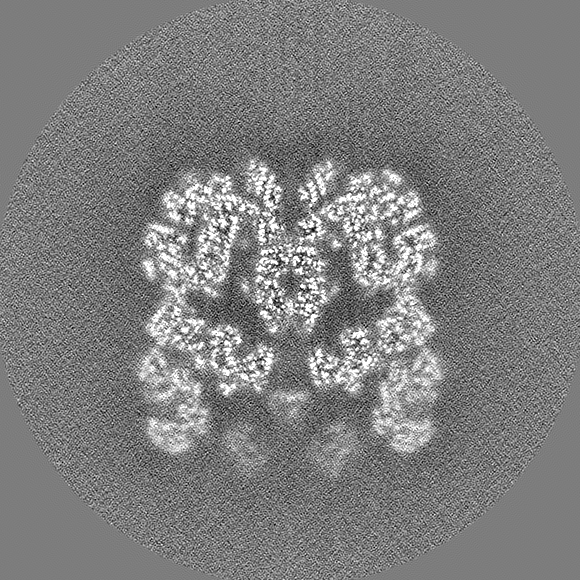
| Title | Un-sharpened map of phycobilisome from Porphyridium purpureum under Middle light | |||||||||
 Map data Map data | Un-sharpened map of phycobilisome under Middle light | |||||||||
 Sample Sample |
| |||||||||
 Keywords Keywords | light-harvesting complex / photosynthesis | |||||||||
| Biological species |  Porphyridium purpureum (eukaryote) Porphyridium purpureum (eukaryote) | |||||||||
| Method | single particle reconstruction / cryo EM / Resolution: 3.6 Å | |||||||||
 Authors Authors | Ma JF / Sui S-F | |||||||||
| Funding support |  China, 2 items China, 2 items
| |||||||||
 Citation Citation | Journal: Commun Biol / Year: 2023 Title: The structural basis for light acclimation in phycobilisome light harvesting systems systems in Porphyridium purpureum. Authors: Emma Joy Dodson / Jianfei Ma / Maayan Suissa Szlejf / Naama Maroudas-Sklare / Yossi Paltiel / Noam Adir / Shan Sun / Sen-Fang Sui / Nir Keren /  Abstract: Photosynthetic organisms adapt to changing light conditions by manipulating their light harvesting complexes. Biophysical, biochemical, physiological and genetic aspects of these processes are ...Photosynthetic organisms adapt to changing light conditions by manipulating their light harvesting complexes. Biophysical, biochemical, physiological and genetic aspects of these processes are studied extensively. The structural basis for these studies is lacking. In this study we address this gap in knowledge by focusing on phycobilisomes (PBS), which are large structures found in cyanobacteria and red algae. In this study we focus on the phycobilisomes (PBS), which are large structures found in cyanobacteria and red algae. Specifically, we examine red algae (Porphyridium purpureum) grown under a low light intensity (LL) and a medium light intensity (ML). Using cryo-electron microscopy, we resolve the structure of ML-PBS and compare it to the LL-PBS structure. The ML-PBS is 13.6 MDa, while the LL-PBS is larger (14.7 MDa). The LL-PBS structure have a higher number of closely coupled chromophore pairs, potentially the source of the red shifted fluorescence emission from LL-PBS. Interestingly, these differences do not significantly affect fluorescence kinetics parameters. This indicates that PBS systems can maintain similar fluorescence quantum yields despite an increase in LL-PBS chromophore numbers. These findings provide a structural basis to the processes by which photosynthetic organisms adapt to changing light conditions. | |||||||||
| History |
|
- Structure visualization
| Supplemental images |
|---|

- Downloads & links
-EMDB archive
| Map data | emd_34534.map.gz | 597.8 MB |  EMDB map data format EMDB map data format | |
|---|---|---|---|---|
| Header (meta data) | emd-34534-v30.xmlemd-34534.xml | 21.4 KB 21.4 KB | Display Display | EMDB header |
| Images | 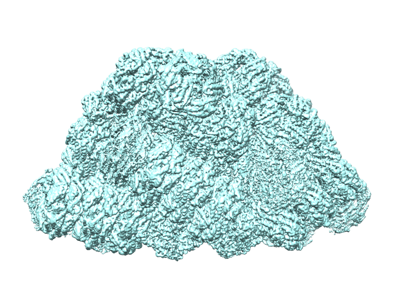 emd_34534.png emd_34534.png | 143 KB | ||
| Filedesc metadata | emd-34534.cif.gz | 4.7 KB | ||
| Others | emd_34534_half_map_1.map.gzemd_34534_half_map_2.map.gz | 697.6 MB 697.6 MB | ||
| Archive directory |  http://ftp.pdbj.org/pub/emdb/structures/EMD-34534ftp://ftp.pdbj.org/pub/emdb/structures/EMD-34534 http://ftp.pdbj.org/pub/emdb/structures/EMD-34534ftp://ftp.pdbj.org/pub/emdb/structures/EMD-34534 | HTTPS FTP |
-Related structure data
| Related structure data |
|---|

-Links
| EMDB pages | EMDB (EBI/PDBe) / EMDataResource |
|---|
-Map
| File | Download / File: emd_34534.map.gz / Format: CCP4 / Size: 744.3 MB / Type: IMAGE STORED AS FLOATING POINT NUMBER (4 BYTES) | ||||||||||||||||||||||||||||||||||||
|---|---|---|---|---|---|---|---|---|---|---|---|---|---|---|---|---|---|---|---|---|---|---|---|---|---|---|---|---|---|---|---|---|---|---|---|---|---|
| Annotation | Un-sharpened map of phycobilisome under Middle light | ||||||||||||||||||||||||||||||||||||
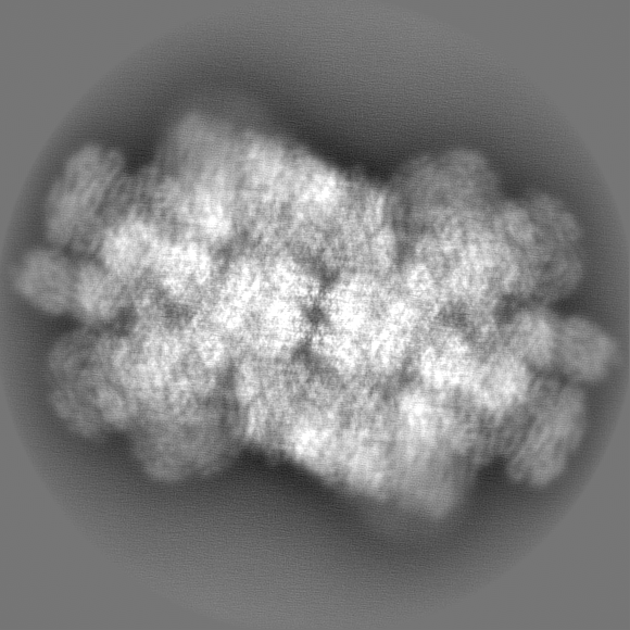
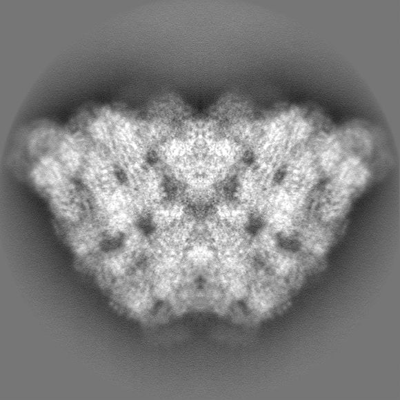
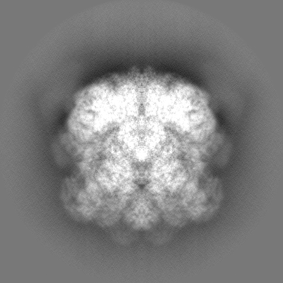
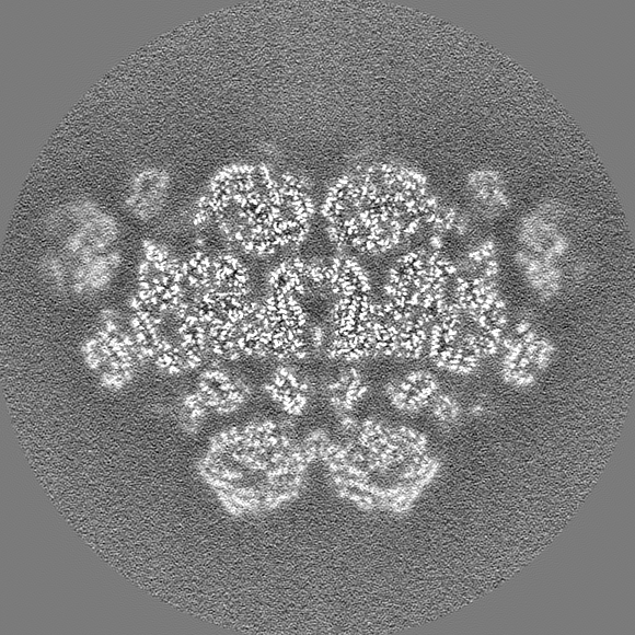
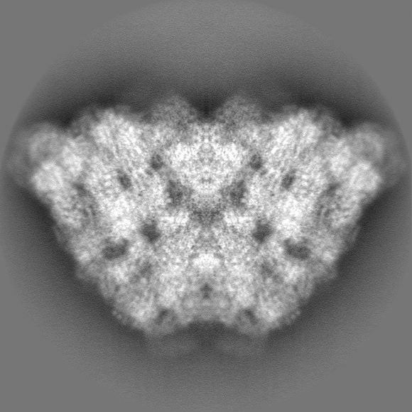
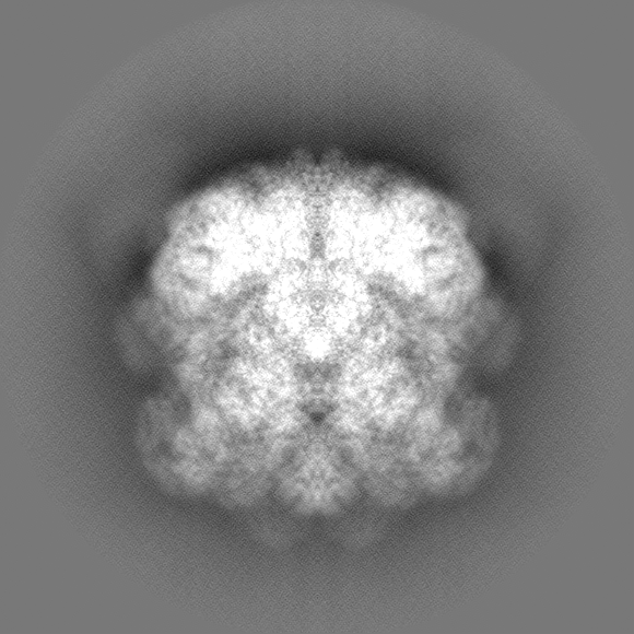
| Projections & slices | Image control
Images are generated by Spider. | ||||||||||||||||||||||||||||||||||||
| Voxel size | X=Y=Z: 1.066 Å | ||||||||||||||||||||||||||||||||||||
| Density |
| ||||||||||||||||||||||||||||||||||||
| Symmetry | Space group: 1 | ||||||||||||||||||||||||||||||||||||
| Details | EMDB XML:
|
 Z (Sec.)
 Y (Row.)
 X (Col.)


-Supplemental data
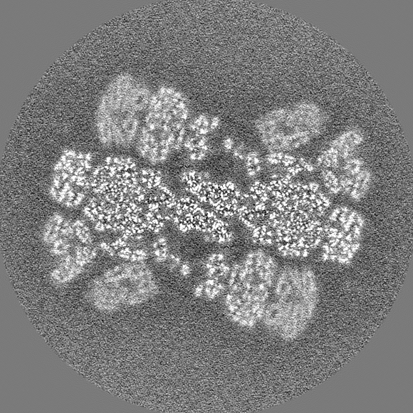
-Half map: Un-sharpened map of phycobilisome under Middle light half2
| File | emd_34534_half_map_1.map | ||||||||||||
|---|---|---|---|---|---|---|---|---|---|---|---|---|---|
| Annotation | Un-sharpened map of phycobilisome under Middle light half2 | ||||||||||||
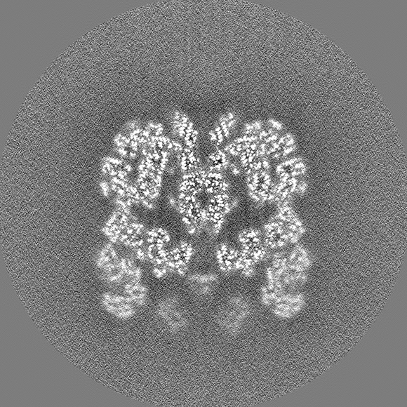
| Projections & Slices |
| ||||||||||||
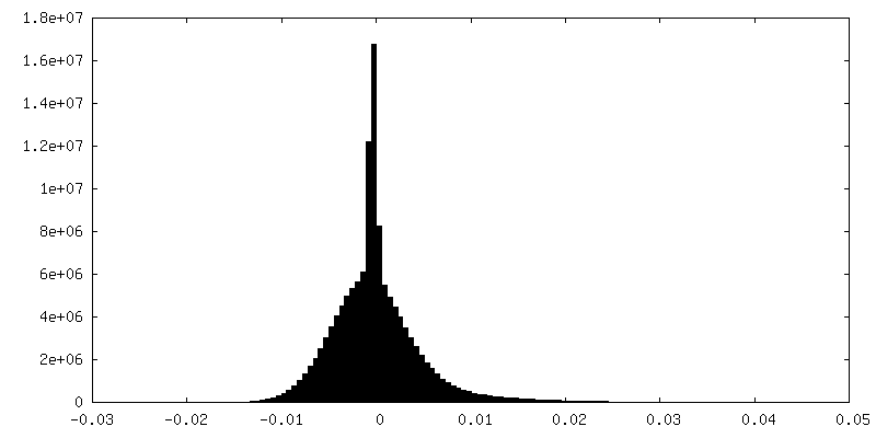
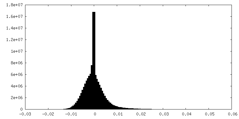
| Density Histograms |

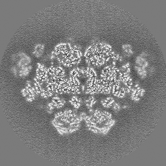
-Half map: Un-sharpened map of phycobilisome under Middle light half1
| File | emd_34534_half_map_2.map | ||||||||||||
|---|---|---|---|---|---|---|---|---|---|---|---|---|---|
| Annotation | Un-sharpened map of phycobilisome under Middle light half1 | ||||||||||||
| Projections & Slices |
| ||||||||||||
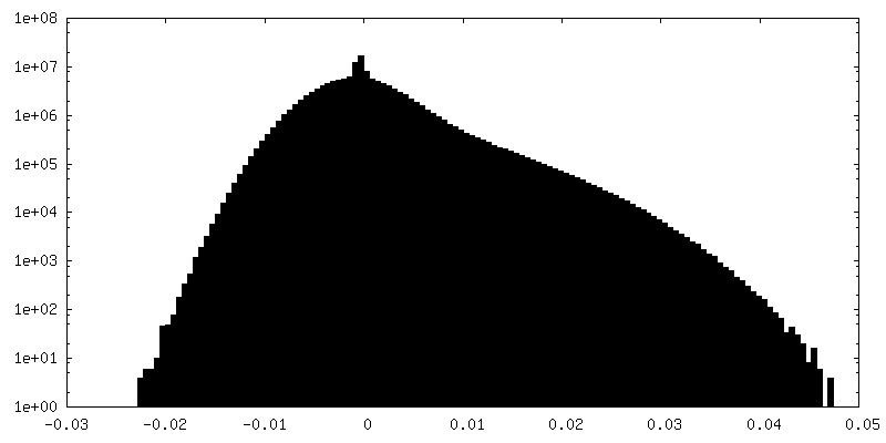
| Density Histograms |


- Sample components
-Entire : phycobilisome
| Entire | Name: phycobilisome |
|---|---|
| Components |
|
-Supramolecule #1: phycobilisome
| Supramolecule | Name: phycobilisome / type: complex / ID: 1 / Parent: 0 / Macromolecule list: #1-#28 |
|---|---|
| Source (natural) | Organism: Porphyridium purpureum (eukaryote) |
| Molecular weight | Theoretical: 14700 kDa/nm |
-Experimental details
-Structure determination
| Method | cryo EM |
|---|---|
 Processing Processing | single particle reconstruction |
| Aggregation state | particle |
-Sample preparation
| Buffer | pH: 7 Component:
| |||||||||
|---|---|---|---|---|---|---|---|---|---|---|
| Grid | Model: PELCO Ultrathin Carbon with Lacey Carbon / Support film - Material: CARBON / Support film - topology: LACEY / Support film - Film thickness: 2 | |||||||||
| Vitrification | Cryogen name: ETHANE / Chamber humidity: 100 % / Chamber temperature: 18 K / Instrument: FEI VITROBOT MARK IV |
- Electron microscopy
| Microscope | FEI TITAN KRIOS |
|---|---|
| Temperature | Min: 70.0 K / Max: 70.0 K |
| Specialist optics | Energy filter - Name: GIF Quantum ER |
| Image recording | Film or detector model: GATAN K2 SUMMIT (4k x 4k) / Detector mode: SUPER-RESOLUTION / Digitization - Dimensions - Width: 4096 pixel / Digitization - Dimensions - Height: 4096 pixel / Digitization - Frames/image: 1-32 / Number grids imaged: 2 / Number real images: 6438 / Average exposure time: 8.2 sec. / Average electron dose: 50.0 e/Å2 |
| Electron beam | Acceleration voltage: 300 kV / Electron source:  FIELD EMISSION GUN FIELD EMISSION GUN |
| Electron optics | C2 aperture diameter: 100.0 µm / Calibrated defocus max: 2.3000000000000003 µm / Calibrated defocus min: 1.3 µm / Calibrated magnification: 130000 / Illumination mode: FLOOD BEAM / Imaging mode: BRIGHT FIELD / Cs: 2.7 mm / Nominal defocus max: 2.3000000000000003 µm / Nominal defocus min: 1.3 µm / Nominal magnification: 130000 |
| Sample stage | Specimen holder model: FEI TITAN KRIOS AUTOGRID HOLDER / Cooling holder cryogen: NITROGEN |
| Experimental equipment |  Model: Titan Krios / Image courtesy: FEI Company |
+Image processing
-Atomic model buiding 1
| Initial model | PDB ID: Chain - Source name: PDB / Chain - Initial model type: experimental model |
|---|---|
| Refinement | Space: REAL / Protocol: RIGID BODY FIT / Overall B value: 62.05 / Target criteria: 0,99 |
