Movie
Movie Controller

[English] 日本語
 Yorodumi
- EMDB-31246: Cryo-electron tomogram of phycobilisome-photosystem II supercompl... -
+ Open data

- Basic information
| Entry |  | |||||||||||||||
|---|---|---|---|---|---|---|---|---|---|---|---|---|---|---|---|---|
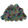
| Title | Cryo-electron tomogram of phycobilisome-photosystem II supercomplex on the thylakoid | |||||||||||||||
 Map data Map data | Cryo-electron tomogram of phycobilisome-photosystem II supercomplex on the thylakoid | |||||||||||||||
 Sample Sample |
| |||||||||||||||
 Keywords Keywords | phycobilisome / photosystem II / thylakoid / PHOTOSYNTHESIS | |||||||||||||||
| Biological species |  Porphyridium purpureum (eukaryote) Porphyridium purpureum (eukaryote) | |||||||||||||||
| Method | electron tomography / cryo EM | |||||||||||||||
 Authors Authors | Sui SF / Li XM / Li MJ / Ma JF | |||||||||||||||
| Funding support |  China, 4 items China, 4 items
| |||||||||||||||
 Citation Citation | Journal: To Be Published Title: In situ cryo-ET structure of phycobilisome-photosystem II supercomplex from red alga Authors: Sui SF / Li XM / Li MJ / Ma JF | |||||||||||||||
| History |
|
- Structure visualization
| Supplemental images |
|---|

- Downloads & links
-EMDB archive
| Map data | emd_31246.map.gz | 463.1 MB |  EMDB map data format EMDB map data format | |
|---|---|---|---|---|
| Header (meta data) | emd-31246-v30.xmlemd-31246.xml | 8.6 KB 8.6 KB | Display Display | EMDB header |
| Images |  emd_31246.png emd_31246.png | 354.1 KB | ||
| Filedesc metadata | emd-31246.cif.gz | 3.7 KB | ||
| Archive directory |  http://ftp.pdbj.org/pub/emdb/structures/EMD-31246ftp://ftp.pdbj.org/pub/emdb/structures/EMD-31246 http://ftp.pdbj.org/pub/emdb/structures/EMD-31246ftp://ftp.pdbj.org/pub/emdb/structures/EMD-31246 | HTTPS FTP |
-Links
| EMDB pages | EMDB (EBI/PDBe) / EMDataResource |
|---|
-Map
| File | Download / File: emd_31246.map.gz / Format: CCP4 / Size: 535.8 MB / Type: IMAGE STORED AS FLOATING POINT NUMBER (4 BYTES) | ||||||||||||||||||||||||||||||||
|---|---|---|---|---|---|---|---|---|---|---|---|---|---|---|---|---|---|---|---|---|---|---|---|---|---|---|---|---|---|---|---|---|---|
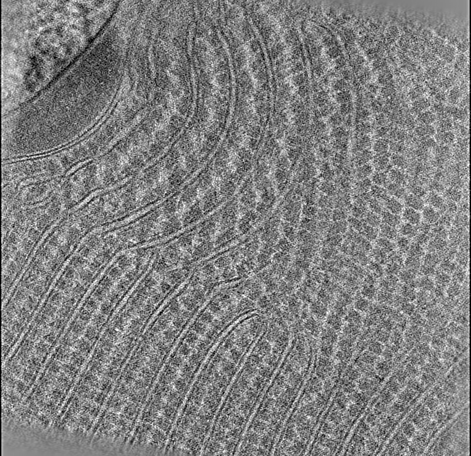
| Annotation | Cryo-electron tomogram of phycobilisome-photosystem II supercomplex on the thylakoid | ||||||||||||||||||||||||||||||||
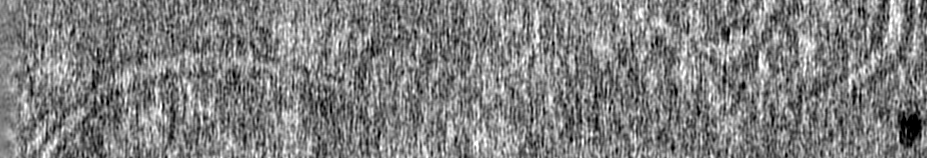
| Projections & slices | Image control
Images are generated by Spider. generated in cubic-lattice coordinate | ||||||||||||||||||||||||||||||||
| Voxel size | X=Y=Z: 13.684 Å | ||||||||||||||||||||||||||||||||
| Density |
| ||||||||||||||||||||||||||||||||
| Symmetry | Space group: 1 | ||||||||||||||||||||||||||||||||
| Details | EMDB XML:
|
 Z (Sec.)
 Y (Row.)
 X (Col.)


-Supplemental data
- Sample components
-Entire : phycobilisome-photosystem II supercomplex
| Entire | Name: phycobilisome-photosystem II supercomplex |
|---|---|
| Components |
|
-Supramolecule #1: phycobilisome-photosystem II supercomplex
| Supramolecule | Name: phycobilisome-photosystem II supercomplex / type: cell / ID: 1 / Parent: 0 / Details: phycobilisome, photosystem II |
|---|---|
| Source (natural) | Organism: Porphyridium purpureum (eukaryote) |
-Experimental details
-Structure determination
| Method | cryo EM |
|---|---|
 Processing Processing | electron tomography |
| Aggregation state | cell |
-Sample preparation
| Buffer | pH: 7 |
|---|---|
| Vitrification | Cryogen name: ETHANE |
| Sectioning | Focused ion beam - Instrument: OTHER / Focused ion beam - Ion: OTHER / Focused ion beam - Voltage: 30 / Focused ion beam - Current: 0.1 / Focused ion beam - Duration: 3600 / Focused ion beam - Temperature: 191 K / Focused ion beam - Initial thickness: 1000 / Focused ion beam - Final thickness: 200 Focused ion beam - Details: The value given for _em_focused_ion_beam.instrument is FEI Quanta FIB. This is not in a list of allowed values {'DB235', 'OTHER'} so OTHER is written into the XML file. |
- Electron microscopy
| Microscope | FEI TITAN KRIOS |
|---|---|
| Image recording | Film or detector model: GATAN K2 SUMMIT (4k x 4k) / Average electron dose: 120.0 e/Å2 |
| Electron beam | Acceleration voltage: 300 kV / Electron source:  FIELD EMISSION GUN FIELD EMISSION GUN |
| Electron optics | Illumination mode: FLOOD BEAM / Imaging mode: BRIGHT FIELD |
| Experimental equipment |  Model: Titan Krios / Image courtesy: FEI Company |
-Image processing
| Final reconstruction | Number images used: 42 |
|---|