National Natural Science Foundation of China (NSFC)
China
Citation
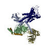
Journal: Nature / Year: 2022 Title: Structural basis for the tethered peptide activation of adhesion GPCRs. Authors: Yu-Qi Ping / Peng Xiao / Fan Yang / Ru-Jia Zhao / Sheng-Chao Guo / Xu Yan / Xiang Wu / Chao Zhang / Yan Lu / Fenghui Zhao / Fulai Zhou / Yue-Tong Xi / Wanchao Yin / Feng-Zhen Liu / Dong-Fang ...Authors: Yu-Qi Ping / Peng Xiao / Fan Yang / Ru-Jia Zhao / Sheng-Chao Guo / Xu Yan / Xiang Wu / Chao Zhang / Yan Lu / Fenghui Zhao / Fulai Zhou / Yue-Tong Xi / Wanchao Yin / Feng-Zhen Liu / Dong-Fang He / Dao-Lai Zhang / Zhong-Liang Zhu / Yi Jiang / Lutao Du / Shi-Qing Feng / Torsten Schöneberg / Ines Liebscher / H Eric Xu / Jin-Peng Sun / Abstract: Adhesion G-protein-coupled receptors (aGPCRs) are important for organogenesis, neurodevelopment, reproduction and other processes. Many aGPCRs are activated by a conserved internal (tethered) agonist ...Adhesion G-protein-coupled receptors (aGPCRs) are important for organogenesis, neurodevelopment, reproduction and other processes. Many aGPCRs are activated by a conserved internal (tethered) agonist sequence known as the Stachel sequence. Here, we report the cryogenic electron microscopy (cryo-EM) structures of two aGPCRs in complex with G: GPR133 and GPR114. The structures indicate that the Stachel sequences of both receptors assume an α-helical-bulge-β-sheet structure and insert into a binding site formed by the transmembrane domain (TMD). A hydrophobic interaction motif (HIM) within the Stachel sequence mediates most of the intramolecular interactions with the TMD. Combined with the cryo-EM structures, biochemical characterization of the HIM motif provides insight into the cross-reactivity and selectivity of the Stachel sequences. Two interconnected mechanisms, the sensing of Stachel sequences by the conserved 'toggle switch' W and the constitution of a hydrogen-bond network formed by Q/Y and the P/VφφG motif (φ indicates a hydrophobic residue), are important in Stachel sequence-mediated receptor activation and G coupling. Notably, this network stabilizes kink formation in TM helices 6 and 7 (TM6 and TM7, respectively). A common G-binding interface is observed between the two aGPCRs, and GPR114 has an extended TM7 that forms unique interactions with G. Our structures reveal the detailed mechanisms of aGPCR activation by Stachel sequences and their G coupling.
In the structure databanks used in Yorodumi, some data are registered as the other names, "COVID-19 virus" and "2019-nCoV". Here are the details of the virus and the list of structure data.
Jan 31, 2019. EMDB accession codes are about to change! (news from PDBe EMDB page)
EMDB accession codes are about to change! (news from PDBe EMDB page)
The allocation of 4 digits for EMDB accession codes will soon come to an end. Whilst these codes will remain in use, new EMDB accession codes will include an additional digit and will expand incrementally as the available range of codes is exhausted. The current 4-digit format prefixed with “EMD-” (i.e. EMD-XXXX) will advance to a 5-digit format (i.e. EMD-XXXXX), and so on. It is currently estimated that the 4-digit codes will be depleted around Spring 2019, at which point the 5-digit format will come into force.
The EM Navigator/Yorodumi systems omit the EMD- prefix.
Related info.:Q: What is EMD? / ID/Accession-code notation in Yorodumi/EM Navigator
Yorodumi is a browser for structure data from EMDB, PDB, SASBDB, etc.
This page is also the successor to EM Navigator detail page, and also detail information page/front-end page for Omokage search.
The word "yorodu" (or yorozu) is an old Japanese word meaning "ten thousand". "mi" (miru) is to see.
Related info.:EMDB / PDB / SASBDB / Comparison of 3 databanks / Yorodumi Search / Aug 31, 2016. New EM Navigator & Yorodumi / Yorodumi Papers / Jmol/JSmol / Function and homology information / Changes in new EM Navigator and Yorodumi
 Movie
Movie Controller

 Yorodumi
 Open data
 Basic information
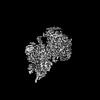
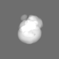
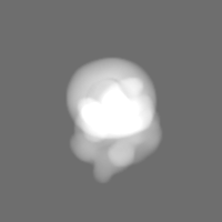
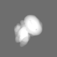
 Map data
 Sample
 Function and homology information
 Homo sapiens (human)
 Authors
 China, 1 items
  Citation
 Structure visualization
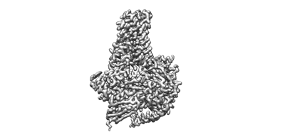
 Downloads & links
 emd_31232.png
 http://ftp.pdbj.org/pub/emdb/structures/EMD-31232
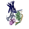


 Z (Sec.)
 Y (Row.)
 X (Col.)


 Sample components
 Spodoptera frugiperda (fall armyworm)
 Processing
 Electron microscopy
 FIELD EMISSION GUN
