- EMDB-30910: Structure of Drosophila melanogaster GlcNAc-1-phosphotransferase -
+
Open data
ID or keywords:
Loading...
-
Basic information
Entry
Database: EMDB / ID: EMD-30910
Title
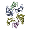
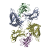
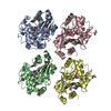
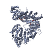
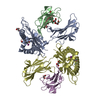
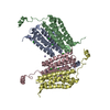
Structure of Drosophila melanogaster GlcNAc-1-phosphotransferase
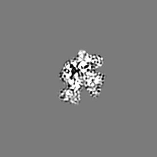
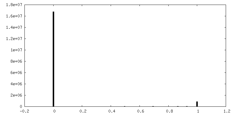
Map data
Sample
Complex: gnpt
Protein or peptide: FI02838p
Ligand: CALCIUM ION
Keywords
GNPTAB / GlcNAc-1-phosphotransferase / SUGAR BINDING PROTEIN / TRANSFERASE
Function / homology
Function and homology information
N-glycan processing to lysosome / UDP-N-acetylglucosamine-lysosomal-enzyme N-acetylglucosaminephosphotransferase activity / Golgi apparatus / membrane Similarity search - Function
Stealth protein CR2, conserved region 2 / Stealth protein CR4, conserved region 4 / Stealth protein CR3, conserved region 3 / Stealth protein CR1, conserved region 1 / : / Stealth protein CR2, conserved region 2 / Stealth protein CR1, conserved region 1 / Stealth protein CR3, conserved region 3 / Stealth protein CR4, conserved region 4 / Notch-like domain superfamily ...Stealth protein CR2, conserved region 2 / Stealth protein CR4, conserved region 4 / Stealth protein CR3, conserved region 3 / Stealth protein CR1, conserved region 1 / : / Stealth protein CR2, conserved region 2 / Stealth protein CR1, conserved region 1 / Stealth protein CR3, conserved region 3 / Stealth protein CR4, conserved region 4 / Notch-like domain superfamily / LNR domain / LNR (Lin-12/Notch) repeat profile. / Notch domain / Domain found in Notch and Lin-12 Similarity search - Domain/homology
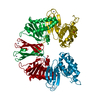
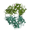
Journal: J Biol Chem / Year: 2022 Title: Structural insights into how GlcNAc-1-phosphotransferase directs lysosomal protein transport. Authors: Shuo Du / Guopeng Wang / Zhiying Zhang / Chengying Ma / Ning Gao / Junyu Xiao / Abstract: GlcNAc-1-phosphotransferase catalyzes the initial step in the formation of the mannose-6-phosphate tag that labels ∼60 lysosomal proteins for transport. Mutations in GlcNAc-1-phosphotransferase are ...GlcNAc-1-phosphotransferase catalyzes the initial step in the formation of the mannose-6-phosphate tag that labels ∼60 lysosomal proteins for transport. Mutations in GlcNAc-1-phosphotransferase are known to cause lysosomal storage disorders such as mucolipidoses. However, the molecular mechanism of GlcNAc-1-phosphotransferase activity remains unclear. Mammalian GlcNAc-1-phosphotransferases are α2β2γ2 hexamers in which the core catalytic α- and β-subunits are derived from the GNPTAB (N-acetylglucosamine-1-phosphate transferase subunits alpha and beta) gene. Here, we present the cryo-electron microscopy structure of the Drosophila melanogaster GNPTAB homolog, DmGNPTAB. We identified four conserved regions located far apart in the sequence that fold into the catalytic domain, which exhibits structural similarity to that of the UDP-glucose glycoprotein glucosyltransferase. Comparison with UDP-glucose glycoprotein glucosyltransferase also revealed a putative donor substrate-binding site, and the functional requirements of critical residues in human GNPTAB were validated using GNPTAB-knockout cells. Finally, we show that DmGNPTAB forms a homodimer that is evolutionarily conserved and that perturbing the dimer interface undermines the maturation and activity of human GNPTAB. These results provide important insights into GlcNAc-1-phosphotransferase function and related diseases.
History
Deposition
Jan 19, 2021
-
Header (metadata) release
Feb 2, 2022
-
Map release
Feb 2, 2022
-
Update
Jun 25, 2025
-
Current status
Jun 25, 2025
Processing site: PDBj / Status: Released
-
Structure visualization
Movie
Surface view with section colored by density value
In the structure databanks used in Yorodumi, some data are registered as the other names, "COVID-19 virus" and "2019-nCoV". Here are the details of the virus and the list of structure data.
Jan 31, 2019. EMDB accession codes are about to change! (news from PDBe EMDB page)
EMDB accession codes are about to change! (news from PDBe EMDB page)
The allocation of 4 digits for EMDB accession codes will soon come to an end. Whilst these codes will remain in use, new EMDB accession codes will include an additional digit and will expand incrementally as the available range of codes is exhausted. The current 4-digit format prefixed with “EMD-” (i.e. EMD-XXXX) will advance to a 5-digit format (i.e. EMD-XXXXX), and so on. It is currently estimated that the 4-digit codes will be depleted around Spring 2019, at which point the 5-digit format will come into force.
The EM Navigator/Yorodumi systems omit the EMD- prefix.
Related info.:Q: What is EMD? / ID/Accession-code notation in Yorodumi/EM Navigator
Yorodumi is a browser for structure data from EMDB, PDB, SASBDB, etc.
This page is also the successor to EM Navigator detail page, and also detail information page/front-end page for Omokage search.
The word "yorodu" (or yorozu) is an old Japanese word meaning "ten thousand". "mi" (miru) is to see.
Related info.:EMDB / PDB / SASBDB / Comparison of 3 databanks / Yorodumi Search / Aug 31, 2016. New EM Navigator & Yorodumi / Yorodumi Papers / Jmol/JSmol / Function and homology information / Changes in new EM Navigator and Yorodumi
 Movie
Movie Controller

 Yorodumi
 Open data
 Basic information
 Map data
 Sample
 Keywords
 Function and homology information
 Authors
 Citation
 Structure visualization
 Downloads & links
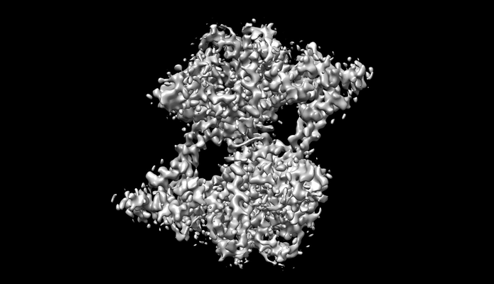 emd_30910.png
 http://ftp.pdbj.org/pub/emdb/structures/EMD-30910
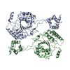


 Z (Sec.)
 Y (Row.)
 X (Col.)


 Sample components
 Processing
 Electron microscopy
 FIELD EMISSION GUN
