 Movie
Movie Controller


+ Open data

- Basic information
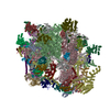
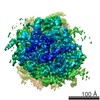
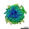
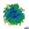
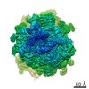
| Entry | Database: EMDB / ID: EMD-30868 | |||||||||
|---|---|---|---|---|---|---|---|---|---|---|
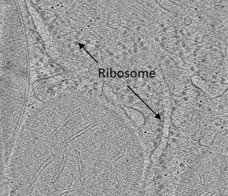
| Title | liver ribosome | |||||||||
 Map data Map data | ||||||||||
 Sample Sample |
| |||||||||
| Biological species | Ribosome display vector pRDV (others) | |||||||||
| Method | subtomogram averaging / cryo EM / Resolution: 18.0 Å | |||||||||
 Authors Authors | Zhang JG / Zhang DY / Sun L / Ji G / Huang XJ / Niu TX / Xu JS / Ma CY / Zhu Y / Gao N ...Zhang JG / Zhang DY / Sun L / Ji G / Huang XJ / Niu TX / Xu JS / Ma CY / Zhu Y / Gao N / Xu W / Sun F | |||||||||
 Citation Citation | Journal: J.Struct.Biol. / Year: 2021 Title: VHUT-cryo-FIB, a method to fabricate frozen hydrated lamellae from tissue specimens for in situ cryo-electron tomography Authors: Zhang J / Zhang D / Sun L / Ji G / Huang X / Niu T / Xu J / Ma C / Zhu Y / Gao N / Xu W / Sun F | |||||||||
| History |
|
- Structure visualization
| Movie |
 Movie viewer Movie viewer |
|---|---|
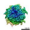
| Structure viewer | EM map: SurfViewMolmilJmol/JSmol |
| Supplemental images |
- Downloads & links
-EMDB archive
| Map data | emd_30868.map.gz | 4.8 MB | EMDB map data format | |
|---|---|---|---|---|
| Header (meta data) | emd-30868-v30.xmlemd-30868.xml | 7.5 KB 7.5 KB | Display Display | EMDB header |
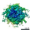
| Images |  emd_30868.png emd_30868.png | 237.7 KB | ||
| Archive directory |  http://ftp.pdbj.org/pub/emdb/structures/EMD-30868ftp://ftp.pdbj.org/pub/emdb/structures/EMD-30868 http://ftp.pdbj.org/pub/emdb/structures/EMD-30868ftp://ftp.pdbj.org/pub/emdb/structures/EMD-30868 | HTTPS FTP |
-Validation report
| Summary document | emd_30868_validation.pdf.gz | 326.8 KB | Display | EMDB validaton report |
|---|---|---|---|---|
| Full document | emd_30868_full_validation.pdf.gz | 326.4 KB | Display | |
| Data in XML | emd_30868_validation.xml.gz | 6.3 KB | Display | |
| Data in CIF | emd_30868_validation.cif.gz | 7.1 KB | Display | |
| Arichive directory | https://ftp.pdbj.org/pub/emdb/validation_reports/EMD-30868ftp://ftp.pdbj.org/pub/emdb/validation_reports/EMD-30868 | HTTPS FTP |
-Related structure data
| Similar structure data | |
|---|---|
| EM raw data | EMPIAR-10713 (Title: VHUT-cryo-FIB, a method to fabricate frozen-hydrated lamella of tissue specimen for in situ cryo-electron tomography Data size: 112.1 Data #1: ungaligned multi-frame micrographs of ribosome [micrographs - multiframe]) |

-Links
| EMDB pages | EMDB (EBI/PDBe) / EMDataResource |
|---|---|
| Related items in Molecule of the Month |

-Map
| File | Download / File: emd_30868.map.gz / Format: CCP4 / Size: 41.7 MB / Type: IMAGE STORED AS FLOATING POINT NUMBER (4 BYTES) | ||||||||||||||||||||||||||||||||||||||||||||||||||||||||||||||||||||
|---|---|---|---|---|---|---|---|---|---|---|---|---|---|---|---|---|---|---|---|---|---|---|---|---|---|---|---|---|---|---|---|---|---|---|---|---|---|---|---|---|---|---|---|---|---|---|---|---|---|---|---|---|---|---|---|---|---|---|---|---|---|---|---|---|---|---|---|---|---|
| Projections & slices | Image control
Images are generated by Spider. | ||||||||||||||||||||||||||||||||||||||||||||||||||||||||||||||||||||
| Voxel size | X=Y=Z: 2.7 Å | ||||||||||||||||||||||||||||||||||||||||||||||||||||||||||||||||||||
| Density |
| ||||||||||||||||||||||||||||||||||||||||||||||||||||||||||||||||||||
| Symmetry | Space group: 1 | ||||||||||||||||||||||||||||||||||||||||||||||||||||||||||||||||||||
| Details | EMDB XML:
CCP4 map header:
| ||||||||||||||||||||||||||||||||||||||||||||||||||||||||||||||||||||
 Z (Sec.)
 Y (Row.)
 X (Col.)


-Supplemental data
- Sample components
-Entire : liver ribosome
| Entire | Name: liver ribosome |
|---|---|
| Components |
|
-Supramolecule #1: liver ribosome
| Supramolecule | Name: liver ribosome / type: complex / ID: 1 / Parent: 0 |
|---|---|
| Source (natural) | Organism: Ribosome display vector pRDV (others) |
-Experimental details
-Structure determination
| Method | cryo EM |
|---|---|
 Processing Processing | subtomogram averaging |
| Aggregation state | tissue |
-Sample preparation
| Buffer | pH: 7 |
|---|---|
| Vitrification | Cryogen name: OTHER |
- Electron microscopy
| Microscope | FEI TITAN |
|---|---|
| Image recording | Film or detector model: GATAN K2 SUMMIT (4k x 4k) / Average electron dose: 3.0 e/Å2 |
| Electron beam | Acceleration voltage: 300 kV / Electron source:  FIELD EMISSION GUN FIELD EMISSION GUN |
| Electron optics | Illumination mode: FLOOD BEAM / Imaging mode: BRIGHT FIELD |
-Image processing
| Final reconstruction | Resolution.type: BY AUTHOR / Resolution: 18.0 Å / Resolution method: FSC 0.143 CUT-OFF / Number subtomograms used: 13 |
|---|---|
| Extraction | Number tomograms: 13 / Number images used: 13 |
| Final angle assignment | Type: MAXIMUM LIKELIHOOD |
