 Movie
Movie Controller

[English] 日本語
 Yorodumi
- EMDB-30712: Cryo-Molecular electron tomography of epidermal growth factor rec... -
+ Open data

- Basic information
| Entry | Database: EMDB / ID: EMD-30712 | |||||||||
|---|---|---|---|---|---|---|---|---|---|---|
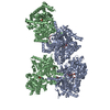
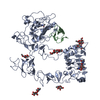
| Title | Cryo-Molecular electron tomography of epidermal growth factor receptor (averaged cluster 1) | |||||||||
 Map data Map data | Unliganded EGFR, averaged cluster 1 | |||||||||
 Sample Sample |
| |||||||||
| Biological species |  EGFR (human) EGFR (human) | |||||||||
| Method | subtomogram averaging / cryo EM / Resolution: 15.0 Å | |||||||||
 Authors Authors | Purba ER / Saita E-I / Akhouri RR / Ofverstedt LG / Wilken G / Skoglund U / Maruyama IN | |||||||||
 Citation Citation | Journal: To Be Published Title: Conformational flexibility transitions of the epidermal growth factor receptor dimer upon activation Authors: Purba ER / Saita E-I / Akhouri RR / Ofverstedt LG / Wilken G / Skoglund U / Maruyama IN | |||||||||
| History |
|
- Structure visualization
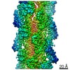
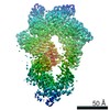
| Movie |
 Movie viewer Movie viewer |
|---|---|
| Structure viewer | EM map: SurfViewMolmilJmol/JSmol |
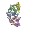
| Supplemental images |

- Downloads & links
-EMDB archive
| Map data | emd_30712.map.gz | 4.2 MB | EMDB map data format | |
|---|---|---|---|---|
| Header (meta data) | emd-30712-v30.xmlemd-30712.xml | 12.8 KB 12.8 KB | Display Display | EMDB header |
| Images |  emd_30712.png emd_30712.png | 12.6 KB | ||
| Archive directory |  http://ftp.pdbj.org/pub/emdb/structures/EMD-30712ftp://ftp.pdbj.org/pub/emdb/structures/EMD-30712 http://ftp.pdbj.org/pub/emdb/structures/EMD-30712ftp://ftp.pdbj.org/pub/emdb/structures/EMD-30712 | HTTPS FTP |
-Related structure data
| Similar structure data |
|---|


-Links
| EMDB pages | EMDB (EBI/PDBe) / EMDataResource |
|---|
-Map
| File | Download / File: emd_30712.map.gz / Format: CCP4 / Size: 5.1 MB / Type: IMAGE STORED AS FLOATING POINT NUMBER (4 BYTES) | ||||||||||||||||||||||||||||||||||||||||||||||||||||||||||||
|---|---|---|---|---|---|---|---|---|---|---|---|---|---|---|---|---|---|---|---|---|---|---|---|---|---|---|---|---|---|---|---|---|---|---|---|---|---|---|---|---|---|---|---|---|---|---|---|---|---|---|---|---|---|---|---|---|---|---|---|---|---|
| Annotation | Unliganded EGFR, averaged cluster 1 | ||||||||||||||||||||||||||||||||||||||||||||||||||||||||||||
| Projections & slices | Image control
Images are generated by Spider. | ||||||||||||||||||||||||||||||||||||||||||||||||||||||||||||
| Voxel size | X=Y=Z: 2.258 Å | ||||||||||||||||||||||||||||||||||||||||||||||||||||||||||||
| Density |
| ||||||||||||||||||||||||||||||||||||||||||||||||||||||||||||
| Symmetry | Space group: 1 | ||||||||||||||||||||||||||||||||||||||||||||||||||||||||||||
| Details | EMDB XML:
CCP4 map header:
| ||||||||||||||||||||||||||||||||||||||||||||||||||||||||||||
 Z (Sec.)
 Y (Row.)
 X (Col.)


-Supplemental data
- Sample components
-Entire : Unliganded EGFR
| Entire | Name: Unliganded EGFR |
|---|---|
| Components |
|
-Supramolecule #1: Unliganded EGFR
| Supramolecule | Name: Unliganded EGFR / type: complex / ID: 1 / Parent: 0 / Macromolecule list: #1 / Details: Unliganded EGFR, subtomogram averaged cluster 1 |
|---|---|
| Source (natural) | Organism: EGFR (human) |
| Recombinant expression | Organism:  |
| Molecular weight | Theoretical: 300 KDa |
-Experimental details
-Structure determination
| Method | cryo EM |
|---|---|
 Processing Processing | subtomogram averaging |
| Aggregation state | particle |
-Sample preparation
| Concentration | 0.5 mg/mL |
|---|---|
| Buffer | pH: 8 / Component - Concentration: 20.0 0.5 / Component - Name: Tris-HCL Details: Solutions were made fresh from concentrated to avoid microbial contamination. |
| Grid | Model: Quantifoil / Material: COPPER / Mesh: 400 / Support film - Material: CARBON / Support film - topology: HOLEY / Pretreatment - Type: PLASMA CLEANING |
| Vitrification | Cryogen name: ETHANE / Chamber humidity: 90 % / Chamber temperature: 298 K / Instrument: FEI VITROBOT MARK I / Details: Blot for 5.5 seconds before plunging. |
| Details | This sample was monodisperse |
- Electron microscopy
| Microscope | FEI TITAN KRIOS |
|---|---|
| Temperature | Min: 70.0 K / Max: 70.0 K |
| Specialist optics | Phase plate: OTHER |
| Image recording | Film or detector model: FEI FALCON II (4k x 4k) / Detector mode: OTHER / Digitization - Dimensions - Width: 2258 pixel / Digitization - Dimensions - Height: 2258 pixel / Average exposure time: 1.8 sec. / Average electron dose: 90.0 e/Å2 |
| Electron beam | Acceleration voltage: 300 kV / Electron source:  FIELD EMISSION GUN FIELD EMISSION GUN |
| Electron optics | C2 aperture diameter: 100.0 µm / Calibrated defocus max: 1.5 µm / Calibrated defocus min: 1.0 µm / Calibrated magnification: 37000 / Illumination mode: FLOOD BEAM / Imaging mode: BRIGHT FIELD / Cs: 2.7 mm / Nominal defocus max: 5.0 µm / Nominal defocus min: -2.0 µm / Nominal magnification: 37000 |
| Sample stage | Specimen holder model: FEI TITAN KRIOS AUTOGRID HOLDER / Cooling holder cryogen: NITROGEN |
| Experimental equipment |  Model: Titan Krios / Image courtesy: FEI Company |
+Image processing
-Atomic model buiding 1
| Initial model | PDB ID: |
|---|---|
| Refinement | Protocol: FLEXIBLE FIT / Target criteria: Correlation coefficient |
-Atomic model buiding 2
| Initial model | PDB ID: |
|---|---|
| Refinement | Protocol: FLEXIBLE FIT / Target criteria: Correlation coefficient |

-Atomic model buiding 3
| Initial model | PDB ID: |
|---|---|
| Refinement | Protocol: FLEXIBLE FIT / Target criteria: Correlation coefficient |

