National Natural Science Foundation of China (NSFC)
31870725
China
Other government
Fundamental Research Funds for the Central Universities (2017EYT19)
China
National Natural Science Foundation of China (NSFC)
31570729
China
National Institutes of Health/National Center for Complementary and Integrative Health (NIH/NCCIH)
R01GM079641
United States
Other government
Memorial Sloan-Kettering Cancer Center Core grant P30CA008748
United States
Other government
Basic Research grant from Shenzhen government to (JCYJ20180302174213122)
China
Citation
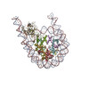
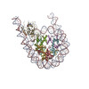
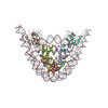
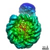
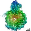
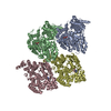
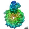
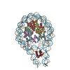
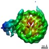
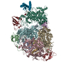
Journal: Nature / Year: 2021 Title: Molecular basis of nucleosomal H3K36 methylation by NSD methyltransferases. Authors: Wanqiu Li / Wei Tian / Gang Yuan / Pujuan Deng / Deepanwita Sengupta / Zhongjun Cheng / Yinghua Cao / Jiahao Ren / Yan Qin / Yuqiao Zhou / Yulin Jia / Or Gozani / Dinshaw J Patel / Zhanxin Wang / Abstract: Histone methyltransferases of the nuclear receptor-binding SET domain protein (NSD) family, including NSD1, NSD2 and NSD3, have crucial roles in chromatin regulation and are implicated in oncogenesis. ...Histone methyltransferases of the nuclear receptor-binding SET domain protein (NSD) family, including NSD1, NSD2 and NSD3, have crucial roles in chromatin regulation and are implicated in oncogenesis. NSD enzymes exhibit an autoinhibitory state that is relieved by binding to nucleosomes, enabling dimethylation of histone H3 at Lys36 (H3K36). However, the molecular basis that underlies this mechanism is largely unknown. Here we solve the cryo-electron microscopy structures of NSD2 and NSD3 bound to mononucleosomes. We find that binding of NSD2 and NSD3 to mononucleosomes causes DNA near the linker region to unwrap, which facilitates insertion of the catalytic core between the histone octamer and the unwrapped segment of DNA. A network of DNA- and histone-specific contacts between NSD2 or NSD3 and the nucleosome precisely defines the position of the enzyme on the nucleosome, explaining the specificity of methylation to H3K36. Intermolecular contacts between NSD proteins and nucleosomes are altered by several recurrent cancer-associated mutations in NSD2 and NSD3. NSDs that contain these mutations are catalytically hyperactive in vitro and in cells, and their ectopic expression promotes the proliferation of cancer cells and the growth of xenograft tumours. Together, our research provides molecular insights into the nucleosome-based recognition and histone-modification mechanisms of NSD2 and NSD3, which could lead to strategies for therapeutic targeting of proteins of the NSD family.
History
Deposition
Aug 14, 2020
-
Header (metadata) release
Oct 21, 2020
-
Map release
Oct 21, 2020
-
Update
Oct 21, 2020
-
Current status
Oct 21, 2020
Processing site: PDBj / Status: Released
-
Structure visualization
Movie
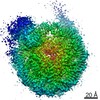
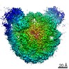
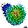
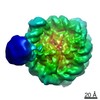
Surface view with section colored by density value
In the structure databanks used in Yorodumi, some data are registered as the other names, "COVID-19 virus" and "2019-nCoV". Here are the details of the virus and the list of structure data.
Jan 31, 2019. EMDB accession codes are about to change! (news from PDBe EMDB page)
EMDB accession codes are about to change! (news from PDBe EMDB page)
The allocation of 4 digits for EMDB accession codes will soon come to an end. Whilst these codes will remain in use, new EMDB accession codes will include an additional digit and will expand incrementally as the available range of codes is exhausted. The current 4-digit format prefixed with “EMD-” (i.e. EMD-XXXX) will advance to a 5-digit format (i.e. EMD-XXXXX), and so on. It is currently estimated that the 4-digit codes will be depleted around Spring 2019, at which point the 5-digit format will come into force.
The EM Navigator/Yorodumi systems omit the EMD- prefix.
Related info.:Q: What is EMD? / ID/Accession-code notation in Yorodumi/EM Navigator
Yorodumi is a browser for structure data from EMDB, PDB, SASBDB, etc.
This page is also the successor to EM Navigator detail page, and also detail information page/front-end page for Omokage search.
The word "yorodu" (or yorozu) is an old Japanese word meaning "ten thousand". "mi" (miru) is to see.
Related info.:EMDB / PDB / SASBDB / Comparison of 3 databanks / Yorodumi Search / Aug 31, 2016. New EM Navigator & Yorodumi / Yorodumi Papers / Jmol/JSmol / Function and homology information / Changes in new EM Navigator and Yorodumi
 Movie
Movie Controller

 Yorodumi
 Open data
 Basic information
 Map data
 Sample
 Homo sapiens (human)
 Authors
 China,
  United States, 6 items
  Citation
 Structure visualization
 Movie viewer
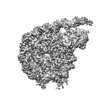
 Downloads & links
 emd_30454.png
 http://ftp.pdbj.org/pub/emdb/structures/EMD-30454

 Z (Sec.)
 Y (Row.)
 X (Col.)


 Sample components
 Baculovirus transfer vector pFASTBAC1
 Processing
 Electron microscopy
 FIELD EMISSION GUN
