 ムービー
ムービー コントローラー


+ データを開く

- 基本情報
基本情報
| 登録情報 | データベース: EMDB / ID: EMD-3027 | |||||||||
|---|---|---|---|---|---|---|---|---|---|---|
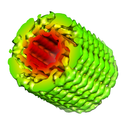
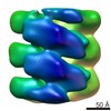
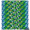
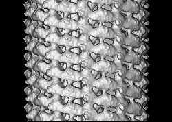
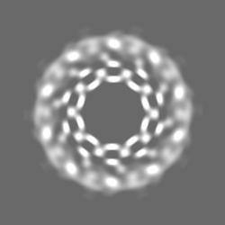
| タイトル | Electron cryo-microscopy of porcine Factor VIII bound to lipid nanotubes and helical reconstruction | |||||||||
 マップデータ マップデータ | Helical reconstruction of porcine Factor VIII bound to LNT as described in the submitted references. | |||||||||
 試料 試料 |
| |||||||||
 キーワード キーワード | Blood coagulation factor VIII / Lipid nanotubes / Cryo-electron microscopy / Membrane-bound organization / Helical reconstruction | |||||||||
| 生物種 |  | |||||||||
| 手法 | らせん対称体再構成法 / クライオ電子顕微鏡法 / ネガティブ染色法 / 解像度: 15.5 Å | |||||||||
 データ登録者 データ登録者 | Dalm D / Galaz-Montoya JG / Miller JL / Grushin K / Villalobos A / Koyfman AY / Schmid MF / Stoilova-McPhie S | |||||||||
 引用 引用 | ジャーナル: J Vis Exp / 年: 2014 タイトル: Helical organization of blood coagulation factor VIII on lipid nanotubes. 著者: Jaimy Miller / Daniela Dalm / Alexey Y Koyfman / Kirill Grushin / Svetla Stoilova-McPhie /  要旨: Cryo-electron microscopy (Cryo-EM)(1) is a powerful approach to investigate the functional structure of proteins and complexes in a hydrated state and membrane environment(2). Coagulation Factor VIII ...Cryo-electron microscopy (Cryo-EM)(1) is a powerful approach to investigate the functional structure of proteins and complexes in a hydrated state and membrane environment(2). Coagulation Factor VIII (FVIII)(3) is a multi-domain blood plasma glycoprotein. Defect or deficiency of FVIII is the cause for Hemophilia type A - a severe bleeding disorder. Upon proteolytic activation, FVIII binds to the serine protease Factor IXa on the negatively charged platelet membrane, which is critical for normal blood clotting(4). Despite the pivotal role FVIII plays in coagulation, structural information for its membrane-bound state is incomplete(5). Recombinant FVIII concentrate is the most effective drug against Hemophilia type A and commercially available FVIII can be expressed as human or porcine, both forming functional complexes with human Factor IXa(6,7). In this study we present a combination of Cryo-electron microscopy (Cryo-EM), lipid nanotechnology and structure analysis applied to resolve the membrane-bound structure of two highly homologous FVIII forms: human and porcine. The methodology developed in our laboratory to helically organize the two functional recombinant FVIII forms on negatively charged lipid nanotubes (LNT) is described. The representative results demonstrate that our approach is sufficiently sensitive to define the differences in the helical organization between the two highly homologous in sequence (86% sequence identity) proteins. Detailed protocols for the helical organization, Cryo-EM and electron tomography (ET) data acquisition are given. The two-dimensional (2D) and three-dimensional (3D) structure analysis applied to obtain the 3D reconstructions of human and porcine FVIII-LNT is discussed. The presented human and porcine FVIII-LNT structures show the potential of the proposed methodology to calculate the functional, membrane-bound organization of blood coagulation Factor VIII at high resolution. | |||||||||
| 履歴 |
|
- 構造の表示
構造の表示
| ムービー |
 ムービービューア ムービービューア |
|---|---|
| 構造ビューア | EMマップ: SurfViewMolmilJmol/JSmol |
| 添付画像 |
- ダウンロードとリンク
-EMDBアーカイブ
| マップデータ | emd_3027.map.gz | 10.7 MB | EMDBマップデータ形式 | |
|---|---|---|---|---|
| ヘッダ (付随情報) | emd-3027-v30.xmlemd-3027.xml | 12.4 KB 12.4 KB | 表示 表示 | EMDBヘッダ |
| 画像 |  emd_3027.png emd_3027.png | 188.6 KB | ||
| アーカイブディレクトリ |  http://ftp.pdbj.org/pub/emdb/structures/EMD-3027ftp://ftp.pdbj.org/pub/emdb/structures/EMD-3027 http://ftp.pdbj.org/pub/emdb/structures/EMD-3027ftp://ftp.pdbj.org/pub/emdb/structures/EMD-3027 | HTTPS FTP |
-検証レポート
| 文書・要旨 | emd_3027_validation.pdf.gz | 225.4 KB | 表示 | EMDB検証レポート |
|---|---|---|---|---|
| 文書・詳細版 | emd_3027_full_validation.pdf.gz | 224.5 KB | 表示 | |
| XML形式データ | emd_3027_validation.xml.gz | 4.1 KB | 表示 | |
| アーカイブディレクトリ | https://ftp.pdbj.org/pub/emdb/validation_reports/EMD-3027ftp://ftp.pdbj.org/pub/emdb/validation_reports/EMD-3027 | HTTPS FTP |
-関連構造データ


-リンク
| EMDBのページ | EMDB (EBI/PDBe) / EMDataResource |
|---|
-マップ
| ファイル | ダウンロード / ファイル: emd_3027.map.gz / 形式: CCP4 / 大きさ: 41.4 MB / タイプ: IMAGE STORED AS FLOATING POINT NUMBER (4 BYTES) | ||||||||||||||||||||||||||||||||||||||||||||||||||||||||||||||||||||
|---|---|---|---|---|---|---|---|---|---|---|---|---|---|---|---|---|---|---|---|---|---|---|---|---|---|---|---|---|---|---|---|---|---|---|---|---|---|---|---|---|---|---|---|---|---|---|---|---|---|---|---|---|---|---|---|---|---|---|---|---|---|---|---|---|---|---|---|---|---|
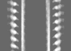
| 注釈 | Helical reconstruction of porcine Factor VIII bound to LNT as described in the submitted references. | ||||||||||||||||||||||||||||||||||||||||||||||||||||||||||||||||||||
| 投影像・断面図 | 画像のコントロール
画像は Spider により作成 これらの図は立方格子座標系で作成されたものです | ||||||||||||||||||||||||||||||||||||||||||||||||||||||||||||||||||||
| ボクセルのサイズ | X=Y=Z: 2.9 Å | ||||||||||||||||||||||||||||||||||||||||||||||||||||||||||||||||||||
| 密度 |
| ||||||||||||||||||||||||||||||||||||||||||||||||||||||||||||||||||||
| 対称性 | 空間群: 1 | ||||||||||||||||||||||||||||||||||||||||||||||||||||||||||||||||||||
| 詳細 | EMDB XML:
CCP4マップ ヘッダ情報:
| ||||||||||||||||||||||||||||||||||||||||||||||||||||||||||||||||||||
 Z (Sec.)
 Y (Row.)
 X (Col.)


-添付データ
- 試料の構成要素
-全体 : Helically organized porcine blood coagulation Factor VIII bound t...
| 全体 | 名称: Helically organized porcine blood coagulation Factor VIII bound to a lipid nanotube |
|---|---|
| 要素 |
|
-超分子 #1000: Helically organized porcine blood coagulation Factor VIII bound t...
| 超分子 | 名称: Helically organized porcine blood coagulation Factor VIII bound to a lipid nanotube タイプ: sample / ID: 1000 / 集合状態: dimer / Number unique components: 2 |
|---|---|
| 分子量 | 実験値: 170 KDa / 理論値: 170 KDa / 手法: SDS gel and amino acid sequence |
-分子 #1: Blood coagulation Factor VIII
| 分子 | 名称: Blood coagulation Factor VIII / タイプ: protein_or_peptide / ID: 1 / Name.synonym: Hemophilia A factor 詳細: Recombinant porcine Factor VIII lacking the B domain was bound to lipid nanotubes and helically organized for structure analysis by cryo-EM. コピー数: 2 / 集合状態: dimer / 組換発現: Yes |
|---|---|
| 由来(天然) | 生物種: |
| 分子量 | 実験値: 170 KDa / 理論値: 170 KDa |
| 組換発現 | 生物種:  Cricetulus griseus (モンゴルキヌゲネズミ) Cricetulus griseus (モンゴルキヌゲネズミ)組換細胞: BHK cells |
-分子 #2: lipid nanotubes
| 分子 | 名称: lipid nanotubes / タイプ: ligand / ID: 2 / 組換発現: No / データベース: NCBI |
|---|---|
| 由来(天然) | 生物種: unidentified (未定義) |
-実験情報
-構造解析
| 手法 | ネガティブ染色法, クライオ電子顕微鏡法 |
|---|---|
 解析 解析 | らせん対称体再構成法 |
| 試料の集合状態 | helical array |
-試料調製
| 濃度 | 1.0 mg/mL |
|---|---|
| 緩衝液 | pH: 7.4 / 詳細: 20 mM HEPES, 150 mM NaCL, 5 mM CaCl2 |
| 染色 | タイプ: NEGATIVE 詳細: samples are adsorbed on quantifoil grids and flash frozen in liquid ethane |
| グリッド | 詳細: carbon coated quantifoil grids Q2x2 were glow discharged for 100 second |
| 凍結 | 凍結剤: ETHANE / チャンバー内温度: 85 K / 装置: FEI VITROBOT MARK IV / 手法: blot for 3.5 seconds, force 1 before plunging |
| 詳細 | The protein and lipid are mixed in 1:1 ration and incubated for 15 minutes |
- 電子顕微鏡法
| 顕微鏡 | JEOL 2100 |
|---|---|
| 温度 | 平均: 99 K |
| 日付 | 2013年9月11日 |
| 撮影 | カテゴリ: CCD フィルム・検出器のモデル: GATAN ULTRASCAN 4000 (4k x 4k) 実像数: 300 / 平均電子線量: 16 e/Å2 |
| 電子線 | 加速電圧: 200 kV / 電子線源: LAB6 |
| 電子光学系 | 倍率(補正後): 52000 / 照射モード: FLOOD BEAM / 撮影モード: BRIGHT FIELD / Cs: 2.0 mm / 最大 デフォーカス(公称値): 0.004 µm / 最小 デフォーカス(公称値): 0.001 µm / 倍率(公称値): 40000 |
| 試料ステージ | 試料ホルダーモデル: GATAN LIQUID NITROGEN |
-画像解析
| 詳細 | Described in supplemental materials and references |
|---|---|
| 最終 再構成 | 想定した対称性 - らせんパラメータ - Δz: 36 Å 想定した対称性 - らせんパラメータ - ΔΦ: 35.5 ° 想定した対称性 - らせんパラメータ - 軸対称性: C5 (5回回転対称) 解像度のタイプ: BY AUTHOR / 解像度: 15.5 Å / 解像度の算出法: OTHER |
| CTF補正 | 詳細: each particle set |
-原子モデル構築 1
| 初期モデル | PDB ID: Chain - #0 - Chain ID: A / Chain - #1 - Chain ID: B |
|---|---|
| ソフトウェア | 名称:  UCSF Chimera UCSF Chimera |
| 精密化 | 空間: REAL / プロトコル: RIGID BODY FIT |

