 Movie
Movie Controller

[English] 日本語
 Yorodumi
- EMDB-30092: The coordinate of the nuclease domain of the apo terminase complex -
+ Open data

- Basic information
| Entry | Database: EMDB / ID: EMD-30092 | |||||||||
|---|---|---|---|---|---|---|---|---|---|---|
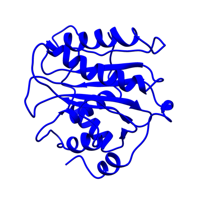
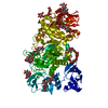
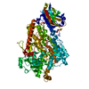
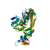
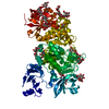
| Title | The coordinate of the nuclease domain of the apo terminase complex | |||||||||
 Map data Map data | ||||||||||
 Sample Sample |
| |||||||||
 Keywords Keywords | HSV-1 / nuclease domain / apo terminase complex / VIRAL PROTEIN | |||||||||
| Function / homology |  Function and homology information Function and homology informationchromosome organization / Hydrolases; Acting on ester bonds / hydrolase activity / host cell nucleus / DNA binding Similarity search - Function | |||||||||
| Biological species |   Human herpesvirus 1 (Herpes simplex virus type 1) / Human alphaherpesvirus 1 strain 17 Human herpesvirus 1 (Herpes simplex virus type 1) / Human alphaherpesvirus 1 strain 17 | |||||||||
| Method | single particle reconstruction / cryo EM / Resolution: 3.6 Å | |||||||||
 Authors Authors | Yang YX / Yang P | |||||||||
| Funding support |  China, 1 items China, 1 items
| |||||||||
 Citation Citation | Journal: Protein Cell / Year: 2020 Title: Architecture of the herpesvirus genome-packaging complex and implications for DNA translocation. Authors: Yunxiang Yang / Pan Yang / Nan Wang / Zhonghao Chen / Dan Su / Z Hong Zhou / Zihe Rao / Xiangxi Wang /  Abstract: Genome packaging is a fundamental process in a viral life cycle and a prime target of antiviral drugs. Herpesviruses use an ATP-driven packaging motor/terminase complex to translocate and cleave ...Genome packaging is a fundamental process in a viral life cycle and a prime target of antiviral drugs. Herpesviruses use an ATP-driven packaging motor/terminase complex to translocate and cleave concatemeric dsDNA into procapsids but its molecular architecture and mechanism are unknown. We report atomic structures of a herpesvirus hexameric terminase complex in both the apo and ADP•BeF3-bound states. Each subunit of the hexameric ring comprises three components-the ATPase/terminase pUL15 and two regulator/fixer proteins, pUL28 and pUL33-unlike bacteriophage terminases. Distal to the nuclease domains, six ATPase domains form a central channel with conserved basic-patches conducive to DNA binding and trans-acting arginine fingers are essential to ATP hydrolysis and sequential DNA translocation. Rearrangement of the nuclease domains mediated by regulatory domains converts DNA translocation mode to cleavage mode. Our structures favor a sequential revolution model for DNA translocation and suggest mechanisms for concerted domain rearrangements leading to DNA cleavage. | |||||||||
| History |
|
- Structure visualization
| Movie |
Movie viewer |
|---|---|
| Structure viewer | EM map: SurfViewMolmilJmol/JSmol |
| Supplemental images |
- Downloads & links
-EMDB archive
| Map data | emd_30092.map.gz | 55.7 MB | EMDB map data format | |
|---|---|---|---|---|
| Header (meta data) | emd-30092-v30.xmlemd-30092.xml | 11 KB 11 KB | Display Display | EMDB header |
| Images |  emd_30092.png emd_30092.png | 78.8 KB | ||
| Filedesc metadata | emd-30092.cif.gz | 5.4 KB | ||
| Archive directory |  http://ftp.pdbj.org/pub/emdb/structures/EMD-30092ftp://ftp.pdbj.org/pub/emdb/structures/EMD-30092 http://ftp.pdbj.org/pub/emdb/structures/EMD-30092ftp://ftp.pdbj.org/pub/emdb/structures/EMD-30092 | HTTPS FTP |
-Related structure data
| Related structure data |  6m5tMC  6m5rC  6m5sC  6m5uC  6m5vC M: atomic model generated by this map C: citing same article ( |
|---|---|
| Similar structure data |


-Links
| EMDB pages | EMDB (EBI/PDBe) / EMDataResource |
|---|
-Map
| File | Download / File: emd_30092.map.gz / Format: CCP4 / Size: 59.6 MB / Type: IMAGE STORED AS FLOATING POINT NUMBER (4 BYTES) | ||||||||||||||||||||||||||||||||||||||||||||||||||||||||||||
|---|---|---|---|---|---|---|---|---|---|---|---|---|---|---|---|---|---|---|---|---|---|---|---|---|---|---|---|---|---|---|---|---|---|---|---|---|---|---|---|---|---|---|---|---|---|---|---|---|---|---|---|---|---|---|---|---|---|---|---|---|---|
| Projections & slices | Image control
Images are generated by Spider. | ||||||||||||||||||||||||||||||||||||||||||||||||||||||||||||
| Voxel size | X=Y=Z: 1.05 Å | ||||||||||||||||||||||||||||||||||||||||||||||||||||||||||||
| Density |
| ||||||||||||||||||||||||||||||||||||||||||||||||||||||||||||
| Symmetry | Space group: 1 | ||||||||||||||||||||||||||||||||||||||||||||||||||||||||||||
| Details | EMDB XML:
CCP4 map header:
| ||||||||||||||||||||||||||||||||||||||||||||||||||||||||||||
 Z (Sec.)
 Y (Row.)
 X (Col.)


-Supplemental data
- Sample components
-Entire : HSV-1 nuclease domain of terminase complex
| Entire | Name: HSV-1 nuclease domain of terminase complex |
|---|---|
| Components |
|
-Supramolecule #1: HSV-1 nuclease domain of terminase complex
| Supramolecule | Name: HSV-1 nuclease domain of terminase complex / type: complex / ID: 1 / Parent: 0 / Macromolecule list: all |
|---|---|
| Source (natural) | Organism: Human herpesvirus 1 (Herpes simplex virus type 1) |
-Macromolecule #1: Tripartite terminase subunit 3
| Macromolecule | Name: Tripartite terminase subunit 3 / type: protein_or_peptide / ID: 1 / Number of copies: 1 / Enantiomer: LEVO / EC number: Hydrolases; Acting on ester bonds |
|---|---|
| Source (natural) | Organism: Human alphaherpesvirus 1 strain 17 / Strain: 17 |
| Molecular weight | Theoretical: 30.918906 KDa |
| Recombinant expression | Organism:   Spodoptera frugiperda (fall armyworm) Spodoptera frugiperda (fall armyworm) |
| Sequence | String: MGSSHHHHHH SSGLVPRGSH MTGDDRPVLT KSAGERFLLY RPSTTTNSGL MAPDLYVYVD PAFTANTRAS GTGVAVVGRY RDDYIIFAL EHFFLRALTG SAPADIARCV VHSLTQVLAL HPGAFRGVRV AVEGNSSQDS AVAIATHVHT EMHRLLASEG A DAGSGPEL ...String: MGSSHHHHHH SSGLVPRGSH MTGDDRPVLT KSAGERFLLY RPSTTTNSGL MAPDLYVYVD PAFTANTRAS GTGVAVVGRY RDDYIIFAL EHFFLRALTG SAPADIARCV VHSLTQVLAL HPGAFRGVRV AVEGNSSQDS AVAIATHVHT EMHRLLASEG A DAGSGPEL LFYHCEPPGS AVLYPFFLLN KQKTPAFEHF IKKFNSGGVM ASQEIVSATV RLQTDPVEYL LEQLNNLTET VS PNTDVRT YSGKRNGASD DLMVAVIMAI YLAAQAGPPH TFAPITRVS UniProtKB: Tripartite terminase subunit 3 |
-Experimental details
-Structure determination
| Method | cryo EM |
|---|---|
 Processing Processing | single particle reconstruction |
| Aggregation state | particle |
-Sample preparation
| Buffer | pH: 7.5 |
|---|---|
| Vitrification | Cryogen name: ETHANE |
- Electron microscopy
| Microscope | FEI TITAN KRIOS |
|---|---|
| Image recording | Film or detector model: GATAN K2 QUANTUM (4k x 4k) / Average electron dose: 2.0 e/Å2 |
| Electron beam | Acceleration voltage: 300 kV / Electron source:  FIELD EMISSION GUN FIELD EMISSION GUN |
| Electron optics | Illumination mode: FLOOD BEAM / Imaging mode: BRIGHT FIELD |
| Experimental equipment |  Model: Titan Krios / Image courtesy: FEI Company |
