 ムービー
ムービー コントローラー


+ データを開く

- 基本情報
基本情報
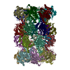
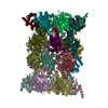
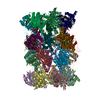
| 登録情報 | データベース: EMDB / ID: EMD-2981 | |||||||||
|---|---|---|---|---|---|---|---|---|---|---|
| タイトル | Cryo-EM reveals the conformation of a substrate analogue in the human 20S proteasome core | |||||||||
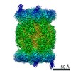
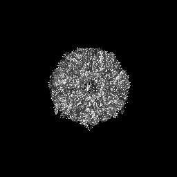
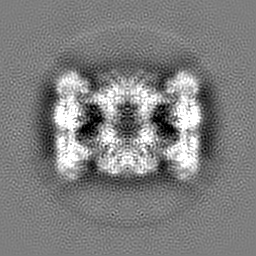
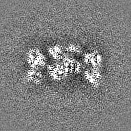
 マップデータ マップデータ | reconstruction of the human 20S proteasome core, as determined by the 3D reconstitution algorithm, without further masking, sharpening or Fourier filtering | |||||||||
 試料 試料 |
| |||||||||
 キーワード キーワード | proteasome / 20S / human / AdaAhx3L3VS / ligand / inhibitor / drug design | |||||||||
| 機能・相同性 |  機能・相同性情報 機能・相同性情報purine ribonucleoside triphosphate binding / Regulation of ornithine decarboxylase (ODC) / Proteasome assembly / Cross-presentation of soluble exogenous antigens (endosomes) / proteasome core complex / Somitogenesis / myofibril / NF-kappaB binding / proteasome endopeptidase complex / proteasome core complex, beta-subunit complex ...purine ribonucleoside triphosphate binding / Regulation of ornithine decarboxylase (ODC) / Proteasome assembly / Cross-presentation of soluble exogenous antigens (endosomes) / proteasome core complex / Somitogenesis / myofibril / NF-kappaB binding / proteasome endopeptidase complex / proteasome core complex, beta-subunit complex / proteasome assembly / threonine-type endopeptidase activity / proteasome core complex, alpha-subunit complex / immune system process / Regulation of activated PAK-2p34 by proteasome mediated degradation / Autodegradation of Cdh1 by Cdh1:APC/C / APC/C:Cdc20 mediated degradation of Securin / Asymmetric localization of PCP proteins / Ubiquitin-dependent degradation of Cyclin D / NIK-->noncanonical NF-kB signaling / SCF-beta-TrCP mediated degradation of Emi1 / proteasome complex / proteolysis involved in protein catabolic process / TNFR2 non-canonical NF-kB pathway / AUF1 (hnRNP D0) binds and destabilizes mRNA / Vpu mediated degradation of CD4 / Assembly of the pre-replicative complex / Ubiquitin-Mediated Degradation of Phosphorylated Cdc25A / Degradation of DVL / Dectin-1 mediated noncanonical NF-kB signaling / sarcomere / Cdc20:Phospho-APC/C mediated degradation of Cyclin A / Degradation of AXIN / Hh mutants are degraded by ERAD / Activation of NF-kappaB in B cells / Degradation of GLI1 by the proteasome / Hedgehog ligand biogenesis / G2/M Checkpoints / Defective CFTR causes cystic fibrosis / GSK3B and BTRC:CUL1-mediated-degradation of NFE2L2 / Autodegradation of the E3 ubiquitin ligase COP1 / Negative regulation of NOTCH4 signaling / Vif-mediated degradation of APOBEC3G / Regulation of RUNX3 expression and activity / Hedgehog 'on' state / Degradation of GLI2 by the proteasome / GLI3 is processed to GLI3R by the proteasome / FBXL7 down-regulates AURKA during mitotic entry and in early mitosis / APC/C:Cdh1 mediated degradation of Cdc20 and other APC/C:Cdh1 targeted proteins in late mitosis/early G1 / MAPK6/MAPK4 signaling / Degradation of beta-catenin by the destruction complex / lipopolysaccharide binding / negative regulation of inflammatory response to antigenic stimulus / ABC-family proteins mediated transport / P-body / Oxygen-dependent proline hydroxylation of Hypoxia-inducible Factor Alpha / CDK-mediated phosphorylation and removal of Cdc6 / CLEC7A (Dectin-1) signaling / SCF(Skp2)-mediated degradation of p27/p21 / Regulation of expression of SLITs and ROBOs / FCERI mediated NF-kB activation / Regulation of PTEN stability and activity / Interleukin-1 signaling / Orc1 removal from chromatin / Regulation of RAS by GAPs / response to virus / Regulation of RUNX2 expression and activity / nuclear matrix / The role of GTSE1 in G2/M progression after G2 checkpoint / Separation of Sister Chromatids / KEAP1-NFE2L2 pathway / UCH proteinases / Downstream TCR signaling / Antigen processing: Ubiquitination & Proteasome degradation / Neddylation / peptidase activity / RUNX1 regulates transcription of genes involved in differentiation of HSCs / ER-Phagosome pathway / response to oxidative stress / regulation of inflammatory response / secretory granule lumen / endopeptidase activity / proteasome-mediated ubiquitin-dependent protein catabolic process / ficolin-1-rich granule lumen / positive regulation of canonical NF-kappaB signal transduction / Ub-specific processing proteases / nuclear body / ciliary basal body / cilium / ribosome / cadherin binding / intracellular membrane-bounded organelle / ubiquitin protein ligase binding / centrosome / Neutrophil degranulation / mitochondrion / proteolysis / RNA binding / extracellular exosome / extracellular region 類似検索 - 分子機能 | |||||||||
| 生物種 |  Homo sapiens (ヒト) Homo sapiens (ヒト) | |||||||||
| 手法 | 単粒子再構成法 / クライオ電子顕微鏡法 / 解像度: 3.5 Å | |||||||||
 データ登録者 データ登録者 | da Fonseca PCA / Morris EP | |||||||||
 引用 引用 | ジャーナル: Nat Commun / 年: 2015 タイトル: Cryo-EM reveals the conformation of a substrate analogue in the human 20S proteasome core. 著者: Paula C A da Fonseca / Edward P Morris /  要旨: The proteasome is a highly regulated protease complex fundamental for cell homeostasis and controlled cell cycle progression. It functions by removing a wide range of specifically tagged proteins, ...The proteasome is a highly regulated protease complex fundamental for cell homeostasis and controlled cell cycle progression. It functions by removing a wide range of specifically tagged proteins, including key cellular regulators. Here we present the structure of the human 20S proteasome core bound to a substrate analogue inhibitor molecule, determined by electron cryo-microscopy (cryo-EM) and single-particle analysis at a resolution of around 3.5 Å. Our map allows the building of protein coordinates as well as defining the location and conformation of the inhibitor at the different active sites. These results open new prospects to tackle the proteasome functional mechanisms. Moreover, they also further demonstrate that cryo-EM is emerging as a realistic approach for general structural studies of protein-ligand interactions. | |||||||||
| 履歴 |
|
- 構造の表示
構造の表示
| ムービー |
ムービービューア |
|---|---|
| 構造ビューア | EMマップ: SurfViewMolmilJmol/JSmol |
| 添付画像 |

- ダウンロードとリンク
-EMDBアーカイブ
| マップデータ | emd_2981.map.gz | 58.3 MB | EMDBマップデータ形式 | |
|---|---|---|---|---|
| ヘッダ (付随情報) | emd-2981-v30.xmlemd-2981.xml | 24.6 KB 24.6 KB | 表示 表示 | EMDBヘッダ |
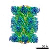
| 画像 | 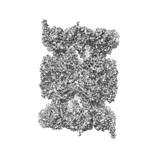 emd_2981.png emd_2981.png | 152.2 KB | ||
| アーカイブディレクトリ |  http://ftp.pdbj.org/pub/emdb/structures/EMD-2981ftp://ftp.pdbj.org/pub/emdb/structures/EMD-2981 http://ftp.pdbj.org/pub/emdb/structures/EMD-2981ftp://ftp.pdbj.org/pub/emdb/structures/EMD-2981 | HTTPS FTP |
-検証レポート
| 文書・要旨 | emd_2981_validation.pdf.gz | 314.1 KB | 表示 | EMDB検証レポート |
|---|---|---|---|---|
| 文書・詳細版 | emd_2981_full_validation.pdf.gz | 313.2 KB | 表示 | |
| XML形式データ | emd_2981_validation.xml.gz | 6.3 KB | 表示 | |
| アーカイブディレクトリ | https://ftp.pdbj.org/pub/emdb/validation_reports/EMD-2981ftp://ftp.pdbj.org/pub/emdb/validation_reports/EMD-2981 | HTTPS FTP |
-関連構造データ
| 関連構造データ |  5a0qMC M: このマップから作成された原子モデル C: 同じ文献を引用 ( |
|---|---|
| 類似構造データ | |
| 電子顕微鏡画像生データ | EMPIAR-10038 (タイトル: Cryo-EM reveals the conformation of a substrate analogue in the human 20S proteasome core Data size: 579.1 Data #1: raw micrographs of the human 20S proteasome core complex bound to the ligand AdaAhx3L3VS [micrographs - multiframe]) |

-リンク
| EMDBのページ | EMDB (EBI/PDBe) / EMDataResource |
|---|---|
| 「今月の分子」の関連する項目 |


-マップ
| ファイル | ダウンロード / ファイル: emd_2981.map.gz / 形式: CCP4 / 大きさ: 62.5 MB / タイプ: IMAGE STORED AS FLOATING POINT NUMBER (4 BYTES) | ||||||||||||||||||||||||||||||||||||||||||||||||||||||||||||
|---|---|---|---|---|---|---|---|---|---|---|---|---|---|---|---|---|---|---|---|---|---|---|---|---|---|---|---|---|---|---|---|---|---|---|---|---|---|---|---|---|---|---|---|---|---|---|---|---|---|---|---|---|---|---|---|---|---|---|---|---|---|
| 注釈 | reconstruction of the human 20S proteasome core, as determined by the 3D reconstitution algorithm, without further masking, sharpening or Fourier filtering | ||||||||||||||||||||||||||||||||||||||||||||||||||||||||||||
| 投影像・断面図 | 画像のコントロール
画像は Spider により作成 | ||||||||||||||||||||||||||||||||||||||||||||||||||||||||||||
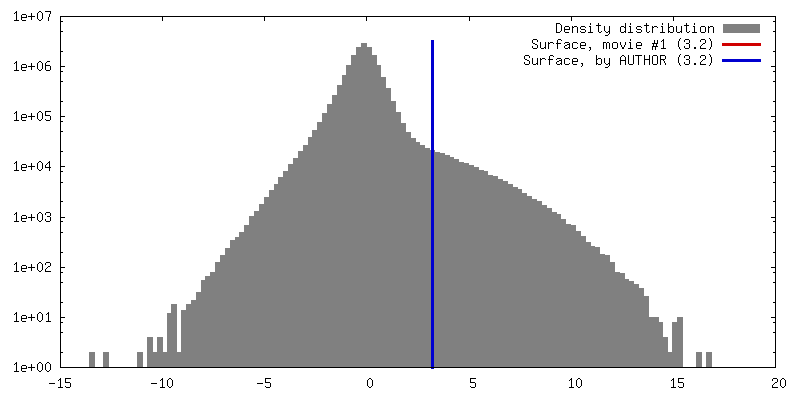
| ボクセルのサイズ | X=Y=Z: 1.04 Å | ||||||||||||||||||||||||||||||||||||||||||||||||||||||||||||
| 密度 |
| ||||||||||||||||||||||||||||||||||||||||||||||||||||||||||||
| 対称性 | 空間群: 1 | ||||||||||||||||||||||||||||||||||||||||||||||||||||||||||||
| 詳細 | EMDB XML:
CCP4マップ ヘッダ情報:
| ||||||||||||||||||||||||||||||||||||||||||||||||||||||||||||
 Z (Sec.)
 Y (Row.)
 X (Col.)


-添付データ
- 試料の構成要素
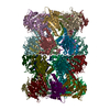
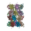
+全体 : human 20S proteasome core
+超分子 #1000: human 20S proteasome core
+分子 #1: Proteasome subunit alpha type-6
+分子 #2: Proteasome subunit alpha type-2
+分子 #3: Proteasome subunit alpha type-4
+分子 #4: Proteasome subunit alpha type-7
+分子 #5: Proteasome subunit alpha type-5
+分子 #6: Proteasome subunit alpha type-1
+分子 #7: Proteasome subunit alpha type-3
+分子 #8: Proteasome subunit beta type-6
+分子 #9: Proteasome subunit beta type-7
+分子 #10: Proteasome subunit beta type-3
+分子 #11: Proteasome subunit beta type-2
+分子 #12: Proteasome subunit beta type-5
+分子 #13: Proteasome subunit beta type-1
+分子 #14: Proteasome subunit beta type-4
-実験情報
-構造解析
| 手法 | クライオ電子顕微鏡法 |
|---|---|
 解析 解析 | 単粒子再構成法 |
| 試料の集合状態 | particle |
-試料調製
| 濃度 | 0.1 mg/mL |
|---|---|
| 緩衝液 | pH: 7.5 / 詳細: 50 mM Tris-HCl, 5 mM MgCl2 and 1mM dithiotreitol |
| グリッド | 詳細: 1.2/1.3 Quantifoil coated with freshly floated thin layer of carbon, glow discharged in amylamine atmosphere |
| 凍結 | 凍結剤: ETHANE / チャンバー内湿度: 95 % / チャンバー内温度: 120 K / 装置: FEI VITROBOT MARK III / 手法: blot 2.5 seconds before plunging |
- 電子顕微鏡法
| 顕微鏡 | FEI TITAN KRIOS |
|---|---|
| 温度 | 平均: 85 K |
| アライメント法 | Legacy - 非点収差: objective lens astigmatism was corrected at the recording magnification |
| 詳細 | Each exposure was recorded as 17 individual frames captured at a rate of 0.056 second/frame, with an electron dose of 2.8 electrons/square angstrom. Data-set recorded using EPU software. |
| 日付 | 2014年2月4日 |
| 撮影 | カテゴリ: CCD フィルム・検出器のモデル: FEI FALCON II (4k x 4k) デジタル化 - サンプリング間隔: 14 µm / 実像数: 960 / 平均電子線量: 48 e/Å2 詳細: each image is the sum of 17 frames recorded by the direct electron detector |
| 電子線 | 加速電圧: 300 kV / 電子線源:  FIELD EMISSION GUN FIELD EMISSION GUN |
| 電子光学系 | 倍率(補正後): 134461 / 照射モード: FLOOD BEAM / 撮影モード: BRIGHT FIELD / Cs: 2.7 mm / 最大 デフォーカス(公称値): 3.0 µm / 最小 デフォーカス(公称値): 1.7 µm / 倍率(公称値): 75000 |
| 試料ステージ | 試料ホルダーモデル: FEI TITAN KRIOS AUTOGRID HOLDER |
| 実験機器 |  モデル: Titan Krios / 画像提供: FEI Company |
-画像解析
| 詳細 | automatic particle picking followed by careful manual removal of false positives and addition of false negatives; high resolution analysis was done using the sum of frames 3-10 |
|---|---|
| CTF補正 | 詳細: full recorded image |
| 最終 再構成 | 想定した対称性 - 点群: C2 (2回回転対称) / アルゴリズム: OTHER / 解像度のタイプ: BY AUTHOR / 解像度: 3.5 Å / 解像度の算出法: OTHER / ソフトウェア - 名称: Spider, Tigris, Imagic 詳細: the analysis was done using a data-set recorded during a single EM session 使用した粒子像数: 76500 |
| 最終 角度割当 | 詳細: Beta 0 degrees, gamma 90 degrees (IMAGIC) |
-原子モデル構築 1
| 初期モデル | PDB ID: Chain - #0 - Chain ID: A / Chain - #1 - Chain ID: B / Chain - #2 - Chain ID: C / Chain - #3 - Chain ID: D / Chain - #4 - Chain ID: E / Chain - #5 - Chain ID: F / Chain - #6 - Chain ID: G / Chain - #7 - Chain ID: H / Chain - #8 - Chain ID: I / Chain - #9 - Chain ID: J / Chain - #10 - Chain ID: K / Chain - #11 - Chain ID: L / Chain - #12 - Chain ID: M / Chain - #13 - Chain ID: N / Chain - #14 - Chain ID: O / Chain - #15 - Chain ID: P / Chain - #16 - Chain ID: Q / Chain - #17 - Chain ID: R / Chain - #18 - Chain ID: S / Chain - #19 - Chain ID: T / Chain - #20 - Chain ID: U / Chain - #21 - Chain ID: V / Chain - #22 - Chain ID: W / Chain - #23 - Chain ID: X / Chain - #24 - Chain ID: Y / Chain - #25 - Chain ID: Z / Chain - #26 - Chain ID: a / Chain - #27 - Chain ID: b |
|---|---|
| ソフトウェア | 名称: Coot, Phenix |
| 詳細 | The model of the human 20S proteasome core was built based on the X-ray crystal structure of the mouse constitutive apo 20S proteasome core (3UNE) using real space refinement in Coot and Phenix |
| 精密化 | 空間: REAL / プロトコル: FLEXIBLE FIT |
| 得られたモデル | PDB-5a0q: |

