Movie
Movie Controller


+ Open data

- Basic information
| Entry |  | |||||||||
|---|---|---|---|---|---|---|---|---|---|---|
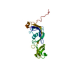
| Title | YES Complex - E. coli MraY, Protein E ID21, E. coli SlyD | |||||||||
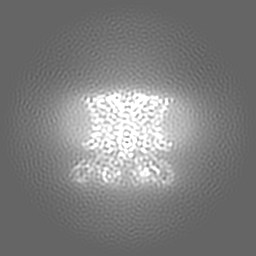
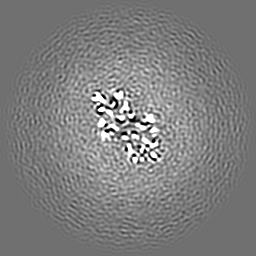
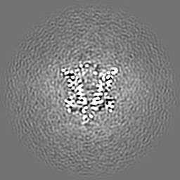
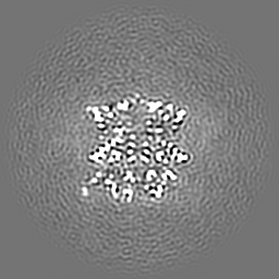
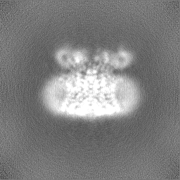
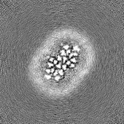
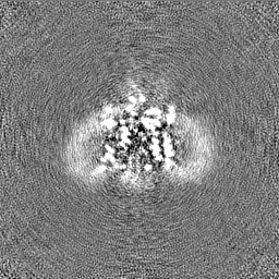
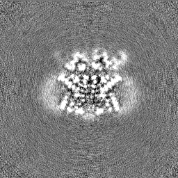
 Map data Map data | CryoEM map of the YES complex (E. coli MraY, Protein E ID21, E. coli SlyD). DeepEMhancer post-processed. | |||||||||
 Sample Sample |
| |||||||||
 Keywords Keywords | inhibitor / antibiotic / chaperone / membrane / bacteriophage / TRANSFERASE-ISOMERASE complex | |||||||||
| Function / homology |  Function and homology information Function and homology informationphospho-N-acetylmuramoyl-pentapeptide-transferase / phospho-N-acetylmuramoyl-pentapeptide-transferase activity / UDP-N-acetylmuramoyl-L-alanyl-D-glutamyl-meso-2,6-diaminopimelyl-D-alanyl-D-alanine:undecaprenyl-phosphate transferase activity / cell wall macromolecule biosynthetic process / cobalt ion binding / nickel cation binding / peptidoglycan biosynthetic process / enzyme inhibitor activity / peptidylprolyl isomerase / peptidyl-prolyl cis-trans isomerase activity ...phospho-N-acetylmuramoyl-pentapeptide-transferase / phospho-N-acetylmuramoyl-pentapeptide-transferase activity / UDP-N-acetylmuramoyl-L-alanyl-D-glutamyl-meso-2,6-diaminopimelyl-D-alanyl-D-alanine:undecaprenyl-phosphate transferase activity / cell wall macromolecule biosynthetic process / cobalt ion binding / nickel cation binding / peptidoglycan biosynthetic process / enzyme inhibitor activity / peptidylprolyl isomerase / peptidyl-prolyl cis-trans isomerase activity / protein maturation / cell wall organization / : / regulation of cell shape / response to heat / protein refolding / killing of cells of another organism / protein stabilization / copper ion binding / cell division / zinc ion binding / membrane / metal ion binding / plasma membrane / cytosol Similarity search - Function | |||||||||
| Biological species |  Escherichia phage ID21 (virus) / Escherichia phage ID21 (virus) /  | |||||||||
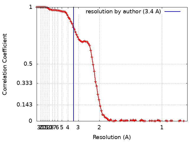
| Method | single particle reconstruction / cryo EM / Resolution: 3.4 Å | |||||||||
 Authors Authors | Orta AK / Clemons WM / Riera N | |||||||||
| Funding support |  United States, 2 items United States, 2 items
| |||||||||
 Citation Citation | Journal: Science / Year: 2023 Title: The mechanism of the phage-encoded protein antibiotic from ΦX174. Authors: Anna K Orta / Nadia Riera / Yancheng E Li / Shiho Tanaka / Hyun Gi Yun / Lada Klaic / William M Clemons / Abstract: The historically important phage ΦX174 kills its host bacteria by encoding a 91-residue protein antibiotic called protein E. Using single-particle electron cryo-microscopy, we demonstrate that ...The historically important phage ΦX174 kills its host bacteria by encoding a 91-residue protein antibiotic called protein E. Using single-particle electron cryo-microscopy, we demonstrate that protein E bridges two bacterial proteins to form the transmembrane YES complex [MraY, protein E, sensitivity to lysis D (SlyD)]. Protein E inhibits peptidoglycan biosynthesis by obstructing the MraY active site leading to loss of lipid I production. We experimentally validate this result for two different viral species, providing a clear model for bacterial lysis and unifying previous experimental data. Additionally, we characterize the MraY structure-revealing features of this essential enzyme-and the structure of the chaperone SlyD bound to a protein. Our structures provide insights into the mechanism of phage-mediated lysis and for structure-based design of phage therapeutics. | |||||||||
| History |
|
- Structure visualization
| Supplemental images |
|---|

- Downloads & links
-EMDB archive
| Map data | emd_29641.map.gz | 54.6 MB | EMDB map data format | |
|---|---|---|---|---|
| Header (meta data) | emd-29641-v30.xmlemd-29641.xml | 26 KB 26 KB | Display Display | EMDB header |
| FSC (resolution estimation) | emd_29641_fsc.xml | 11.7 KB | Display | FSC data file |
| Images |  emd_29641.png emd_29641.png | 162.7 KB | ||
| Filedesc metadata | emd-29641.cif.gz | 6.7 KB | ||
| Others | emd_29641_additional_1.map.gzemd_29641_half_map_1.map.gzemd_29641_half_map_2.map.gz | 59.6 MB 59.1 MB 59.1 MB | ||
| Archive directory |  http://ftp.pdbj.org/pub/emdb/structures/EMD-29641ftp://ftp.pdbj.org/pub/emdb/structures/EMD-29641 http://ftp.pdbj.org/pub/emdb/structures/EMD-29641ftp://ftp.pdbj.org/pub/emdb/structures/EMD-29641 | HTTPS FTP |
-Related structure data
| Related structure data |  8g01MC  8g02C C: citing same article ( M: atomic model generated by this map |
|---|---|
| Similar structure data |
-Links
| EMDB pages | EMDB (EBI/PDBe) / EMDataResource |
|---|---|
| Related items in Molecule of the Month |


-Map
| File | Download / File: emd_29641.map.gz / Format: CCP4 / Size: 64 MB / Type: IMAGE STORED AS FLOATING POINT NUMBER (4 BYTES) | ||||||||||||||||||||||||||||||||||||
|---|---|---|---|---|---|---|---|---|---|---|---|---|---|---|---|---|---|---|---|---|---|---|---|---|---|---|---|---|---|---|---|---|---|---|---|---|---|
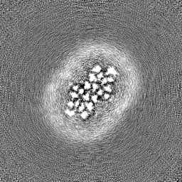
| Annotation | CryoEM map of the YES complex (E. coli MraY, Protein E ID21, E. coli SlyD). DeepEMhancer post-processed. | ||||||||||||||||||||||||||||||||||||
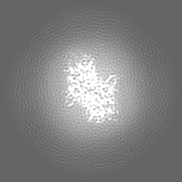
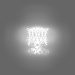
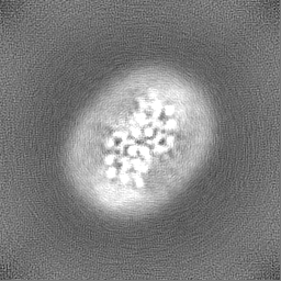
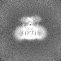
| Projections & slices | Image control
Images are generated by Spider. | ||||||||||||||||||||||||||||||||||||
| Voxel size | X=Y=Z: 0.832 Å | ||||||||||||||||||||||||||||||||||||
| Density |
| ||||||||||||||||||||||||||||||||||||
| Symmetry | Space group: 1 | ||||||||||||||||||||||||||||||||||||
| Details | EMDB XML:
|
 Z (Sec.)
 Y (Row.)
 X (Col.)


-Supplemental data
-Additional map: CryoEM map of the YES complex (E. coli...
| File | emd_29641_additional_1.map | ||||||||||||
|---|---|---|---|---|---|---|---|---|---|---|---|---|---|
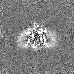
| Annotation | CryoEM map of the YES complex (E. coli MraY, Protein E ID21, E. coli SlyD). | ||||||||||||
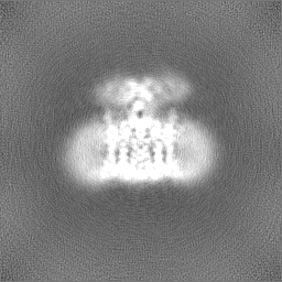
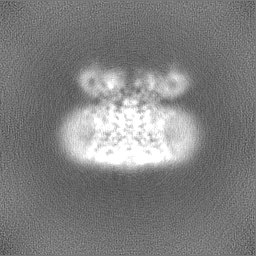
| Projections & Slices |
| ||||||||||||
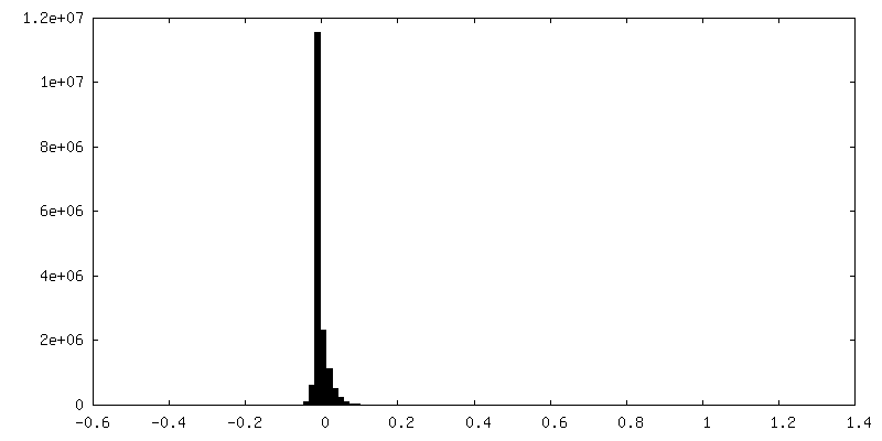
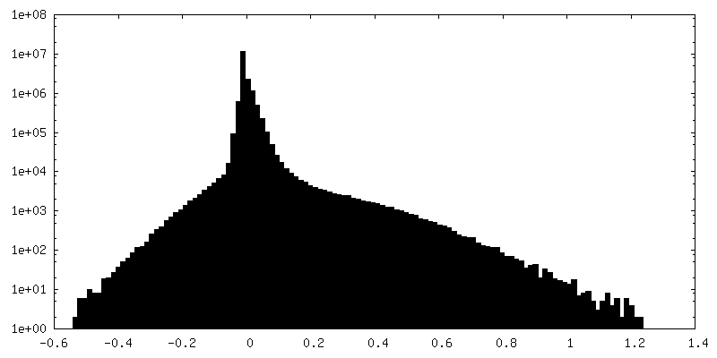
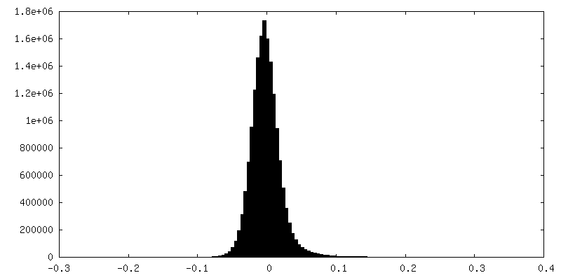
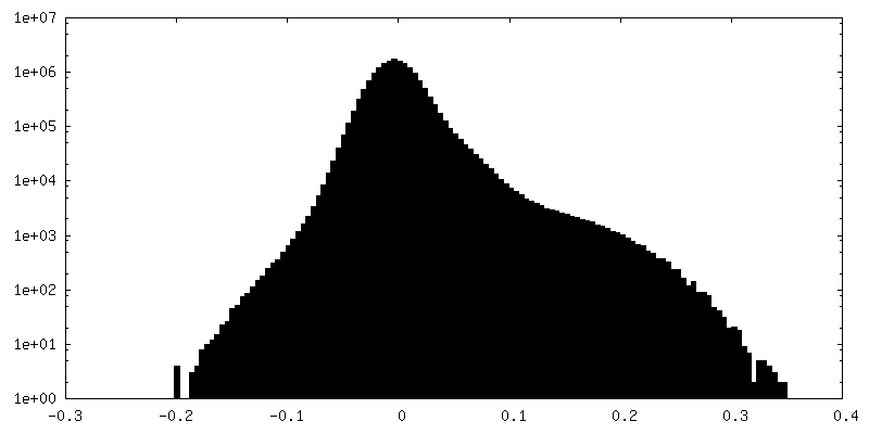
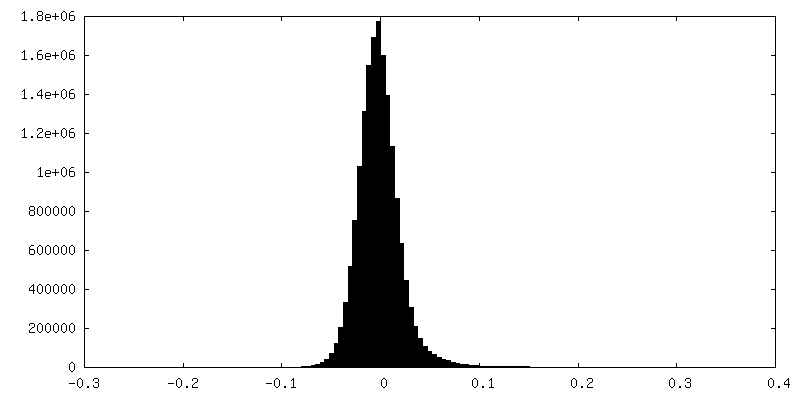
| Density Histograms |
-Half map: #1
| File | emd_29641_half_map_1.map | ||||||||||||
|---|---|---|---|---|---|---|---|---|---|---|---|---|---|
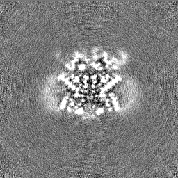
| Projections & Slices |
| ||||||||||||
| Density Histograms |
-Half map: #2
| File | emd_29641_half_map_2.map | ||||||||||||
|---|---|---|---|---|---|---|---|---|---|---|---|---|---|
| Projections & Slices |
| ||||||||||||
| Density Histograms |


- Sample components
-Entire : YES complex
| Entire | Name: YES complex |
|---|---|
| Components |
|
-Supramolecule #1: YES complex
| Supramolecule | Name: YES complex / type: complex / ID: 1 / Parent: 0 / Macromolecule list: all Details: Hexameric complex of E. coli MraY dimer bound to two molecules of Protein E (ID21), stabilized by E. coli SlyD |
|---|---|
| Molecular weight | Theoretical: 140.32 KDa |
-Supramolecule #2: Lysis Protein E
| Supramolecule | Name: Lysis Protein E / type: complex / ID: 2 / Parent: 1 / Macromolecule list: #2 / Details: Protein E from ID21 phage |
|---|---|
| Source (natural) | Organism: Escherichia phage ID21 (virus) |
| Molecular weight | Theoretical: 8.65 KDa |
-Supramolecule #3: Dimeric structure of MraY
| Supramolecule | Name: Dimeric structure of MraY / type: complex / ID: 3 / Parent: 1 / Macromolecule list: #1 |
|---|---|
| Source (natural) | Organism: |
| Molecular weight | Theoretical: 39.88 KDa |
-Supramolecule #4: SlyD
| Supramolecule | Name: SlyD / type: complex / ID: 4 / Parent: 1 / Macromolecule list: #3 / Details: E. coli SlyD truncated at residue 154 |
|---|---|
| Source (natural) | Organism: |
| Molecular weight | Theoretical: 20.86 KDa |
-Macromolecule #1: Phospho-N-acetylmuramoyl-pentapeptide-transferase
| Macromolecule | Name: Phospho-N-acetylmuramoyl-pentapeptide-transferase / type: protein_or_peptide / ID: 1 / Number of copies: 2 / Enantiomer: LEVO EC number: phospho-N-acetylmuramoyl-pentapeptide-transferase |
|---|---|
| Source (natural) | Organism: |
| Molecular weight | Theoretical: 39.909539 KDa |
| Recombinant expression | Organism: |
| Sequence | String: MLVWLAEHLV KYYSGFNVFS YLTFRAIVSL LTALFISLWM GPRMIAHLQK LSFGQVVRND GPESHFSKRG TPTMGGIMIL TAIVISVLL WAYPSNPYVW CVLVVLVGYG VIGFVDDYRK VVRKDTKGLI ARWKYFWMSV IALGVAFALY LAGKDTPATQ L VVPFFKDV ...String: MLVWLAEHLV KYYSGFNVFS YLTFRAIVSL LTALFISLWM GPRMIAHLQK LSFGQVVRND GPESHFSKRG TPTMGGIMIL TAIVISVLL WAYPSNPYVW CVLVVLVGYG VIGFVDDYRK VVRKDTKGLI ARWKYFWMSV IALGVAFALY LAGKDTPATQ L VVPFFKDV MPQLGLFYIL LAYFVIVGTG NAVNLTDGLD GLAIMPTVFV AGGFALVAWA TGNMNFASYL HIPYLRHAGE LV IVCTAIV GAGLGFLWFN TYPAQVFMGD VGSLALGGAL GIIAVLLRQE FLLVIMGGVF VVETLSVILQ VGSFKLRGQR IFR MAPIHH HYELKGWPEP RVIVRFWIIS LMLVLIGLAT LKVR UniProtKB: Phospho-N-acetylmuramoyl-pentapeptide-transferase |
-Macromolecule #2: GPE
| Macromolecule | Name: GPE / type: protein_or_peptide / ID: 2 / Number of copies: 2 / Enantiomer: LEVO |
|---|---|
| Source (natural) | Organism: Escherichia phage ID21 (virus) |
| Molecular weight | Theoretical: 9.489507 KDa |
| Recombinant expression | Organism: |
| Sequence | String: MGHWTLSGIL AFLLLLSLLL PSLLIMFIPL TFRRPASSWK ARSLQKILLM ASSVRLKPLS SSRIPCVLRP DSKRRFHHHH HH UniProtKB: GPE |
-Macromolecule #3: FKBP-type peptidyl-prolyl cis-trans isomerase SlyD
| Macromolecule | Name: FKBP-type peptidyl-prolyl cis-trans isomerase SlyD / type: protein_or_peptide / ID: 3 / Number of copies: 2 / Enantiomer: LEVO / EC number: peptidylprolyl isomerase |
|---|---|
| Source (natural) | Organism: |
| Molecular weight | Theoretical: 16.659486 KDa |
| Recombinant expression | Organism: |
| Sequence | String: MKVAKDLVVS LAYQVRTEDG VLVDESPVSA PLDYLHGHGS LISGLETALE GHEVGDKFDV AVGANDAYGQ YDENLVQRVP KDVFMGVDE LQVGMRFLAE TDQGPVPVEI TAVEDDHVVV DGNHMLAGQN LKFNVEVVAI REATEEELAH GHVHG UniProtKB: FKBP-type peptidyl-prolyl cis-trans isomerase SlyD |
-Experimental details
-Structure determination
| Method | cryo EM |
|---|---|
 Processing Processing | single particle reconstruction |
| Aggregation state | particle |
-Sample preparation
| Concentration | 5 mg/mL | ||||||||||||||||||
|---|---|---|---|---|---|---|---|---|---|---|---|---|---|---|---|---|---|---|---|
| Buffer | pH: 7.5 Component:
Details: Supplemented with 2mM E. coli lipid extract | ||||||||||||||||||
| Grid | Model: Quantifoil R1.2/1.3 / Material: COPPER / Mesh: 300 / Support film - Material: CARBON / Support film - topology: HOLEY | ||||||||||||||||||
| Vitrification | Cryogen name: ETHANE / Chamber humidity: 100 % / Chamber temperature: 277.15 K / Instrument: FEI VITROBOT MARK IV |
- Electron microscopy
| Microscope | FEI TITAN KRIOS |
|---|---|
| Image recording | Film or detector model: GATAN K3 (6k x 4k) / Number grids imaged: 1 / Number real images: 12070 / Average exposure time: 2.0 sec. / Average electron dose: 60.0 e/Å2 |
| Electron beam | Acceleration voltage: 300 kV / Electron source:  FIELD EMISSION GUN FIELD EMISSION GUN |
| Electron optics | Illumination mode: OTHER / Imaging mode: BRIGHT FIELD / Nominal defocus max: 5.0 µm / Nominal defocus min: 0.5 µm / Nominal magnification: 105000 |
| Sample stage | Specimen holder model: FEI TITAN KRIOS AUTOGRID HOLDER / Cooling holder cryogen: NITROGEN |
| Experimental equipment |  Model: Titan Krios / Image courtesy: FEI Company |
+Image processing
-Atomic model buiding 1
| Initial model |
| ||||||
|---|---|---|---|---|---|---|---|
| Refinement | Space: REAL / Protocol: RIGID BODY FIT | ||||||
| Output model | PDB-8g01: |