 Movie
Movie Controller

[English] 日本語
 Yorodumi
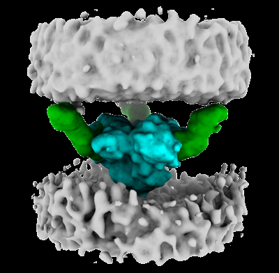
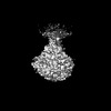
- EMDB-29294: HIV-1 envelope trimer bound to three CD4 molecules on membranes -
+ Open data

- Basic information
| Entry |  | |||||||||
|---|---|---|---|---|---|---|---|---|---|---|
| Title | HIV-1 envelope trimer bound to three CD4 molecules on membranes | |||||||||
 Map data Map data | ||||||||||
 Sample Sample |
| |||||||||
 Keywords Keywords | Virus-Host Complex / Glycoprotein / Cell receptor / VIRAL PROTEIN | |||||||||
| Biological species |   Human immunodeficiency virus 1 Human immunodeficiency virus 1 | |||||||||
| Method | subtomogram averaging / cryo EM / Resolution: 15.0 Å | |||||||||
 Authors Authors | Li W / Qin Z / Nand E / Grunst MW / Grover JR / Uchil PD / Mothes W | |||||||||
| Funding support |  United States, 1 items United States, 1 items
| |||||||||
 Citation Citation | Journal: Nature / Year: 2023 Title: HIV-1 Env trimers asymmetrically engage CD4 receptors in membranes. Authors: Wenwei Li / Zhuan Qin / Elizabeth Nand / Michael W Grunst / Jonathan R Grover / Julian W Bess / Jeffrey D Lifson / Michael B Zwick / Hemant D Tagare / Pradeep D Uchil / Walther Mothes / Abstract: Human immunodeficiency virus 1 (HIV-1) infection is initiated by binding of the viral envelope glycoprotein (Env) to the cell-surface receptor CD4. Although high-resolution structures of Env in a ...Human immunodeficiency virus 1 (HIV-1) infection is initiated by binding of the viral envelope glycoprotein (Env) to the cell-surface receptor CD4. Although high-resolution structures of Env in a complex with the soluble domains of CD4 have been determined, the binding process is less understood in native membranes. Here we used cryo-electron tomography to monitor Env-CD4 interactions at the membrane-membrane interfaces formed between HIV-1 and CD4-presenting virus-like particles. Env-CD4 complexes organized into clusters and rings, bringing the opposing membranes closer together. Env-CD4 clustering was dependent on capsid maturation. Subtomogram averaging and classification revealed that Env bound to one, two and finally three CD4 molecules, after which Env adopted an open state. Our data indicate that asymmetric HIV-1 Env trimers bound to one and two CD4 molecules are detectable intermediates during virus binding to host cell membranes, which probably has consequences for antibody-mediated immune responses and vaccine immunogen design. | |||||||||
| History |
|
- Structure visualization
| Supplemental images |
|---|
- Downloads & links
-EMDB archive
| Map data | emd_29294.map.gz | 14.4 MB |  EMDB map data format EMDB map data format | |
|---|---|---|---|---|
| Header (meta data) | emd-29294-v30.xmlemd-29294.xml | 13.3 KB 13.3 KB | Display Display | EMDB header |
| FSC (resolution estimation) | emd_29294_fsc.xml | 5.8 KB | Display | FSC data file |
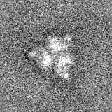
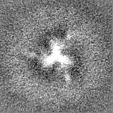
| Images |  emd_29294.png emd_29294.png | 96 KB | ||
| Filedesc metadata | emd-29294.cif.gz | 4.2 KB | ||
| Others | emd_29294_half_map_1.map.gzemd_29294_half_map_2.map.gz | 14.4 MB 14.4 MB | ||
| Archive directory |  http://ftp.pdbj.org/pub/emdb/structures/EMD-29294ftp://ftp.pdbj.org/pub/emdb/structures/EMD-29294 http://ftp.pdbj.org/pub/emdb/structures/EMD-29294ftp://ftp.pdbj.org/pub/emdb/structures/EMD-29294 | HTTPS FTP |
-Related structure data

-Links
| EMDB pages | EMDB (EBI/PDBe) / EMDataResource |
|---|
-Map
| File | Download / File: emd_29294.map.gz / Format: CCP4 / Size: 15.6 MB / Type: IMAGE STORED AS FLOATING POINT NUMBER (4 BYTES) | ||||||||||||||||||||||||||||||||||||
|---|---|---|---|---|---|---|---|---|---|---|---|---|---|---|---|---|---|---|---|---|---|---|---|---|---|---|---|---|---|---|---|---|---|---|---|---|---|
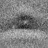
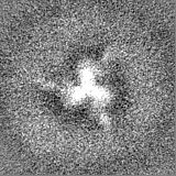
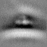
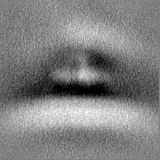
| Projections & slices | Image control
Images are generated by Spider. | ||||||||||||||||||||||||||||||||||||
| Voxel size | X=Y=Z: 2.7 Å | ||||||||||||||||||||||||||||||||||||
| Density |
| ||||||||||||||||||||||||||||||||||||
| Symmetry | Space group: 1 | ||||||||||||||||||||||||||||||||||||
| Details | EMDB XML:
|
 Z (Sec.)
 Y (Row.)
 X (Col.)


-Supplemental data
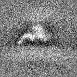
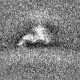
-Half map: #1
| File | emd_29294_half_map_1.map | ||||||||||||
|---|---|---|---|---|---|---|---|---|---|---|---|---|---|
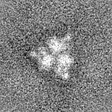
| Projections & Slices |
| ||||||||||||
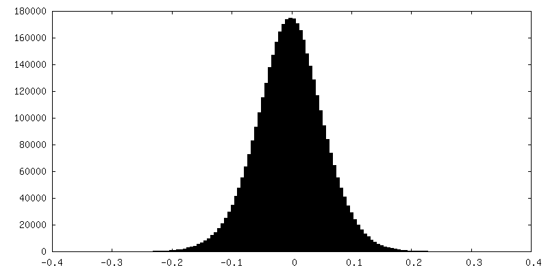
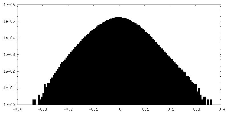
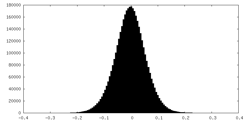
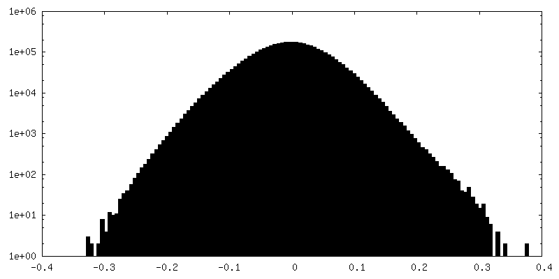
| Density Histograms |

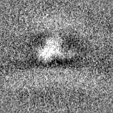
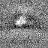
-Half map: #2
| File | emd_29294_half_map_2.map | ||||||||||||
|---|---|---|---|---|---|---|---|---|---|---|---|---|---|
| Projections & Slices |
| ||||||||||||
| Density Histograms |

- Sample components
-Entire : HIV-1 envelope trimer bound to three CD4 molecules on membranes
| Entire | Name: HIV-1 envelope trimer bound to three CD4 molecules on membranes |
|---|---|
| Components |
|
-Supramolecule #1: HIV-1 envelope trimer bound to three CD4 molecules on membranes
| Supramolecule | Name: HIV-1 envelope trimer bound to three CD4 molecules on membranes type: complex / ID: 1 / Parent: 0 / Macromolecule list: #1-#2 |
|---|---|
| Source (natural) | Organism: Human immunodeficiency virus 1 |
-Experimental details
-Structure determination
| Method | cryo EM |
|---|---|
 Processing Processing | subtomogram averaging |
| Aggregation state | particle |
-Sample preparation
| Buffer | pH: 7.4 |
|---|---|
| Vitrification | Cryogen name: ETHANE |
- Electron microscopy
| Microscope | FEI TITAN KRIOS |
|---|---|
| Specialist optics | Phase plate: VOLTA PHASE PLATE / Energy filter - Name: GIF Quantum LS / Energy filter - Slit width: 20 eV |
| Image recording | Film or detector model: GATAN K3 BIOQUANTUM (6k x 4k) / Average electron dose: 3.0 e/Å2 |
| Electron beam | Acceleration voltage: 300 kV / Electron source:  FIELD EMISSION GUN FIELD EMISSION GUN |
| Electron optics | C2 aperture diameter: 50.0 µm / Illumination mode: OTHER / Imaging mode: BRIGHT FIELD / Cs: 2.7 mm / Nominal defocus max: 0.5 µm / Nominal defocus min: 0.0 µm |
| Experimental equipment |  Model: Titan Krios / Image courtesy: FEI Company |
-Image processing
| Final reconstruction | Number classes used: 1953 / Applied symmetry - Point group: C1 (asymmetric) / Resolution.type: BY AUTHOR / Resolution: 15.0 Å / Resolution method: FSC 0.143 CUT-OFF / Number subtomograms used: 168 |
|---|---|
| Extraction | Number tomograms: 168 / Number images used: 1953 |
| Final angle assignment | Type: OTHER |
| FSC plot (resolution estimation) | 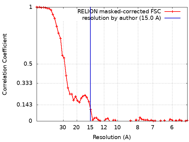 |
