 Movie
Movie Controller

[English] 日本語
 Yorodumi
- EMDB-29007: Human L-type voltage-gated calcium channel Cav1.2 complexed with ... -
+ Open data

- Basic information
| Entry | 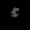 | |||||||||
|---|---|---|---|---|---|---|---|---|---|---|
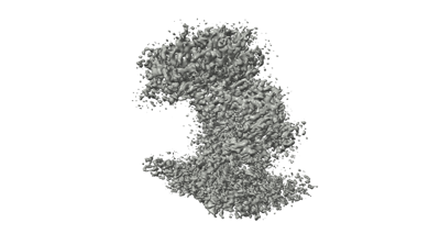
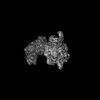
| Title | Human L-type voltage-gated calcium channel Cav1.2 complexed with gabapentin (Segment Map) | |||||||||
 Map data Map data | ||||||||||
 Sample Sample |
| |||||||||
 Keywords Keywords | voltage-gated calcium channel / CaV alpha2delta / drug binding / gabapentinoid / MEMBRANE PROTEIN / gabapentin | |||||||||
| Biological species |  | |||||||||
| Method | single particle reconstruction / cryo EM / Resolution: 3.1 Å | |||||||||
 Authors Authors | Chen Z / Mondal A / Minor DL | |||||||||
| Funding support |  United States, 2 items United States, 2 items
| |||||||||
 Citation Citation | Journal: Nat Struct Mol Biol / Year: 2023 Title: Structural basis for Caαδ:gabapentin binding. Authors: Zhou Chen / Abhisek Mondal / Daniel L Minor / Abstract: Gabapentinoid drugs for pain and anxiety act on the Caαδ-1 and Caαδ-2 subunits of high-voltage-activated calcium channels (Ca1s and Ca2s). Here we present the cryo-EM structure of the gabapentin- ...Gabapentinoid drugs for pain and anxiety act on the Caαδ-1 and Caαδ-2 subunits of high-voltage-activated calcium channels (Ca1s and Ca2s). Here we present the cryo-EM structure of the gabapentin-bound brain and cardiac Ca1.2/Caβ/Caαδ-1 channel. The data reveal a binding pocket in the Caαδ-1 dCache1 domain that completely encapsulates gabapentin and define Caαδ isoform sequence variations that explain the gabapentin binding selectivity of Caαδ-1 and Caαδ-2. | |||||||||
| History |
|
- Structure visualization
| Supplemental images |
|---|
- Downloads & links
-EMDB archive
| Map data | emd_29007.map.gz | 17.9 MB |  EMDB map data format EMDB map data format | |
|---|---|---|---|---|
| Header (meta data) | emd-29007-v30.xmlemd-29007.xml | 14 KB 14 KB | Display Display | EMDB header |
| Images |  emd_29007.png emd_29007.png | 52.7 KB | ||
| Others | emd_29007_half_map_1.map.gzemd_29007_half_map_2.map.gz | 238.2 MB 238.3 MB | ||
| Archive directory |  http://ftp.pdbj.org/pub/emdb/structures/EMD-29007ftp://ftp.pdbj.org/pub/emdb/structures/EMD-29007 http://ftp.pdbj.org/pub/emdb/structures/EMD-29007ftp://ftp.pdbj.org/pub/emdb/structures/EMD-29007 | HTTPS FTP |
-Validation report
| Summary document | emd_29007_validation.pdf.gz | 781.5 KB | Display | EMDB validaton report |
|---|---|---|---|---|
| Full document | emd_29007_full_validation.pdf.gz | 781.1 KB | Display | |
| Data in XML | emd_29007_validation.xml.gz | 16.7 KB | Display | |
| Data in CIF | emd_29007_validation.cif.gz | 19.9 KB | Display | |
| Arichive directory | https://ftp.pdbj.org/pub/emdb/validation_reports/EMD-29007ftp://ftp.pdbj.org/pub/emdb/validation_reports/EMD-29007 | HTTPS FTP |
-Related structure data


-Links
| EMDB pages | EMDB (EBI/PDBe) / EMDataResource |
|---|
-Map
| File | Download / File: emd_29007.map.gz / Format: CCP4 / Size: 325 MB / Type: IMAGE STORED AS FLOATING POINT NUMBER (4 BYTES) | ||||||||||||||||||||||||||||||||||||
|---|---|---|---|---|---|---|---|---|---|---|---|---|---|---|---|---|---|---|---|---|---|---|---|---|---|---|---|---|---|---|---|---|---|---|---|---|---|
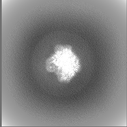
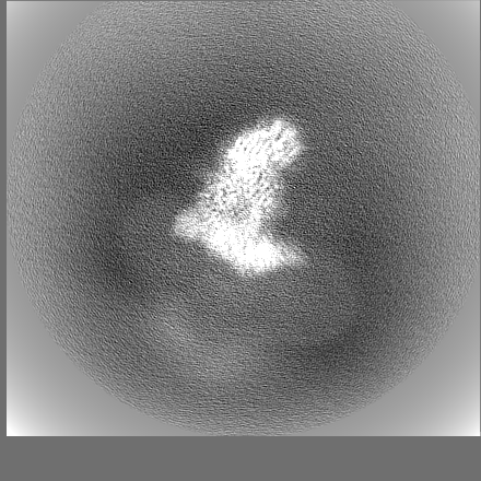
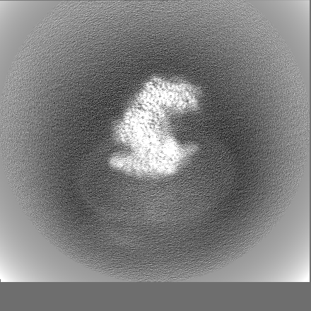
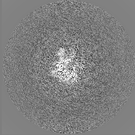
| Projections & slices | Image control
Images are generated by Spider. | ||||||||||||||||||||||||||||||||||||
| Voxel size | X=Y=Z: 0.835 Å | ||||||||||||||||||||||||||||||||||||
| Density |
| ||||||||||||||||||||||||||||||||||||
| Symmetry | Space group: 1 | ||||||||||||||||||||||||||||||||||||
| Details | EMDB XML:
|
 Z (Sec.)
 Y (Row.)
 X (Col.)


-Supplemental data
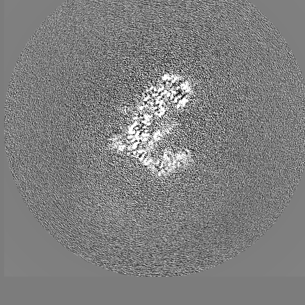
-Half map: #1
| File | emd_29007_half_map_1.map | ||||||||||||
|---|---|---|---|---|---|---|---|---|---|---|---|---|---|
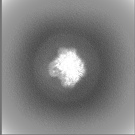
| Projections & Slices |
| ||||||||||||
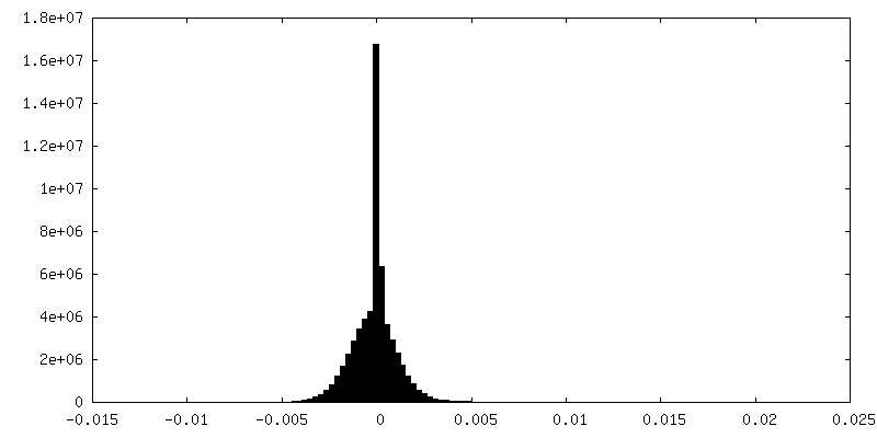
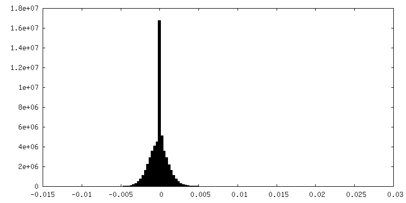
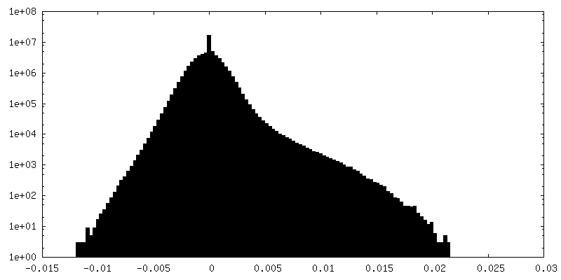
| Density Histograms |

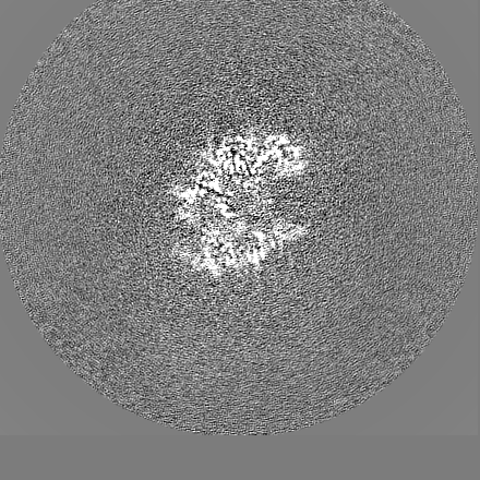
-Half map: #2
| File | emd_29007_half_map_2.map | ||||||||||||
|---|---|---|---|---|---|---|---|---|---|---|---|---|---|
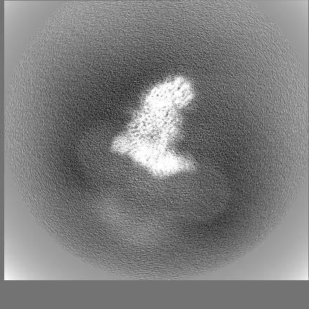
| Projections & Slices |
| ||||||||||||
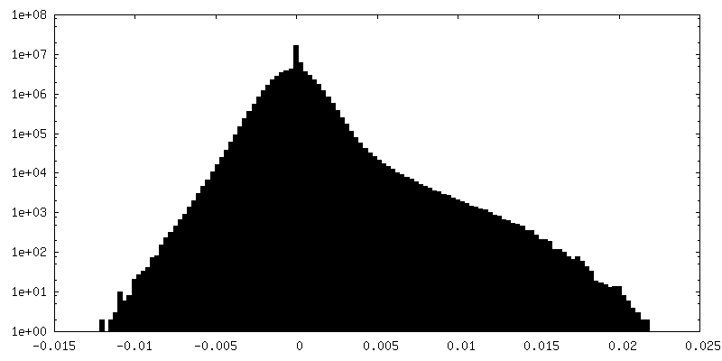
| Density Histograms |

- Sample components
-Entire : Ternary complex of human CaV alpha1C with rabbit CaV alpha2delta-1
| Entire | Name: Ternary complex of human CaV alpha1C with rabbit CaV alpha2delta-1 |
|---|---|
| Components |
|
-Supramolecule #1: Ternary complex of human CaV alpha1C with rabbit CaV alpha2delta-1
| Supramolecule | Name: Ternary complex of human CaV alpha1C with rabbit CaV alpha2delta-1 type: complex / ID: 1 / Parent: 0 / Macromolecule list: #1-#2 |
|---|
-Supramolecule #2: Rabbit CaV alpha2delta-1
| Supramolecule | Name: Rabbit CaV alpha2delta-1 / type: complex / ID: 2 / Parent: 1 / Macromolecule list: #1 |
|---|---|
| Source (natural) | Organism: |
| Molecular weight | Theoretical: 125 KDa |
-Experimental details
-Structure determination
| Method | cryo EM |
|---|---|
 Processing Processing | single particle reconstruction |
| Aggregation state | particle |
-Sample preparation
| Concentration | 2.7 mg/mL |
|---|---|
| Buffer | pH: 8 |
| Grid | Model: Quantifoil R1.2/1.3 / Support film - Material: CARBON / Support film - topology: HOLEY |
| Vitrification | Cryogen name: ETHANE / Chamber humidity: 100 % / Chamber temperature: 277 K / Instrument: FEI VITROBOT MARK IV |
- Electron microscopy
| Microscope | FEI TITAN KRIOS |
|---|---|
| Image recording | Film or detector model: GATAN K3 (6k x 4k) / Average electron dose: 46.0 e/Å2 |
| Electron beam | Acceleration voltage: 300 kV / Electron source:  FIELD EMISSION GUN FIELD EMISSION GUN |
| Electron optics | Illumination mode: FLOOD BEAM / Imaging mode: BRIGHT FIELD / Cs: 2.7 mm / Nominal defocus max: 1.7 µm / Nominal defocus min: 0.9 µm / Nominal magnification: 105000 |
| Experimental equipment |  Model: Titan Krios / Image courtesy: FEI Company |
-Image processing
| Startup model | Type of model: PDB ENTRY PDB model - PDB ID: |
|---|---|
| Final reconstruction | Resolution.type: BY AUTHOR / Resolution: 3.1 Å / Resolution method: FSC 0.143 CUT-OFF / Number images used: 259107 |
| Initial angle assignment | Type: MAXIMUM LIKELIHOOD |
| Final angle assignment | Type: MAXIMUM LIKELIHOOD |

