 Movie
Movie Controller

[English] 日本語
 Yorodumi
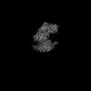
- PDB-8fd7: Structure of the human L-type voltage-gated calcium channel Cav1.... -
+ Open data

- Basic information
| Entry | Database: PDB / ID: 8fd7 | |||||||||
|---|---|---|---|---|---|---|---|---|---|---|
| Title | Structure of the human L-type voltage-gated calcium channel Cav1.2 complexed with gabapentin | |||||||||
 Components Components |
| |||||||||
 Keywords Keywords | MEMBRANE PROTEIN / voltage-gated calcium channel / CaV alpha2delta / drug binding / gabapentin | |||||||||
| Function / homology |  Function and homology information Function and homology informationpositive regulation of high voltage-gated calcium channel activity / voltage-gated calcium channel activity involved in AV node cell action potential / voltage-gated calcium channel activity involved in cardiac muscle cell action potential / immune system development / positive regulation of adenylate cyclase activity / membrane depolarization during atrial cardiac muscle cell action potential / calcium ion transmembrane transport via high voltage-gated calcium channel / Phase 2 - plateau phase / high voltage-gated calcium channel activity / cardiac conduction ...positive regulation of high voltage-gated calcium channel activity / voltage-gated calcium channel activity involved in AV node cell action potential / voltage-gated calcium channel activity involved in cardiac muscle cell action potential / immune system development / positive regulation of adenylate cyclase activity / membrane depolarization during atrial cardiac muscle cell action potential / calcium ion transmembrane transport via high voltage-gated calcium channel / Phase 2 - plateau phase / high voltage-gated calcium channel activity / cardiac conduction / membrane depolarization during AV node cell action potential / L-type voltage-gated calcium channel complex / membrane depolarization during cardiac muscle cell action potential / positive regulation of muscle contraction / cell communication by electrical coupling involved in cardiac conduction / NCAM1 interactions / camera-type eye development / regulation of ventricular cardiac muscle cell action potential / cardiac muscle cell action potential involved in contraction / embryonic forelimb morphogenesis / calcium ion transport into cytosol / voltage-gated calcium channel complex / Phase 0 - rapid depolarisation / regulation of heart rate by cardiac conduction / alpha-actinin binding / calcium ion import across plasma membrane / voltage-gated calcium channel activity / regulation of cardiac muscle contraction by regulation of the release of sequestered calcium ion / T-tubule / calcium channel regulator activity / Regulation of insulin secretion / postsynaptic density membrane / Z disc / calcium ion transmembrane transport / Adrenaline,noradrenaline inhibits insulin secretion / heart development / positive regulation of cytosolic calcium ion concentration / perikaryon / calmodulin binding / postsynaptic density / cilium / dendrite / nucleoplasm / membrane / metal ion binding / plasma membrane / cytoplasm Similarity search - Function | |||||||||
| Biological species |   Homo sapiens (human) Homo sapiens (human) | |||||||||
| Method | ELECTRON MICROSCOPY / single particle reconstruction / cryo EM / Resolution: 3.1 Å | |||||||||
 Authors Authors | Chen, Z. / Mondal, A. / Minor, D.L. | |||||||||
| Funding support |  United States, 2items United States, 2items
| |||||||||
 Citation Citation | Journal: Nat Struct Mol Biol / Year: 2023 Title: Structural basis for Caαδ:gabapentin binding. Authors: Zhou Chen / Abhisek Mondal / Daniel L Minor / Abstract: Gabapentinoid drugs for pain and anxiety act on the Caαδ-1 and Caαδ-2 subunits of high-voltage-activated calcium channels (Ca1s and Ca2s). Here we present the cryo-EM structure of the gabapentin- ...Gabapentinoid drugs for pain and anxiety act on the Caαδ-1 and Caαδ-2 subunits of high-voltage-activated calcium channels (Ca1s and Ca2s). Here we present the cryo-EM structure of the gabapentin-bound brain and cardiac Ca1.2/Caβ/Caαδ-1 channel. The data reveal a binding pocket in the Caαδ-1 dCache1 domain that completely encapsulates gabapentin and define Caαδ isoform sequence variations that explain the gabapentin binding selectivity of Caαδ-1 and Caαδ-2. | |||||||||
| History |
|
- Structure visualization
| Structure viewer | Molecule: MolmilJmol/JSmol |
|---|
- Downloads & links
-Download
| PDBx/mmCIF format | 8fd7.cif.gz | 516.6 KB | Display | PDBx/mmCIF format |
|---|---|---|---|---|
| PDB format | pdb8fd7.ent.gz | 403.1 KB | Display | PDB format |
| PDBx/mmJSON format | 8fd7.json.gz | Tree view | PDBx/mmJSON format | |
| Others |  Other downloads Other downloads |
-Validation report
| Arichive directory | https://data.pdbj.org/pub/pdb/validation_reports/fd/8fd7ftp://data.pdbj.org/pub/pdb/validation_reports/fd/8fd7 | HTTPS FTP |
|---|
-Related structure data
| Related structure data | 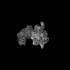 29004MC M: map data used to model this data C: citing same article ( |
|---|---|
| Similar structure data |


-Links
 PDBj
PDBj


- Assembly
| Deposited unit | 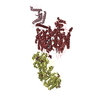
|
|---|---|
| 1 |
|
-Components
-Protein , 1 types, 1 molecules D
| #1: Protein | Mass: 125082.906 Da / Num. of mol.: 1 Source method: isolated from a genetically manipulated source Source: (gene. exp.) Homo sapiens (human) / References: UniProt: P13806 |
|---|
-Voltage-dependent L-type calcium channel subunit ... , 2 types, 2 molecules KC
| #2: Protein | Mass: 187082.188 Da / Num. of mol.: 1 Source method: isolated from a genetically manipulated source Source: (gene. exp.) Homo sapiens (human) / Gene: CACNA1C, CACH2, CACN2, CACNL1A1, CCHL1A1 / Production host: Homo sapiens (human) / References: UniProt: Q13936 |
|---|---|
| #3: Protein | Mass: 53889.152 Da / Num. of mol.: 1 Source method: isolated from a genetically manipulated source Source: (gene. exp.) Homo sapiens (human) / References: UniProt: P54286 |
-Sugars , 2 types, 9 molecules 
| #4: Polysaccharide | 2-acetamido-2-deoxy-beta-D-glucopyranose-(1-4)-2-acetamido-2-deoxy-beta-D-glucopyranose Source method: isolated from a genetically manipulated source |
|---|---|
| #5: Sugar | ChemComp-NAG /  Type: D-saccharide, beta linking / Mass: 221.208 Da / Num. of mol.: 8 Type: D-saccharide, beta linking / Mass: 221.208 Da / Num. of mol.: 8Source method: isolated from a genetically manipulated source Formula: C8H15NO6 |
-Non-polymers , 7 types, 13 molecules 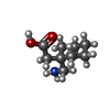


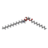


| #6: Chemical | ChemComp-GBN / [ Mass: 171.237 Da / Num. of mol.: 1 / Source method: obtained synthetically / Formula: C9H17NO2 Mass: 171.237 Da / Num. of mol.: 1 / Source method: obtained synthetically / Formula: C9H17NO2 | ||||||||||
|---|---|---|---|---|---|---|---|---|---|---|---|
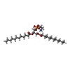
| #7: Chemical |  Mass: 40.078 Da / Num. of mol.: 3 / Source method: obtained synthetically / Formula: Ca Mass: 40.078 Da / Num. of mol.: 3 / Source method: obtained synthetically / Formula: Ca#8: Chemical | ChemComp-NA / |  Mass: 22.990 Da / Num. of mol.: 1 / Source method: obtained synthetically / Formula: Na Mass: 22.990 Da / Num. of mol.: 1 / Source method: obtained synthetically / Formula: Na#9: Chemical | ChemComp-WO9 / ( |  Mass: 649.879 Da / Num. of mol.: 1 / Source method: obtained synthetically / Formula: C34H68NO8P Mass: 649.879 Da / Num. of mol.: 1 / Source method: obtained synthetically / Formula: C34H68NO8P#10: Chemical | ChemComp-YSW / ( |  Mass: 579.746 Da / Num. of mol.: 1 / Source method: obtained synthetically / Formula: C29H58NO8P Mass: 579.746 Da / Num. of mol.: 1 / Source method: obtained synthetically / Formula: C29H58NO8P#11: Chemical |  Mass: 386.654 Da / Num. of mol.: 3 / Source method: obtained synthetically / Formula: C27H46O Mass: 386.654 Da / Num. of mol.: 3 / Source method: obtained synthetically / Formula: C27H46O#12: Water | ChemComp-HOH / | Mass: 18.015 Da / Num. of mol.: 3 / Source method: isolated from a natural source / Formula: H2O |
-Details
| Has ligand of interest | N |
|---|---|
| Has protein modification | Y |
-Experimental details
-Experiment
| Experiment | Method: ELECTRON MICROSCOPY |
|---|---|
| EM experiment | Aggregation state: PARTICLE / 3D reconstruction method: single particle reconstruction |
- Sample preparation
| Component |
| ||||||||||||||||||||||||||||||
|---|---|---|---|---|---|---|---|---|---|---|---|---|---|---|---|---|---|---|---|---|---|---|---|---|---|---|---|---|---|---|---|
| Molecular weight |
| ||||||||||||||||||||||||||||||
| Source (natural) |
| ||||||||||||||||||||||||||||||
| Source (recombinant) |
| ||||||||||||||||||||||||||||||
| Buffer solution | pH: 8 | ||||||||||||||||||||||||||||||
| Specimen | Conc.: 2 mg/ml / Embedding applied: NO / Shadowing applied: NO / Staining applied: NO / Vitrification applied: YES | ||||||||||||||||||||||||||||||
| Specimen support | Grid material: GOLD / Grid mesh size: 300 divisions/in. / Grid type: Quantifoil R1.2/1.3 | ||||||||||||||||||||||||||||||
| Vitrification | Instrument: FEI VITROBOT MARK IV / Cryogen name: ETHANE / Humidity: 100 % / Chamber temperature: 277 K |
- Electron microscopy imaging
| Experimental equipment |  Model: Titan Krios / Image courtesy: FEI Company |
|---|---|
| Microscopy | Model: FEI TITAN KRIOS |
| Electron gun | Electron source:  FIELD EMISSION GUN / Accelerating voltage: 300 kV / Illumination mode: FLOOD BEAM FIELD EMISSION GUN / Accelerating voltage: 300 kV / Illumination mode: FLOOD BEAM |
| Electron lens | Mode: BRIGHT FIELD / Nominal magnification: 105000 X / Nominal defocus max: 1700 nm / Nominal defocus min: 900 nm / Cs: 2.7 mm |
| Image recording | Electron dose: 46 e/Å2 / Film or detector model: GATAN K3 (6k x 4k) |
- Processing
| Software | Name: PHENIX / Version: 1.20.1_4487: / Classification: refinement | ||||||||||||||||||||||||
|---|---|---|---|---|---|---|---|---|---|---|---|---|---|---|---|---|---|---|---|---|---|---|---|---|---|
| CTF correction | Type: PHASE FLIPPING AND AMPLITUDE CORRECTION | ||||||||||||||||||||||||
| Symmetry | Point symmetry: C1 (asymmetric) | ||||||||||||||||||||||||
| 3D reconstruction | Resolution: 3.1 Å / Resolution method: FSC 0.143 CUT-OFF / Num. of particles: 259107 / Symmetry type: POINT | ||||||||||||||||||||||||
| Refine LS restraints |
|
