 Movie
Movie Controller

[English] 日本語
 Yorodumi
- EMDB-28896: Human olfactory receptor OR51E2 bound to propionate in complex wi... -
+ Open data

- Basic information
| Entry |  | |||||||||
|---|---|---|---|---|---|---|---|---|---|---|
| Title | Human olfactory receptor OR51E2 bound to propionate in complex with miniGs399 | |||||||||
 Map data Map data | Sharpened map from cryoSPARC | |||||||||
 Sample Sample |
| |||||||||
 Keywords Keywords | Human olfactory receptor / G protein-coupled receptor / odorant-binding / sensory transduction / MEMBRANE PROTEIN | |||||||||
| Function / homology |  Function and homology information Function and homology informationmelanocyte proliferation / positive regulation of renin secretion into blood stream / Expression and translocation of olfactory receptors / olfactory receptor activity / intracellular organelle / positive regulation of blood pressure / melanocyte differentiation / cellular response to fatty acid / steroid hormone receptor signaling pathway / nuclear steroid receptor activity ...melanocyte proliferation / positive regulation of renin secretion into blood stream / Expression and translocation of olfactory receptors / olfactory receptor activity / intracellular organelle / positive regulation of blood pressure / melanocyte differentiation / cellular response to fatty acid / steroid hormone receptor signaling pathway / nuclear steroid receptor activity / PKA activation in glucagon signalling / developmental growth / hair follicle placode formation / D1 dopamine receptor binding / intracellular transport / vascular endothelial cell response to laminar fluid shear stress / activation of adenylate cyclase activity / renal water homeostasis / Hedgehog 'off' state / adenylate cyclase-activating adrenergic receptor signaling pathway / cellular response to glucagon stimulus / regulation of insulin secretion / adenylate cyclase activator activity / trans-Golgi network membrane / negative regulation of inflammatory response to antigenic stimulus / G protein-coupled receptor activity / bone development / platelet aggregation / G-protein beta/gamma-subunit complex binding / Olfactory Signaling Pathway / cognition / Activation of the phototransduction cascade / G beta:gamma signalling through PLC beta / Presynaptic function of Kainate receptors / Thromboxane signalling through TP receptor / G protein-coupled acetylcholine receptor signaling pathway / G-protein activation / Activation of G protein gated Potassium channels / Inhibition of voltage gated Ca2+ channels via Gbeta/gamma subunits / adenylate cyclase-activating G protein-coupled receptor signaling pathway / Prostacyclin signalling through prostacyclin receptor / G beta:gamma signalling through CDC42 / Glucagon signaling in metabolic regulation / G beta:gamma signalling through BTK / Synthesis, secretion, and inactivation of Glucagon-like Peptide-1 (GLP-1) / ADP signalling through P2Y purinoceptor 12 / Sensory perception of sweet, bitter, and umami (glutamate) taste / photoreceptor disc membrane / Glucagon-type ligand receptors / Adrenaline,noradrenaline inhibits insulin secretion / Vasopressin regulates renal water homeostasis via Aquaporins / G alpha (z) signalling events / Glucagon-like Peptide-1 (GLP1) regulates insulin secretion / ADP signalling through P2Y purinoceptor 1 / cellular response to catecholamine stimulus / ADORA2B mediated anti-inflammatory cytokines production / G beta:gamma signalling through PI3Kgamma / sensory perception of smell / Cooperation of PDCL (PhLP1) and TRiC/CCT in G-protein beta folding / adenylate cyclase-activating dopamine receptor signaling pathway / cell migration / GPER1 signaling / Inactivation, recovery and regulation of the phototransduction cascade / cellular response to prostaglandin E stimulus / G-protein beta-subunit binding / heterotrimeric G-protein complex / G alpha (12/13) signalling events / sensory perception of taste / extracellular vesicle / signaling receptor activity / signaling receptor complex adaptor activity / Thrombin signalling through proteinase activated receptors (PARs) / positive regulation of cold-induced thermogenesis / retina development in camera-type eye / G protein activity / GTPase binding / Ca2+ pathway / fibroblast proliferation / High laminar flow shear stress activates signaling by PIEZO1 and PECAM1:CDH5:KDR in endothelial cells / early endosome membrane / G alpha (i) signalling events / G alpha (s) signalling events / phospholipase C-activating G protein-coupled receptor signaling pathway / Hydrolases; Acting on acid anhydrides; Acting on GTP to facilitate cellular and subcellular movement / G alpha (q) signalling events / Ras protein signal transduction / Extra-nuclear estrogen signaling / cell population proliferation / G protein-coupled receptor signaling pathway / lysosomal membrane / GTPase activity / synapse / GTP binding / protein-containing complex binding / signal transduction / extracellular exosome / metal ion binding / membrane / plasma membrane / cytosol Similarity search - Function | |||||||||
| Biological species |  Homo sapiens (human) / Homo sapiens (human) /  | |||||||||
| Method | single particle reconstruction / cryo EM / Resolution: 3.1 Å | |||||||||
 Authors Authors | Billesboelle CB / Manglik A | |||||||||
| Funding support |  United States, 1 items United States, 1 items
| |||||||||
 Citation Citation | Journal: Nature / Year: 2023 Title: Structural basis of odorant recognition by a human odorant receptor. Authors: Christian B Billesbølle / Claire A de March / Wijnand J C van der Velden / Ning Ma / Jeevan Tewari / Claudia Llinas Del Torrent / Linus Li / Bryan Faust / Nagarajan Vaidehi / Hiroaki ...Authors: Christian B Billesbølle / Claire A de March / Wijnand J C van der Velden / Ning Ma / Jeevan Tewari / Claudia Llinas Del Torrent / Linus Li / Bryan Faust / Nagarajan Vaidehi / Hiroaki Matsunami / Aashish Manglik /   Abstract: Our sense of smell enables us to navigate a vast space of chemically diverse odour molecules. This task is accomplished by the combinatorial activation of approximately 400 odorant G protein-coupled ...Our sense of smell enables us to navigate a vast space of chemically diverse odour molecules. This task is accomplished by the combinatorial activation of approximately 400 odorant G protein-coupled receptors encoded in the human genome. How odorants are recognized by odorant receptors remains unclear. Here we provide mechanistic insight into how an odorant binds to a human odorant receptor. Using cryo-electron microscopy, we determined the structure of the active human odorant receptor OR51E2 bound to the fatty acid propionate. Propionate is bound within an occluded pocket in OR51E2 and makes specific contacts critical to receptor activation. Mutation of the odorant-binding pocket in OR51E2 alters the recognition spectrum for fatty acids of varying chain length, suggesting that odorant selectivity is controlled by tight packing interactions between an odorant and an odorant receptor. Molecular dynamics simulations demonstrate that propionate-induced conformational changes in extracellular loop 3 activate OR51E2. Together, our studies provide a high-resolution view of chemical recognition of an odorant by a vertebrate odorant receptor, providing insight into how this large family of G protein-coupled receptors enables our olfactory sense. | |||||||||
| History |
|
- Structure visualization
| Supplemental images |
|---|

- Downloads & links
-EMDB archive
| Map data | emd_28896.map.gz | 85.8 MB | EMDB map data format | |
|---|---|---|---|---|
| Header (meta data) | emd-28896-v30.xmlemd-28896.xml | 25.6 KB 25.6 KB | Display Display | EMDB header |
| FSC (resolution estimation) | emd_28896_fsc.xml | 10.8 KB | Display | FSC data file |
| Images | 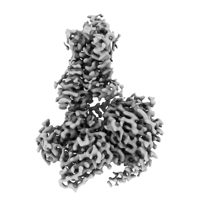 emd_28896.png emd_28896.png | 82.2 KB | ||
| Masks | emd_28896_msk_1.map | 91.1 MB | Mask map | |
| Filedesc metadata | emd-28896.cif.gz | 7.4 KB | ||
| Others | emd_28896_additional_1.map.gzemd_28896_half_map_1.map.gzemd_28896_half_map_2.map.gz | 44.1 MB 84.5 MB 84.5 MB | ||
| Archive directory |  http://ftp.pdbj.org/pub/emdb/structures/EMD-28896ftp://ftp.pdbj.org/pub/emdb/structures/EMD-28896 http://ftp.pdbj.org/pub/emdb/structures/EMD-28896ftp://ftp.pdbj.org/pub/emdb/structures/EMD-28896 | HTTPS FTP |
-Validation report
| Summary document | emd_28896_validation.pdf.gz | 846 KB | Display | EMDB validaton report |
|---|---|---|---|---|
| Full document | emd_28896_full_validation.pdf.gz | 845.5 KB | Display | |
| Data in XML | emd_28896_validation.xml.gz | 17.5 KB | Display | |
| Data in CIF | emd_28896_validation.cif.gz | 22.3 KB | Display | |
| Arichive directory | https://ftp.pdbj.org/pub/emdb/validation_reports/EMD-28896ftp://ftp.pdbj.org/pub/emdb/validation_reports/EMD-28896 | HTTPS FTP |
-Related structure data
| Related structure data |  8f76MC M: atomic model generated by this map C: citing same article ( |
|---|---|
| Similar structure data |

-Links
| EMDB pages | EMDB (EBI/PDBe) / EMDataResource |
|---|---|
| Related items in Molecule of the Month |


-Map
| File | Download / File: emd_28896.map.gz / Format: CCP4 / Size: 91.1 MB / Type: IMAGE STORED AS FLOATING POINT NUMBER (4 BYTES) | ||||||||||||||||||||||||||||||||||||
|---|---|---|---|---|---|---|---|---|---|---|---|---|---|---|---|---|---|---|---|---|---|---|---|---|---|---|---|---|---|---|---|---|---|---|---|---|---|
| Annotation | Sharpened map from cryoSPARC | ||||||||||||||||||||||||||||||||||||
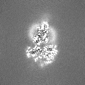
| Projections & slices | Image control
Images are generated by Spider. | ||||||||||||||||||||||||||||||||||||
| Voxel size | X=Y=Z: 0.81 Å | ||||||||||||||||||||||||||||||||||||
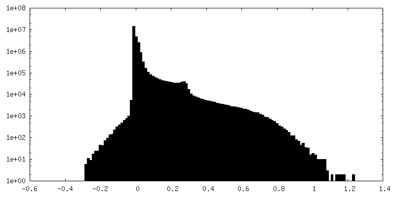
| Density |
| ||||||||||||||||||||||||||||||||||||
| Symmetry | Space group: 1 | ||||||||||||||||||||||||||||||||||||
| Details | EMDB XML:
|
 Z (Sec.)
 Y (Row.)
 X (Col.)


-Supplemental data
-Mask #1
| File | emd_28896_msk_1.map | ||||||||||||
|---|---|---|---|---|---|---|---|---|---|---|---|---|---|
| Projections & Slices |
| ||||||||||||
| Density Histograms |


-Additional map: Unsharpened map from cryoSPARC
| File | emd_28896_additional_1.map | ||||||||||||
|---|---|---|---|---|---|---|---|---|---|---|---|---|---|
| Annotation | Unsharpened map from cryoSPARC | ||||||||||||
| Projections & Slices |
| ||||||||||||
| Density Histograms |


-Half map: Half map A from cryoSPARC
| File | emd_28896_half_map_1.map | ||||||||||||
|---|---|---|---|---|---|---|---|---|---|---|---|---|---|
| Annotation | Half map A from cryoSPARC | ||||||||||||
| Projections & Slices |
| ||||||||||||
| Density Histograms |


-Half map: Half map B from cryoSPARC
| File | emd_28896_half_map_2.map | ||||||||||||
|---|---|---|---|---|---|---|---|---|---|---|---|---|---|
| Annotation | Half map B from cryoSPARC | ||||||||||||
| Projections & Slices |
| ||||||||||||
| Density Histograms |


- Sample components
-Entire : OR51E2-Gs complex
| Entire | Name: OR51E2-Gs complex |
|---|---|
| Components |
|
-Supramolecule #1: OR51E2-Gs complex
| Supramolecule | Name: OR51E2-Gs complex / type: complex / ID: 1 / Parent: 0 / Macromolecule list: #1-#5 |
|---|---|
| Source (natural) | Organism: Homo sapiens (human) |
| Molecular weight | Theoretical: 100 KDa |
-Macromolecule #1: Olfactory receptor 51E2
| Macromolecule | Name: Olfactory receptor 51E2 / type: protein_or_peptide / ID: 1 / Number of copies: 1 / Enantiomer: LEVO |
|---|---|
| Source (natural) | Organism: Homo sapiens (human) |
| Molecular weight | Theoretical: 36.651543 KDa |
| Recombinant expression | Organism: Homo sapiens (human) |
| Sequence | String: DYKDDDDASI DSSCNFTHAT FVLIGIPGLE KAHFWVGFPL LSMYVVAMFG NCIVVFIVRT ERSLHAPMYL FLCMLAAIDL ALSTSTMPK ILALFWFDSR EISFEACLTQ MFFIHALSAI ESTILLAMAF DRYVAICHPL RHAAVLNNTV TAQIGIVAVV R GSLFFFPL ...String: DYKDDDDASI DSSCNFTHAT FVLIGIPGLE KAHFWVGFPL LSMYVVAMFG NCIVVFIVRT ERSLHAPMYL FLCMLAAIDL ALSTSTMPK ILALFWFDSR EISFEACLTQ MFFIHALSAI ESTILLAMAF DRYVAICHPL RHAAVLNNTV TAQIGIVAVV R GSLFFFPL PLLIKRLAFC HSNVLSHSYC VHQDVMKLAY ADTLPNVVYG LTAILLVMGV DVMFISLSYF LIIRTVLQLP SK SERAKAF GTCVSHIGVV LAFYVPLIGL SVVHRFGNSL HPIVRVVMGD IYLLLPPVIN PIIYGAKTKQ IRTRVLAMFK ISC DKDLQA VGGK UniProtKB: Olfactory receptor 51E2 |
-Macromolecule #2: Nanobody 35
| Macromolecule | Name: Nanobody 35 / type: protein_or_peptide / ID: 2 / Number of copies: 1 / Enantiomer: LEVO |
|---|---|
| Source (natural) | Organism: |
| Molecular weight | Theoretical: 15.931698 KDa |
| Recombinant expression | Organism:  |
| Sequence | String: QVQLQESGGG LVQPGGSLRL SCAASGFTFS NYKMNWVRQA PGKGLEWVSD ISQSGASISY TGSVKGRFTI SRDNAKNTLY LQMNSLKPE DTAVYYCARC PAPFTRDCFD VTSTTYAYRG QGTQVTVSSL EVLFQGPGHH HHHHHH |
-Macromolecule #3: Guanine nucleotide-binding protein G(s) subunit alpha isoforms short
| Macromolecule | Name: Guanine nucleotide-binding protein G(s) subunit alpha isoforms short type: protein_or_peptide / ID: 3 / Number of copies: 1 / Enantiomer: LEVO |
|---|---|
| Source (natural) | Organism: Homo sapiens (human) |
| Molecular weight | Theoretical: 30.137025 KDa |
| Recombinant expression | Organism: Homo sapiens (human) |
| Sequence | String: GGSLEVLFQG PSGNSKTEDQ RNEEKAQREA NKKIEKQLQK DKQVYRATHR LLLLGADNSG KSTIVKQMRI LHGGSGGSGG TSGIFETKF QVDKVNFHMF DVGGQRDERR KWIQCFNDVT AIIFVVDSSD YNRLQEALNL FKSIWNNRWL RTISVILFLN K QDLLAEKV ...String: GGSLEVLFQG PSGNSKTEDQ RNEEKAQREA NKKIEKQLQK DKQVYRATHR LLLLGADNSG KSTIVKQMRI LHGGSGGSGG TSGIFETKF QVDKVNFHMF DVGGQRDERR KWIQCFNDVT AIIFVVDSSD YNRLQEALNL FKSIWNNRWL RTISVILFLN K QDLLAEKV LAGKSKIEDY FPEFARYTTP EDATPEPGED PRVTRAKYFI RDEFLRISTA SGDGRHYCYP HFTCAVDTEN AR RIFNDCR DIIQRMHLRQ YELL UniProtKB: Guanine nucleotide-binding protein G(s) subunit alpha isoforms short, Guanine nucleotide-binding protein G(s) subunit alpha isoforms short |
-Macromolecule #4: Guanine nucleotide-binding protein G(I)/G(S)/G(T) subunit beta-1
| Macromolecule | Name: Guanine nucleotide-binding protein G(I)/G(S)/G(T) subunit beta-1 type: protein_or_peptide / ID: 4 / Number of copies: 1 / Enantiomer: LEVO |
|---|---|
| Source (natural) | Organism: Homo sapiens (human) |
| Molecular weight | Theoretical: 40.786566 KDa |
| Recombinant expression | Organism:  Trichoplusia ni (cabbage looper) Trichoplusia ni (cabbage looper) |
| Sequence | String: MHHHHHHLEV LFQGPEDQVD PRLIDGKGSS GSELDQLRQE AEQLKNQIRD ARKACADATL SQITNNIDPV GRIQMRTRRT LRGHLAKIY AMHWGTDSRL LVSASQDGKL IIWDSYTTNK VHAIPLRSSW VMTCAYAPSG NYVACGGLDN ICSIYNLKTR E GNVRVSRE ...String: MHHHHHHLEV LFQGPEDQVD PRLIDGKGSS GSELDQLRQE AEQLKNQIRD ARKACADATL SQITNNIDPV GRIQMRTRRT LRGHLAKIY AMHWGTDSRL LVSASQDGKL IIWDSYTTNK VHAIPLRSSW VMTCAYAPSG NYVACGGLDN ICSIYNLKTR E GNVRVSRE LAGHTGYLSC CRFLDDNQIV TSSGDTTCAL WDIETGQQTT TFTGHTGDVM SLSLAPDTRL FVSGACDASA KL WDVREGM CRQTFTGHES DINAICFFPN GNAFATGSDD ATCRLFDLRA DQELMTYSHD NIICGITSVS FSKSGRLLLA GYD DFNCNV WDALKADRAG VLAGHDNRVS CLGVTDDGMA VATGSWDSFL KIWN UniProtKB: Guanine nucleotide-binding protein G(I)/G(S)/G(T) subunit beta-1 |
-Macromolecule #5: Guanine nucleotide-binding protein G(I)/G(S)/G(O) subunit gamma-2
| Macromolecule | Name: Guanine nucleotide-binding protein G(I)/G(S)/G(O) subunit gamma-2 type: protein_or_peptide / ID: 5 / Number of copies: 1 / Enantiomer: LEVO |
|---|---|
| Source (natural) | Organism: Homo sapiens (human) |
| Molecular weight | Theoretical: 7.861143 KDa |
| Recombinant expression | Organism: Trichoplusia ni (cabbage looper) |
| Sequence | String: MASNNTASIA QARKLVEQLK MEANIDRIKV SKAAADLMAY CEAHAKEDPL LTPVPASENP FREKKFFCAI L UniProtKB: Guanine nucleotide-binding protein G(I)/G(S)/G(O) subunit gamma-2 |
-Macromolecule #6: PROPANOIC ACID
| Macromolecule | Name: PROPANOIC ACID / type: ligand / ID: 6 / Number of copies: 1 / Formula: PPI |
|---|---|
| Molecular weight | Theoretical: 74.079 Da |
| Chemical component information |  ChemComp-PPI: |
-Experimental details
-Structure determination
| Method | cryo EM |
|---|---|
 Processing Processing | single particle reconstruction |
| Aggregation state | particle |
-Sample preparation
| Buffer | pH: 7.5 |
|---|---|
| Grid | Model: UltrAuFoil R1.2/1.3 / Material: GOLD / Mesh: 300 |
| Vitrification | Cryogen name: ETHANE |
- Electron microscopy
| Microscope | FEI TITAN KRIOS |
|---|---|
| Image recording | Film or detector model: GATAN K3 BIOQUANTUM (6k x 4k) / Average electron dose: 50.0 e/Å2 |
| Electron beam | Acceleration voltage: 300 kV / Electron source:  FIELD EMISSION GUN FIELD EMISSION GUN |
| Electron optics | Illumination mode: FLOOD BEAM / Imaging mode: BRIGHT FIELD / Nominal defocus max: 2.2 µm / Nominal defocus min: 0.8 µm / Nominal magnification: 105000 |
| Sample stage | Cooling holder cryogen: NITROGEN |
| Experimental equipment |  Model: Titan Krios / Image courtesy: FEI Company |

