Movie
Movie Controller


+ Open data

- Basic information
| Entry |  | |||||||||
|---|---|---|---|---|---|---|---|---|---|---|
| Title | PolyC9 in complex with aE11 Fab | |||||||||
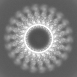
 Map data Map data | Local resolution filtered, sharpened combined map. Real-space cropped from box size 450px to 296px. | |||||||||
 Sample Sample |
| |||||||||
| Biological species |  Homo sapiens (human) / Homo sapiens (human) /  | |||||||||
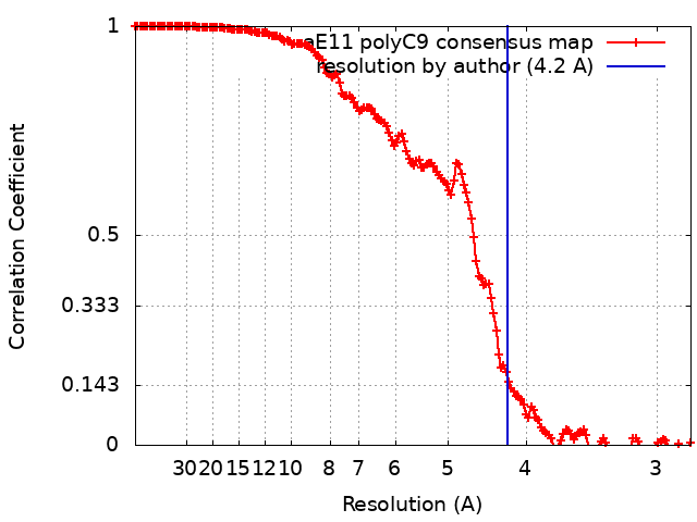
| Method | single particle reconstruction / cryo EM / Resolution: 4.2 Å | |||||||||
 Authors Authors | Bayly-Jones C | |||||||||
| Funding support |  Australia, 2 items Australia, 2 items
| |||||||||
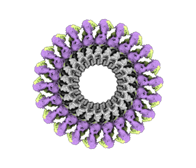
 Citation Citation | Journal: Commun Biol / Year: 2023 Title: The neoepitope of the complement C5b-9 Membrane Attack Complex is formed by proximity of adjacent ancillary regions of C9. Authors: Charles Bayly-Jones / Bill H T Ho / Corinna Lau / Eleanor W W Leung / Laura D'Andrea / Christopher J Lupton / Susan M Ekkel / Hariprasad Venugopal / James C Whisstock / Tom E Mollnes / ...Authors: Charles Bayly-Jones / Bill H T Ho / Corinna Lau / Eleanor W W Leung / Laura D'Andrea / Christopher J Lupton / Susan M Ekkel / Hariprasad Venugopal / James C Whisstock / Tom E Mollnes / Bradley A Spicer / Michelle A Dunstone /  Abstract: The Membrane Attack Complex (MAC) is responsible for forming large β-barrel channels in the membranes of pathogens, such as gram-negative bacteria. Off-target MAC assembly on endogenous tissue is ...The Membrane Attack Complex (MAC) is responsible for forming large β-barrel channels in the membranes of pathogens, such as gram-negative bacteria. Off-target MAC assembly on endogenous tissue is associated with inflammatory diseases and cancer. Accordingly, a human C5b-9 specific antibody, aE11, has been developed that detects a neoepitope exposed in C9 when it is incorporated into the C5b-9 complex, but not present in the plasma native C9. For nearly four decades aE11 has been routinely used to study complement, MAC-related inflammation, and pathophysiology. However, the identity of C9 neoepitope remains unknown. Here, we determined the cryo-EM structure of aE11 in complex with polyC9 at 3.2 Å resolution. The aE11 binding site is formed by two separate surfaces of the oligomeric C9 periphery and is therefore a discontinuous quaternary epitope. These surfaces are contributed by portions of the adjacent TSP1, LDLRA, and MACPF domains of two neighbouring C9 protomers. By substituting key antibody interacting residues to the murine orthologue, we validated the unusual binding modality of aE11. Furthermore, aE11 can recognise a partial epitope in purified monomeric C9 in vitro, albeit weakly. Taken together, our results reveal the structural basis for MAC recognition by aE11. | |||||||||
| History |
|
- Structure visualization
| Supplemental images |
|---|
- Downloads & links
-EMDB archive
| Map data | emd_28863.map.gz | 91.3 MB |  EMDB map data format EMDB map data format | |
|---|---|---|---|---|
| Header (meta data) | emd-28863-v30.xmlemd-28863.xml | 20.2 KB 20.2 KB | Display Display | EMDB header |
| FSC (resolution estimation) | emd_28863_fsc.xml | 16.2 KB | Display | FSC data file |
| Images |  emd_28863.png emd_28863.png | 108.3 KB | ||
| Masks | emd_28863_msk_1.map | 347.6 MB | Mask map | |
| Others | emd_28863_additional_1.map.gzemd_28863_half_map_1.map.gzemd_28863_half_map_2.map.gz | 89.9 MB 322.7 MB 322.6 MB | ||
| Archive directory |  http://ftp.pdbj.org/pub/emdb/structures/EMD-28863ftp://ftp.pdbj.org/pub/emdb/structures/EMD-28863 http://ftp.pdbj.org/pub/emdb/structures/EMD-28863ftp://ftp.pdbj.org/pub/emdb/structures/EMD-28863 | HTTPS FTP |
-Related structure data

-Links
| EMDB pages | EMDB (EBI/PDBe) / EMDataResource |
|---|
-Map
| File | Download / File: emd_28863.map.gz / Format: CCP4 / Size: 98.9 MB / Type: IMAGE STORED AS FLOATING POINT NUMBER (4 BYTES) | ||||||||||||||||||||||||||||||||||||
|---|---|---|---|---|---|---|---|---|---|---|---|---|---|---|---|---|---|---|---|---|---|---|---|---|---|---|---|---|---|---|---|---|---|---|---|---|---|
| Annotation | Local resolution filtered, sharpened combined map. Real-space cropped from box size 450px to 296px. | ||||||||||||||||||||||||||||||||||||
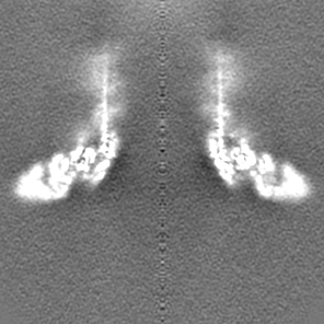
| Projections & slices | Image control
Images are generated by Spider. | ||||||||||||||||||||||||||||||||||||
| Voxel size | X=Y=Z: 1.4 Å | ||||||||||||||||||||||||||||||||||||
| Density |
| ||||||||||||||||||||||||||||||||||||
| Symmetry | Space group: 1 | ||||||||||||||||||||||||||||||||||||
| Details | EMDB XML:
|
 Z (Sec.)
 Y (Row.)
 X (Col.)


-Supplemental data
-Mask #1
| File | emd_28863_msk_1.map | ||||||||||||
|---|---|---|---|---|---|---|---|---|---|---|---|---|---|
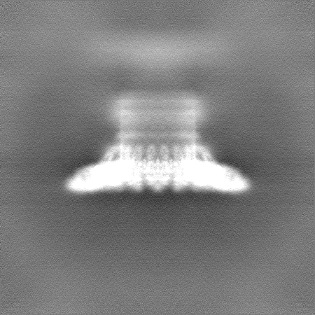
| Projections & Slices |
| ||||||||||||
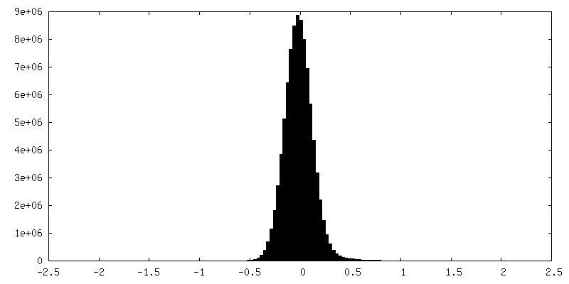
| Density Histograms |


-Additional map: Unsharpened, local resolution filtered combined map. Real-space cropped...
| File | emd_28863_additional_1.map | ||||||||||||
|---|---|---|---|---|---|---|---|---|---|---|---|---|---|
| Annotation | Unsharpened, local resolution filtered combined map. Real-space cropped from box size 450px to 296px. | ||||||||||||
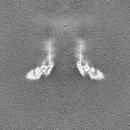
| Projections & Slices |
| ||||||||||||
| Density Histograms |


-Half map: Half map 1
| File | emd_28863_half_map_1.map | ||||||||||||
|---|---|---|---|---|---|---|---|---|---|---|---|---|---|
| Annotation | Half map 1 | ||||||||||||
| Projections & Slices |
| ||||||||||||
| Density Histograms |


-Half map: Half map 2
| File | emd_28863_half_map_2.map | ||||||||||||
|---|---|---|---|---|---|---|---|---|---|---|---|---|---|
| Annotation | Half map 2 | ||||||||||||
| Projections & Slices |
| ||||||||||||
| Density Histograms |


- Sample components
-Entire : Quaternary complex of aE11 Fab and polyC9
| Entire | Name: Quaternary complex of aE11 Fab and polyC9 |
|---|---|
| Components |
|
-Supramolecule #1: Quaternary complex of aE11 Fab and polyC9
| Supramolecule | Name: Quaternary complex of aE11 Fab and polyC9 / type: complex / ID: 1 / Chimera: Yes / Parent: 0 / Macromolecule list: all Details: Fab fragment of aE11 generated by proteolytic cleavage of aE11 IgG antibody, in complex with recombinant human C9 incubated at 37 degrees Celsius to form homo-oligomeric C9. |
|---|---|
| Source (natural) | Organism: Homo sapiens (human) |
| Molecular weight | Theoretical: 2.6 MDa |
-Macromolecule #1: Complement component 9
| Macromolecule | Name: Complement component 9 / type: protein_or_peptide / ID: 1 / Enantiomer: LEVO |
|---|---|
| Source (natural) | Organism: Homo sapiens (human) |
| Recombinant expression | Organism: Homo sapiens (human) |
| Sequence | String: QYTTSYDPEL TESSGSASHI DCRMSPWSEW SQCDPCLRQM FRSRSIEVFG QFNGKRCTDA VGDRRQCVPT EPCEDAEDDC GNDFQCSTGR CIKMRLRCNG DNDCGDFSDE DDCESEPRPP CRDRVVEESE LARTAGYGIN ILGMDPLSTP FDNEFYNGLC NRDRDGNTLT ...String: QYTTSYDPEL TESSGSASHI DCRMSPWSEW SQCDPCLRQM FRSRSIEVFG QFNGKRCTDA VGDRRQCVPT EPCEDAEDDC GNDFQCSTGR CIKMRLRCNG DNDCGDFSDE DDCESEPRPP CRDRVVEESE LARTAGYGIN ILGMDPLSTP FDNEFYNGLC NRDRDGNTLT YYRRPWNVAS LIYETKGEKN FRTEHYEEQI EAFKSIIQEK TSNFNAAISL KFTPTETNKA EQCCEETASS ISLHGKGSFR FSYSKNETYQ LFLSYSSKKE KMFLHVKGEI HLGRFVMRNR DVVLTTTFVD DIKALPTTYE KGEYFAFLET YGTHYSSSGS LGGLYELIYV LDKASMKRKG VELKDIKRCL GYHLDVSLAF SEISVGAEFN KDDCVKRGEG RAVNITSENL IDDVVSLIRG GTRKYAFELK EKLLRGTVID VTDFVNWASS INDAPVLISQ KLSPIYNLVP VKMKNAHLKK QNLERAIEDY INEFSVRKCH TCQNGGTVIL MDGKCLCACP FKFEGIACEI SKQKISEGLP ALEFPNEK |
-Macromolecule #2: Monoclonal antibody aE11 Fab (heavy chain)
| Macromolecule | Name: Monoclonal antibody aE11 Fab (heavy chain) / type: protein_or_peptide / ID: 2 / Enantiomer: LEVO |
|---|---|
| Source (natural) | Organism: |
| Sequence | String: VQLKESGPGL VAPSQSLSIT CTVSGFSLTV YGVNWIRQPP GKGLEWLGMI WGDGSTDYNS ALKSRLSITK DNSKSQVFLK MNSLQTDDTA RYYCARDRSY GGSSAWFGYW GQGTLVTVSA |
-Macromolecule #3: Monoclonal antibody aE11 Fab (light chain)
| Macromolecule | Name: Monoclonal antibody aE11 Fab (light chain) / type: protein_or_peptide / ID: 3 / Enantiomer: LEVO |
|---|---|
| Source (natural) | Organism: |
| Sequence | String: QMTQTTSSLS ASLGDRVTIS CRASHDISNY LNWYQQKPDG TLKLLIYYTS RLHSGVPSRF SGSGSGTDYS LTISNLEQED VATYFCQQGN YLPYTFGGGT KLEIK |
-Experimental details
-Structure determination
| Method | cryo EM |
|---|---|
 Processing Processing | single particle reconstruction |
| Aggregation state | particle |
-Sample preparation
| Buffer | pH: 7.4 Component:
| |||||||||
|---|---|---|---|---|---|---|---|---|---|---|
| Vitrification | Cryogen name: ETHANE / Chamber humidity: 100 % / Chamber temperature: 277 K / Instrument: FEI VITROBOT MARK IV |
- Electron microscopy
| Microscope | FEI TITAN KRIOS |
|---|---|
| Image recording | Film or detector model: GATAN K2 SUMMIT (4k x 4k) / Detector mode: COUNTING / Digitization - Frames/image: 0-40 / Average exposure time: 16.0 sec. / Average electron dose: 52.4 e/Å2 |
| Electron beam | Acceleration voltage: 300 kV / Electron source:  FIELD EMISSION GUN FIELD EMISSION GUN |
| Electron optics | C2 aperture diameter: 50.0 µm / Illumination mode: FLOOD BEAM / Imaging mode: BRIGHT FIELD / Cs: 2.7 mm / Nominal defocus max: 2.0 µm / Nominal defocus min: 0.5 µm |
| Sample stage | Specimen holder model: FEI TITAN KRIOS AUTOGRID HOLDER |
| Experimental equipment |  Model: Titan Krios / Image courtesy: FEI Company |