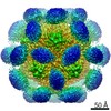
[日本語] English
- EMDB-2823: Structure determination of feline calicivirus virus-like particle... -

+
データを開く


IDまたはキーワード:

読み込み中...

-
基本情報

登録情報
データベース: EMDB / ID: EMD-2823
タイトルStructure determination of feline calicivirus virus-like particles in the context of a pseudo-octahedral arrangement
マップデータReconstruction of feline calicivirus VLP T=1.
試料
  • 試料: feline calicivirus virus-like particles
  • ウイルス: Canine calicivirus (ウイルス)
キーワードvirus-like particle / VLP / VP1 / calicivirus / FCV / icosahedral symmetry / octahedral symmetry / non-crystallographic symmetry / feline calici virus / vaccine / subviral particle
機能・相同性T=3 icosahedral viral capsid / Calicivirus coat protein / Calicivirus coat protein / Picornavirus/Calicivirus coat protein / Viral coat protein subunit / host cell cytoplasm / Capsid protein VP1
機能・相同性情報
生物種Canine calicivirus (ウイルス)
手法単粒子再構成法 / クライオ電子顕微鏡法 / ネガティブ染色法 / 解像度: 14.0 Å
データ登録者Burmeister WP / Buisson M / Estrozi LF / Schoehn G / Billet O / Hannas Z / Sigoillot-Claude C / Poulet H
引用ジャーナル: PLoS One / : 2015
タイトル: Structure determination of feline calicivirus virus-like particles in the context of a pseudo-octahedral arrangement.
著者: Wim P Burmeister / Marlyse Buisson / Leandro F Estrozi / Guy Schoehn / Olivier Billet / Zahia Hannas / Cécile Sigoillot / Hervé Poulet /
要旨: The vesivirus feline calicivirus (FCV) is a positive strand RNA virus encapsidated by an icosahedral T=3 shell formed by the viral VP1 protein. Upon its expression in the insect cell - baculovirus ...The vesivirus feline calicivirus (FCV) is a positive strand RNA virus encapsidated by an icosahedral T=3 shell formed by the viral VP1 protein. Upon its expression in the insect cell - baculovirus system in the context of vaccine development, two types of virus-like particles (VLPs) were formed, a majority built of 60 subunits (T=1) and a minority probably built of 180 subunits (T=3). The structure of the small particles was determined by x-ray crystallography at 0.8 nm resolution helped by cryo-electron microscopy in order to understand their formation. Cubic crystals belonged to space group P213. Their self-rotation function showed the presence of an octahedral pseudo-symmetry similar to the one described previously by Agerbandje and co-workers for human parvovirus VLPs. The crystal structure could be solved starting from the published VP1 structure in the context of the T=3 viral capsid. In contrast to viral capsids, where the capsomers are interlocked by the exchange of the N-terminal arm (NTA) domain, this domain is disordered in the T=1 capsid of the VLPs. Furthermore it is prone to proteolytic cleavage. The relative orientation of P (protrusion) and S (shell) domains is alerted so as to fit VP1 to the smaller T=1 particle whereas the intermolecular contacts around 2-fold, 3-fold and 5-fold axes are conserved. By consequence the surface of the VLP is very similar compared to the viral capsid and suggests a similar antigenicity. The knowledge of the structure of the VLPs will help to improve their stability, in respect to a use for vaccination.
履歴
登録2014年11月18日-
ヘッダ(付随情報) 公開2014年11月26日-
マップ公開2015年4月1日-
更新2015年4月15日-
現状2015年4月15日処理サイト: PDBe / 状態: 公開

-
構造の表示

ムービー
  • 表面図(断面を密度値に従い着色)
  • 表面レベル: 1.2
  • UCSF Chimeraによる作画
  • ダウンロード
  • 表面図(半径に従い着色)
  • 表面レベル: 1.2
  • UCSF Chimeraによる作画
  • ダウンロード
ムービービューア
構造ビューアEMマップ:
SurfViewMolmilJmol/JSmol

ダウンロードとリンク

-
マップ

ファイルダウンロード / ファイル: emd_2823.map.gz / 形式: CCP4 / 大きさ: 12.6 MB / タイプ: IMAGE STORED AS FLOATING POINT NUMBER (4 BYTES)
注釈Reconstruction of feline calicivirus VLP T=1.
投影像・断面図

画像のコントロール

大きさ
明度
コントラスト
その他
Z (Sec.)Y (Row.)X (Col.)
2.33 Å/pix.
x 150 pix.
= 350. Å
2.33 Å/pix.
x 150 pix.
= 350. Å
2.33 Å/pix.
x 150 pix.
= 350. Å

表面

投影像

断面 (1/3)

断面 (1/2)

断面 (2/3)

画像は Spider により作成

ボクセルのサイズX=Y=Z: 2.33333 Å
密度
表面レベル登録者による: 1.2 / ムービー #1: 1.2
最小 - 最大-5.52515793 - 5.53812361
平均 (標準偏差)0.0 (±0.99999994)
対称性空間群: 1
詳細

EMDB XML:

マップ形状
Axis orderXYZ
Origin-75-75-75
サイズ150150150
Spacing150150150
セルA=B=C: 350.0 Å
α=β=γ: 90.0 °

CCP4マップ ヘッダ情報:

modeImage stored as Reals
Å/pix. X/Y/Z2.33333333333332.33333333333332.3333333333333
M x/y/z150150150
origin x/y/z0.0000.0000.000
length x/y/z350.000350.000350.000
α/β/γ90.00090.00090.000
MAP C/R/S123
start NC/NR/NS-75-75-75
NC/NR/NS150150150
D min/max/mean-5.5255.538-0.000

-
添付データ

-
試料の構成要素

-
全体 : feline calicivirus virus-like particles

全体名称: feline calicivirus virus-like particles
要素
  • 試料: feline calicivirus virus-like particles
  • ウイルス: Canine calicivirus (ウイルス)

-
超分子 #1000: feline calicivirus virus-like particles

超分子名称: feline calicivirus virus-like particles / タイプ: sample / ID: 1000 / 集合状態: icosahedral / Number unique components: 1
分子量理論値: 3.552 MDa

-
超分子 #1: Canine calicivirus

超分子名称: Canine calicivirus / タイプ: virus / ID: 1 / NCBI-ID: 74724 / 生物種: Canine calicivirus / ウイルスタイプ: VIRUS-LIKE PARTICLE / ウイルス・単離状態: OTHER / ウイルス・エンベロープ: No / ウイルス・中空状態: Yes
宿主生物種: Felis catus (イエネコ) / 別称: VERTEBRATES
Host system生物種: Spodoptera frugiperda (ツマジロクサヨトウ)
組換細胞: Sf9 / 組換プラスミド: Merial100869
分子量理論値: 3.5 MDa
ウイルス殻Shell ID: 1 / 直径: 280 Å / T番号(三角分割数): 1

-
実験情報

-
構造解析

手法ネガティブ染色法, クライオ電子顕微鏡法
解析単粒子再構成法
試料の集合状態particle

-
試料調製

緩衝液pH: 7 / 詳細: 20 mM MES pH 6, 200 mM NaCl
染色タイプ: NEGATIVE / 詳細: no stain (cryo-EM)
グリッド詳細: Quantifoil R2/1 holey grid
凍結凍結剤: ETHANE / 装置: FEI VITROBOT MARK IV

-
電子顕微鏡法

顕微鏡FEI POLARA 300
日付2010年9月1日
撮影カテゴリ: FILM / フィルム・検出器のモデル: KODAK SO-163 FILM / デジタル化 - スキャナー: ZEISS SCAI / デジタル化 - サンプリング間隔: 7 µm / 実像数: 20 / 平均電子線量: 20 e/Å2 / ビット/ピクセル: 8
電子線加速電圧: 300 kV / 電子線源: FIELD EMISSION GUN
電子光学系照射モード: FLOOD BEAM / 撮影モード: BRIGHT FIELD / Cs: 2.0 mm / 最大 デフォーカス(公称値): 3.0 µm / 最小 デフォーカス(公称値): 1.0 µm / 倍率(公称値): 39000
試料ステージ試料ホルダー: Top-entry polara / 試料ホルダーモデル: OTHER
実験機器
モデル: Tecnai Polara / 画像提供: FEI Company

-
画像解析

詳細Particles semi-automatically picked by the boxer program (EMAN)
CTF補正詳細: Phase-flipping
最終 再構成想定した対称性 - 点群: I (正20面体型対称) / アルゴリズム: OTHER / 解像度のタイプ: BY AUTHOR / 解像度: 14.0 Å / 解像度の算出法: OTHER / ソフトウェア - 名称: RIco, Bsoft, ctffind3, FPM / 使用した粒子像数: 11158

+
万見について

-
お知らせ

-
2022年2月9日: EMDBエントリの付随情報ファイルのフォーマットが新しくなりました

EMDBエントリの付随情報ファイルのフォーマットが新しくなりました

  • EMDBのヘッダファイルのバージョン3が、公式のフォーマットとなりました。
  • これまでは公式だったバージョン1.9は、アーカイブから削除されます。

関連情報:EMDBヘッダ

外部リンク:wwPDBはEMDBデータモデルのバージョン3へ移行します

-
2020年8月12日: 新型コロナ情報

新型コロナ情報

URL: https://pdbj.org/emnavi/covid19.php

新ページ: EM Navigatorに新型コロナウイルスの特設ページを開設しました。

関連情報:Covid-19情報 / 2020年3月5日: 新型コロナウイルスの構造データ

+
2020年3月5日: 新型コロナウイルスの構造データ

新型コロナウイルスの構造データ

関連情報:万見生物種 / 2020年8月12日: 新型コロナ情報

外部リンク:COVID-19特集ページ - PDBj / 今月の分子2020年2月:コロナウイルスプロテーアーゼ

+
2019年1月31日: EMDBのIDの桁数の変更

EMDBのIDの桁数の変更

  • EMDBエントリに付与されているアクセスコード(EMDB-ID)は4桁の数字(例、EMD-1234)でしたが、間もなく枯渇します。これまでの4桁のID番号は4桁のまま変更されませんが、4桁の数字を使い切った後に発行されるIDは5桁以上の数字(例、EMD-12345)になります。5桁のIDは2019年の春頃から発行される見通しです。
  • EM Navigator/万見では、接頭語「EMD-」は省略されています。

関連情報:Q: 「EMD」とは何ですか? / 万見/EM NavigatorにおけるID/アクセスコードの表記

外部リンク:EMDB Accession Codes are Changing Soon! / PDBjへお問い合わせ

+
2017年7月12日: PDB大規模アップデート

PDB大規模アップデート

  • 新バージョンのPDBx/mmCIF辞書形式に基づくデータがリリースされました。
  • 今回の更新はバージョン番号が4から5になる大規模なもので、全エントリデータの書き換えが行われる「Remediation」というアップデートに該当します。
  • このバージョンアップで、電子顕微鏡の実験手法に関する多くの項目の書式が改定されました(例:em_softwareなど)。
  • EM NavigatorとYorodumiでも、この改定に基づいた表示内容になります。

外部リンク:wwPDB Remediation / OneDepデータ基準に準拠した、より強化された内容のモデル構造ファイルが、PDBアーカイブで公開されました。

-
万見 (Yorodumi)

幾万の構造データを、幾万の視点から

  • 万見(Yorodumi)は、EMDB/PDB/SASBDBなどの構造データを閲覧するためのページです。
  • EM Navigatorの詳細ページの後継、Omokage検索のフロントエンドも兼ねています。

関連情報:EMDB / PDB / SASBDB / 3つのデータバンクの比較 / 万見検索 / 2016年8月31日: 新しいEM Navigatorと万見 / 万見文献 / Jmol/JSmol / 機能・相同性情報 / 新しいEM Navigatorと万見の変更点

他の情報も見る