National Institutes of Health/National Cancer Institute (NIH/NCI)
United States
Citation
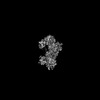
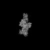
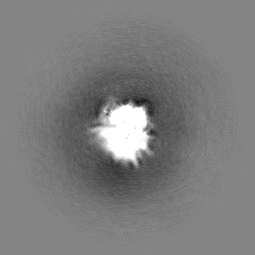
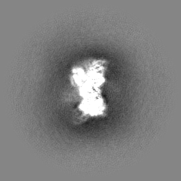
Journal: Nucleic Acids Res / Year: 2023 Title: Structural basis for the allosteric regulation and dynamic assembly of DNMT3B. Authors: Jiuwei Lu / Jian Fang / Hongtao Zhu / Kimberly Lu Liang / Nelli Khudaverdyan / Jikui Song / Abstract: Oligomerization of DNMT3B, a mammalian de novo DNA methyltransferase, critically regulates its chromatin targeting and DNA methylation activities. However, how the N-terminal PWWP and ADD domains ...Oligomerization of DNMT3B, a mammalian de novo DNA methyltransferase, critically regulates its chromatin targeting and DNA methylation activities. However, how the N-terminal PWWP and ADD domains interplay with the C-terminal methyltransferase (MTase) domain in regulating the dynamic assembly of DNMT3B remains unclear. Here, we report the cryo-EM structure of DNMT3B under various oligomerization states. The ADD domain of DNMT3B interacts with the MTase domain to form an autoinhibitory conformation, resembling the previously observed DNMT3A autoinhibition. Our combined structural and biochemical study further identifies a role for the PWWP domain and its associated ICF mutation in the allosteric regulation of DNMT3B tetramer, and a differential functional impact on DNMT3B by potential ADD-H3K4me0 and PWWP-H3K36me3 bindings. In addition, our comparative structural analysis reveals a coupling between DNMT3B oligomerization and folding of its substrate-binding sites. Together, this study provides mechanistic insights into the allosteric regulation and dynamic assembly of DNMT3B.
In the structure databanks used in Yorodumi, some data are registered as the other names, "COVID-19 virus" and "2019-nCoV". Here are the details of the virus and the list of structure data.
Jan 31, 2019. EMDB accession codes are about to change! (news from PDBe EMDB page)
EMDB accession codes are about to change! (news from PDBe EMDB page)
The allocation of 4 digits for EMDB accession codes will soon come to an end. Whilst these codes will remain in use, new EMDB accession codes will include an additional digit and will expand incrementally as the available range of codes is exhausted. The current 4-digit format prefixed with “EMD-” (i.e. EMD-XXXX) will advance to a 5-digit format (i.e. EMD-XXXXX), and so on. It is currently estimated that the 4-digit codes will be depleted around Spring 2019, at which point the 5-digit format will come into force.
The EM Navigator/Yorodumi systems omit the EMD- prefix.
Related info.:Q: What is EMD? / ID/Accession-code notation in Yorodumi/EM Navigator
Yorodumi is a browser for structure data from EMDB, PDB, SASBDB, etc.
This page is also the successor to EM Navigator detail page, and also detail information page/front-end page for Omokage search.
The word "yorodu" (or yorozu) is an old Japanese word meaning "ten thousand". "mi" (miru) is to see.
Related info.:EMDB / PDB / SASBDB / Comparison of 3 databanks / Yorodumi Search / Aug 31, 2016. New EM Navigator & Yorodumi / Yorodumi Papers / Jmol/JSmol / Function and homology information / Changes in new EM Navigator and Yorodumi
 Movie
Movie Controller


 Open data
 Basic information
 Map data
 Sample
 Keywords
 Function and homology information
 Homo sapiens (human)
 Authors
 United States, 1 items
  Citation
 Structure visualization
 Downloads & links
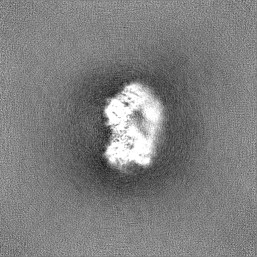
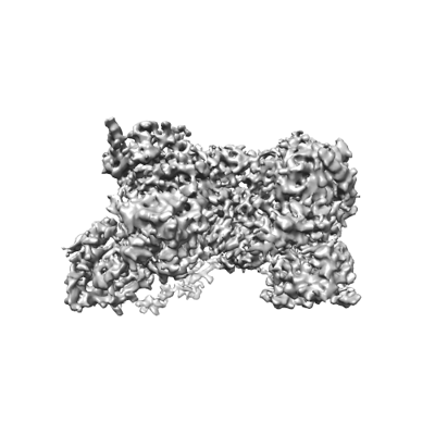 emd_28158.png
 http://ftp.pdbj.org/pub/emdb/structures/EMD-28158


 Z (Sec.)
 Y (Row.)
 X (Col.)


 Sample components

 Processing
 Electron microscopy
 FIELD EMISSION GUN
