 ムービー
ムービー コントローラー


+ データを開く

- 基本情報
基本情報
| 登録情報 | データベース: EMDB / ID: EMD-2801 | |||||||||
|---|---|---|---|---|---|---|---|---|---|---|
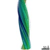
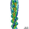
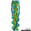
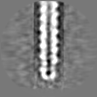
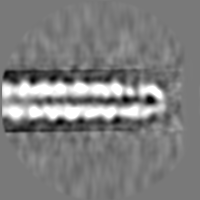
| タイトル | Negative stain electron microscopy reconstruction of the wild type tip complex from the type III secretion system of Shigella flexneri | |||||||||
 マップデータ マップデータ | Negative stain electron microscopy reconstruction of the wild type tip complex and needle of the type III secretion system from Shigella flexneri. | |||||||||
 試料 試料 |
| |||||||||
 キーワード キーワード | Tip complex / type III secretion system / Shigella flexneri / wild type | |||||||||
| 機能・相同性 | effector-mediated activation of host programmed cell death by symbiont / Type III secretion systems tip complex components / BipD-like superfamily / Type III secretion systems tip complex components / extracellular region / Invasin IpaD 機能・相同性情報 機能・相同性情報 | |||||||||
| 生物種 |  Shigella flexneri (フレクスナー赤痢菌) Shigella flexneri (フレクスナー赤痢菌) | |||||||||
| 手法 | 単粒子再構成法 / ネガティブ染色法 / 解像度: 24.0 Å | |||||||||
 データ登録者 データ登録者 | Cheung M / Shen D / Makino F / Kato T / Roehrich D / Martinez-Argudo I / Walker ML / Murillo I / Liu X / Pain M ...Cheung M / Shen D / Makino F / Kato T / Roehrich D / Martinez-Argudo I / Walker ML / Murillo I / Liu X / Pain M / Brown J / Frazer G / Mantell J / Mina P / Todd T / Sessions RB / Namba K / Blocker AJ | |||||||||
 引用 引用 | ジャーナル: Proc Natl Acad Sci U S A / 年: 2008 タイトル: What's the point of the type III secretion system needle? 著者: Ariel J Blocker / Janet E Deane / Andreas K J Veenendaal / Pietro Roversi / Julie L Hodgkinson / Steven Johnson / Susan M Lea /  要旨: Recent work by several groups has significantly expanded our knowledge of the structure, regulation of assembly, and function of components of the extracellular portion of the type III secretion ...Recent work by several groups has significantly expanded our knowledge of the structure, regulation of assembly, and function of components of the extracellular portion of the type III secretion system (T3SS) of Gram-negative bacteria. This perspective presents a structure-informed analysis of functional data and discusses three nonmutually exclusive models of how a key aspect of T3SS biology, the sensing of host cells, may be performed. | |||||||||
| 履歴 |
|
- 構造の表示
構造の表示
| ムービー |
ムービービューア |
|---|---|
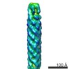
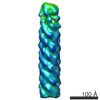
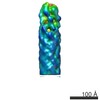
| 構造ビューア | EMマップ: SurfViewMolmilJmol/JSmol |
| 添付画像 |

- ダウンロードとリンク
-EMDBアーカイブ
| マップデータ | emd_2801.map.gz | 28.1 MB | EMDBマップデータ形式 | |
|---|---|---|---|---|
| ヘッダ (付随情報) | emd-2801-v30.xmlemd-2801.xml | 17.5 KB 17.5 KB | 表示 表示 | EMDBヘッダ |
| 画像 | 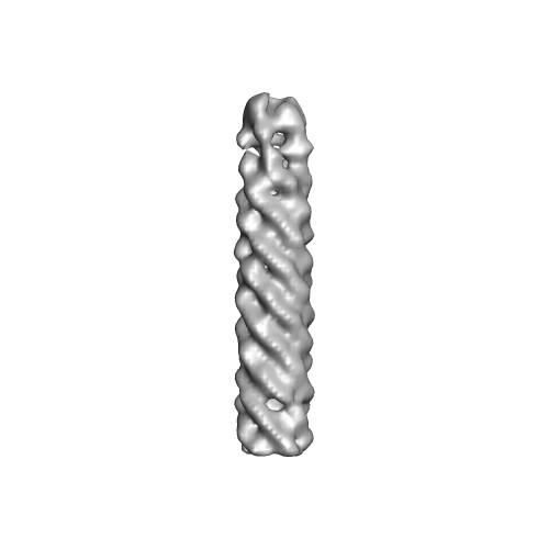 EMD-2801.png EMD-2801.png | 32.1 KB | ||
| アーカイブディレクトリ |  http://ftp.pdbj.org/pub/emdb/structures/EMD-2801ftp://ftp.pdbj.org/pub/emdb/structures/EMD-2801 http://ftp.pdbj.org/pub/emdb/structures/EMD-2801ftp://ftp.pdbj.org/pub/emdb/structures/EMD-2801 | HTTPS FTP |
-検証レポート
| 文書・要旨 | emd_2801_validation.pdf.gz | 213.3 KB | 表示 | EMDB検証レポート |
|---|---|---|---|---|
| 文書・詳細版 | emd_2801_full_validation.pdf.gz | 212.5 KB | 表示 | |
| XML形式データ | emd_2801_validation.xml.gz | 5.8 KB | 表示 | |
| アーカイブディレクトリ | https://ftp.pdbj.org/pub/emdb/validation_reports/EMD-2801ftp://ftp.pdbj.org/pub/emdb/validation_reports/EMD-2801 | HTTPS FTP |
-関連構造データ


-リンク
| EMDBのページ | EMDB (EBI/PDBe) / EMDataResource |
|---|
-マップ
| ファイル | ダウンロード / ファイル: emd_2801.map.gz / 形式: CCP4 / 大きさ: 29.8 MB / タイプ: IMAGE STORED AS FLOATING POINT NUMBER (4 BYTES) | ||||||||||||||||||||||||||||||||||||||||||||||||||||||||||||||||||||
|---|---|---|---|---|---|---|---|---|---|---|---|---|---|---|---|---|---|---|---|---|---|---|---|---|---|---|---|---|---|---|---|---|---|---|---|---|---|---|---|---|---|---|---|---|---|---|---|---|---|---|---|---|---|---|---|---|---|---|---|---|---|---|---|---|---|---|---|---|---|
| 注釈 | Negative stain electron microscopy reconstruction of the wild type tip complex and needle of the type III secretion system from Shigella flexneri. | ||||||||||||||||||||||||||||||||||||||||||||||||||||||||||||||||||||
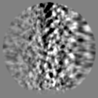
| 投影像・断面図 | 画像のコントロール
画像は Spider により作成 | ||||||||||||||||||||||||||||||||||||||||||||||||||||||||||||||||||||
| ボクセルのサイズ | X=Y=Z: 2.12 Å | ||||||||||||||||||||||||||||||||||||||||||||||||||||||||||||||||||||
| 密度 |
| ||||||||||||||||||||||||||||||||||||||||||||||||||||||||||||||||||||
| 対称性 | 空間群: 1 | ||||||||||||||||||||||||||||||||||||||||||||||||||||||||||||||||||||
| 詳細 | EMDB XML:
CCP4マップ ヘッダ情報:
| ||||||||||||||||||||||||||||||||||||||||||||||||||||||||||||||||||||
 Z (Sec.)
 Y (Row.)
 X (Col.)


-添付データ
- 試料の構成要素
-全体 : Wild type conformation of the tip complex from the type III secre...
| 全体 | 名称: Wild type conformation of the tip complex from the type III secretion system of Shigella flexneri, bound to the needle |
|---|---|
| 要素 |
|
-超分子 #1000: Wild type conformation of the tip complex from the type III secre...
| 超分子 | 名称: Wild type conformation of the tip complex from the type III secretion system of Shigella flexneri, bound to the needle タイプ: sample / ID: 1000 詳細: Tip complexes were analysed from purified needle complexes and therefore when still bound to the needle tip 集合状態: Five subunits of the tip complex bound to fifty subunits of the needle Number unique components: 2 |
|---|---|
| 分子量 | 理論値: 680 KDa |
-超分子 #1: Tip complex
| 超分子 | 名称: Tip complex / タイプ: organelle_or_cellular_component / ID: 1 / コピー数: 1 / 集合状態: Heteropentameric / 組換発現: Yes |
|---|---|
| Ref GO | 0: GO:0030257 |
| Ref INTERPRO | 0: IPR006135 |
| 由来(天然) | 生物種: Shigella flexneri (フレクスナー赤痢菌) / 株: serotype 5a / Organelle: Type III secretion system 細胞中の位置: Extracellular component of membrane embedded complex |
| 分子量 | 理論値: 210 KDa |
| 組換発現 | 生物種: Shigella flexneri (フレクスナー赤痢菌) / 組換株: M90T derivative / 組換プラスミド: pACT3, pBAD |
-超分子 #2: Needle
| 超分子 | 名称: Needle / タイプ: organelle_or_cellular_component / ID: 2 / コピー数: 1 / 集合状態: helical polymer / 組換発現: Yes |
|---|---|
| Ref GO | 0: GO:0030257 |
| Ref INTERPRO | 0: IPR006135 |
| 由来(天然) | 生物種: Shigella flexneri (フレクスナー赤痢菌) / 株: serotype 5a / Organelle: Type III secretion system 細胞中の位置: Extracellular component of membrane embedded complex |
| 分子量 | 理論値: 470 KDa |
| 組換発現 | 生物種: Shigella flexneri (フレクスナー赤痢菌) / 組換株: M90T derivative / 組換プラスミド: pACT3, pBAD |
-実験情報
-構造解析
| 手法 | ネガティブ染色法 |
|---|---|
 解析 解析 | 単粒子再構成法 |
| 試料の集合状態 | particle |
-試料調製
| 濃度 | 0.1 mg/mL |
|---|---|
| 緩衝液 | pH: 8 詳細: 0.1% (w/v) DDM, 150 mM NaCl, 25 mM Tris pH 8, 5 mM EDTA |
| 染色 | タイプ: NEGATIVE 詳細: Carbon coating mica sheets were submerged in a drop of 0.01% (w/v) DDM until the carbon sheet partially detached from the mica. The mica was then submerged in the sample for 5 s, then ...詳細: Carbon coating mica sheets were submerged in a drop of 0.01% (w/v) DDM until the carbon sheet partially detached from the mica. The mica was then submerged in the sample for 5 s, then submerged in a drop of water for 5 s. The mica was then placed onto the surface of a drop of 1% uranyl acetate and allowed to sink, causing the carbon to float on the surface of the drop. After 10 s, the carbon sheet was adsorbed onto a grid. |
| グリッド | 詳細: 300 mesh copper grid with a thin carbon film to which the specimen was adsorbed to |
| 凍結 | 凍結剤: NONE / 装置: OTHER |
- 電子顕微鏡法
| 顕微鏡 | FEI TECNAI 20 |
|---|---|
| アライメント法 | Legacy - 非点収差: Thon rings visualised at 50-100,000 times magnification using the fast FT and corrected accordingly Legacy - Electron beam tilt params: 0 |
| 日付 | 2011年8月31日 |
| 撮影 | カテゴリ: CCD / フィルム・検出器のモデル: FEI EAGLE (4k x 4k) / デジタル化 - サンプリング間隔: 15 µm / 実像数: 300 / 平均電子線量: 9 e/Å2 / カメラ長: 460 / Od range: 3.56 / ビット/ピクセル: 16 |
| 電子線 | 加速電圧: 200 kV / 電子線源: LAB6 |
| 電子光学系 | 倍率(補正後): 70754 / 照射モード: FLOOD BEAM / 撮影モード: BRIGHT FIELD / Cs: 2 mm / 最大 デフォーカス(公称値): 1.4 µm / 最小 デフォーカス(公称値): 1.4 µm / 倍率(公称値): 50000 |
| 試料ステージ | 試料ホルダー: FEI single tilt holder / 試料ホルダーモデル: SIDE ENTRY, EUCENTRIC |
+画像解析
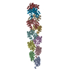
-原子モデル構築 1
| 初期モデル | PDB ID: Chain - Chain ID: A |
|---|---|
| ソフトウェア | 名称:  Chimera Chimera |
| 詳細 | Residues comprising the N domain were removed. The 10 residues at the C terminus (not resolved in the crystal structure) were built into an alpha helix |
| 精密化 | 空間: REAL / プロトコル: RIGID BODY FIT |
| 得られたモデル |  PDB-4d3e: |

