National Institutes of Health/National Institute of General Medical Sciences (NIH/NIGMS)
R01-GM099989
米国
National Institutes of Health/National Institute of General Medical Sciences (NIH/NIGMS)
U24GM129547
米国
引用
ジャーナル: Nat Commun / 年: 2022 タイトル: Visualization of conformational changes and membrane remodeling leading to genome delivery by viral class-II fusion machinery. 著者: Vidya Mangala Prasad / Jelle S Blijleven / Jolanda M Smit / Kelly K Lee / 要旨: Chikungunya virus (CHIKV) is a human pathogen that delivers its genome to the host cell cytoplasm through endocytic low pH-activated membrane fusion mediated by class-II fusion proteins. Though ...Chikungunya virus (CHIKV) is a human pathogen that delivers its genome to the host cell cytoplasm through endocytic low pH-activated membrane fusion mediated by class-II fusion proteins. Though structures of prefusion, icosahedral CHIKV are available, structural characterization of virion interaction with membranes has been limited. Here, we have used cryo-electron tomography to visualize CHIKV's complete membrane fusion pathway, identifying key intermediary glycoprotein conformations coupled to membrane remodeling events. Using sub-tomogram averaging, we elucidate features of the low pH-exposed virion, nucleocapsid and full-length E1-glycoprotein's post-fusion structure. Contrary to class-I fusion systems, CHIKV achieves membrane apposition by protrusion of extended E1-glycoprotein homotrimers into the target membrane. The fusion process also features a large hemifusion diaphragm that transitions to a wide pore for intact nucleocapsid delivery. Our analyses provide comprehensive ultrastructural insights into the class-II virus fusion system function and direct mechanistic characterization of the fundamental process of protein-mediated membrane fusion.
名称: Spike glycoprotein E1 / タイプ: protein_or_peptide / ID: 1 詳細: The previously determined X-ray crystal structure PDB ID 1RER was rigidly docked into the sub-tomogram averaged map but no further refinement was performed コピー数: 3 / 光学異性体: LEVO
 ムービー
ムービー コントローラー


 データを開く
 基本情報
基本情報
 マップデータ
 試料
試料 キーワード
キーワード 機能・相同性情報
 Chikungunya virus strain S27-African prototype (ウイルス)
 データ登録者
 米国, 2件
  引用
引用

 構造の表示
構造の表示
 ダウンロードとリンク
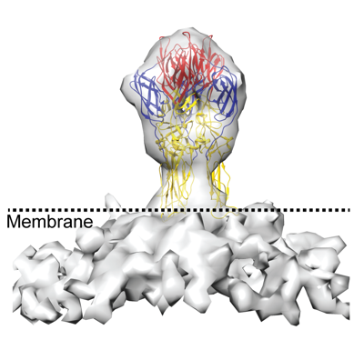 emd_27248.png
 http://ftp.pdbj.org/pub/emdb/structures/EMD-27248


 Z (Sec.)
 Y (Row.)
 X (Col.)


 試料の構成要素


 解析
解析 電子顕微鏡法
 FIELD EMISSION GUN


