- EMDB-2714: Negative stain reconstruction of AcrB/SMALP complex -
+
データを開く
IDまたはキーワード:
読み込み中...
-
基本情報
登録情報
データベース: EMDB / ID: EMD-2714
タイトル
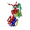
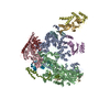
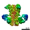
Negative stain reconstruction of AcrB/SMALP complex
マップデータ
Reconstruction of AcrB encapsulated in a SMALP polymer
試料
試料: E. coli AcrB in a SMALP scaffold
タンパク質・ペプチド: Acridine resistance protein B
キーワード
AcrB / negative stain / SMALP
機能・相同性
機能・相同性情報
alkane transmembrane transporter activity / alkane transport / enterobactin transport / enterobactin transmembrane transporter activity / xenobiotic detoxification by transmembrane export across the cell outer membrane / periplasmic side of plasma membrane / efflux pump complex / bile acid transmembrane transporter activity / xenobiotic transport / bile acid and bile salt transport ...alkane transmembrane transporter activity / alkane transport / enterobactin transport / enterobactin transmembrane transporter activity / xenobiotic detoxification by transmembrane export across the cell outer membrane / periplasmic side of plasma membrane / efflux pump complex / bile acid transmembrane transporter activity / xenobiotic transport / bile acid and bile salt transport / efflux transmembrane transporter activity / xenobiotic transmembrane transporter activity / fatty acid transport / response to toxic substance / response to xenobiotic stimulus / response to antibiotic / identical protein binding / membrane / plasma membrane 類似検索 - 分子機能
ジャーナル: Biochim Biophys Acta / 年: 2015 タイトル: The use of SMALPs as a novel membrane protein scaffold for structure study by negative stain electron microscopy. 著者: Vincent Postis / Shaun Rawson / Jennifer K Mitchell / Sarah C Lee / Rosemary A Parslow / Tim R Dafforn / Stephen A Baldwin / Stephen P Muench / 要旨: Despite the great progress recently made in resolving their structures, investigation of the structural biology of membrane proteins still presents major challenges. Even with new technical advances ...Despite the great progress recently made in resolving their structures, investigation of the structural biology of membrane proteins still presents major challenges. Even with new technical advances such as lipidic cubic phase crystallisation, obtaining well-ordered crystals remains a significant hurdle in membrane protein X-ray crystallographic studies. As an alternative, electron microscopy has been shown to be capable of resolving >3.5Å resolution detail in membrane proteins of modest (~300 kDa) size, without the need for crystals. However, the conventional use of detergents for either approach presents several issues, including the possible effects on structure of removing the proteins from their natural membrane environment. As an alternative, it has recently been demonstrated that membrane proteins can be effectively isolated, in the absence of detergents, using a styrene maleic acid co-polymer (SMA). This approach yields SMA lipid particles (SMALPs) in which the membrane proteins are surrounded by a small disk of lipid bilayer encircled by polymer. Here we use the Escherichia coli secondary transporter AcrB as a model membrane protein to demonstrate how a SMALP scaffold can be used to visualise membrane proteins, embedded in a near-native lipid environment, by negative stain electron microscopy, yielding structures at a modest resolution in a short (days) timeframe. Moreover, we show that AcrB within a SMALP scaffold is significantly more active than the equivalent DDM stabilised form. The advantages of SMALP scaffolds within electron microscopy are discussed and we conclude that they may prove to be an important tool in studying membrane protein structure and function.
Particles were picked manually using EMAN2 boxer program
CTF補正
詳細: Each micrograph CTF-find 3
最終 再構成
想定した対称性 - 点群: C3 (3回回転対称) / アルゴリズム: OTHER / 解像度のタイプ: BY AUTHOR / 解像度: 23.0 Å / 解像度の算出法: OTHER / ソフトウェア - 名称: RELION 詳細: Gold standard FSC used in RELION for resolution determination. A simple elipsoid used as a starting model 使用した粒子像数: 6884
 ムービー
ムービー コントローラー


 データを開く
 基本情報
基本情報 マップデータ
 試料
試料 キーワード
キーワード 機能・相同性情報
 データ登録者
 引用
引用
 構造の表示
構造の表示 UCSF Chimera
 ダウンロードとリンク
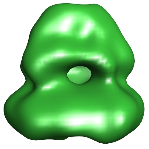 EMD-2714.png
 http://ftp.pdbj.org/pub/emdb/structures/EMD-2714
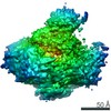

 Z (Sec.)
 Y (Row.)
 X (Col.)


 試料の構成要素
 解析
解析 電子顕微鏡法