 Movie
Movie Controller

[English] 日本語
 Yorodumi
- EMDB-2669: Spa33 mutant structure of Shigella Type III secretion injectisome -
+ Open data

- Basic information
| Entry | Database: EMDB / ID: EMD-2669 | |||||||||
|---|---|---|---|---|---|---|---|---|---|---|
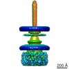
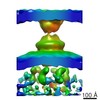
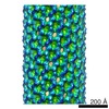
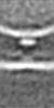
| Title | Spa33 mutant structure of Shigella Type III secretion injectisome | |||||||||
 Map data Map data | Spa33 mutant structure | |||||||||
 Sample Sample |
| |||||||||
 Keywords Keywords | Spa33 mutant / T3SS / Secretion | |||||||||
| Biological species |  Shigella flexneri (bacteria) Shigella flexneri (bacteria) | |||||||||
| Method | electron tomography / cryo EM | |||||||||
 Authors Authors | Hu B / Margolin W / Rohde J / Picking WL / Picking WD / Liu J | |||||||||
 Citation Citation | Journal: Proc Natl Acad Sci U S A / Year: 2015 Title: Visualization of the type III secretion sorting platform of Shigella flexneri. Authors: Bo Hu / Dustin R Morado / William Margolin / John R Rohde / Olivia Arizmendi / Wendy L Picking / William D Picking / Jun Liu /   Abstract: Bacterial type III secretion machines are widely used to inject virulence proteins into eukaryotic host cells. These secretion machines are evolutionarily related to bacterial flagella and consist of ...Bacterial type III secretion machines are widely used to inject virulence proteins into eukaryotic host cells. These secretion machines are evolutionarily related to bacterial flagella and consist of a large cytoplasmic complex, a transmembrane basal body, and an extracellular needle. The cytoplasmic complex forms a sorting platform essential for effector selection and needle assembly, but it remains largely uncharacterized. Here we use high-throughput cryoelectron tomography (cryo-ET) to visualize intact machines in a virulent Shigella flexneri strain genetically modified to produce minicells capable of interaction with host cells. A high-resolution in situ structure of the intact machine determined by subtomogram averaging reveals the cytoplasmic sorting platform, which consists of a central hub and six spokes, with a pod-like structure at the terminus of each spoke. Molecular modeling of wild-type and mutant machines allowed us to propose a model of the sorting platform in which the hub consists mainly of a hexamer of the Spa47 ATPase, whereas the MxiN protein comprises the spokes and the Spa33 protein forms the pods. Multiple contacts among those components are essential to align the Spa47 ATPase with the central channel of the MxiA protein export gate to form a unique nanomachine. The molecular architecture of the Shigella type III secretion machine and its sorting platform provide the structural foundation for further dissecting the mechanisms underlying type III secretion and pathogenesis and also highlight the major structural distinctions from bacterial flagella. | |||||||||
| History |
|
- Structure visualization
| Movie |
 Movie viewer Movie viewer |
|---|---|
| Structure viewer | EM map: SurfViewMolmilJmol/JSmol |
| Supplemental images |

- Downloads & links
-EMDB archive
| Map data | emd_2669.map.gz | 875.8 KB | EMDB map data format | |
|---|---|---|---|---|
| Header (meta data) | emd-2669-v30.xmlemd-2669.xml | 8.4 KB 8.4 KB | Display Display | EMDB header |
| Images | 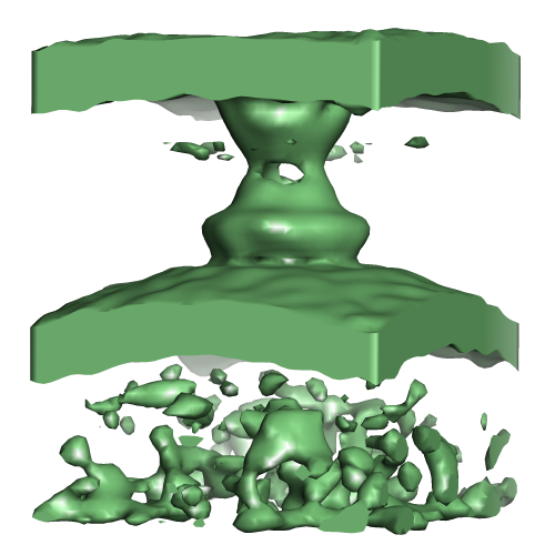 EMD-2669.png EMD-2669.png | 111.2 KB | ||
| Archive directory |  http://ftp.pdbj.org/pub/emdb/structures/EMD-2669ftp://ftp.pdbj.org/pub/emdb/structures/EMD-2669 http://ftp.pdbj.org/pub/emdb/structures/EMD-2669ftp://ftp.pdbj.org/pub/emdb/structures/EMD-2669 | HTTPS FTP |
-Related structure data
-Links
| EMDB pages | EMDB (EBI/PDBe) / EMDataResource |
|---|
-Map
| File | Download / File: emd_2669.map.gz / Format: CCP4 / Size: 955.1 KB / Type: IMAGE STORED AS FLOATING POINT NUMBER (4 BYTES) | ||||||||||||||||||||||||||||||||||||||||||||||||||||||||||||||||||||
|---|---|---|---|---|---|---|---|---|---|---|---|---|---|---|---|---|---|---|---|---|---|---|---|---|---|---|---|---|---|---|---|---|---|---|---|---|---|---|---|---|---|---|---|---|---|---|---|---|---|---|---|---|---|---|---|---|---|---|---|---|---|---|---|---|---|---|---|---|---|
| Annotation | Spa33 mutant structure | ||||||||||||||||||||||||||||||||||||||||||||||||||||||||||||||||||||
| Projections & slices | Image control
Images are generated by Spider. generated in cubic-lattice coordinate | ||||||||||||||||||||||||||||||||||||||||||||||||||||||||||||||||||||
| Voxel size | X=Y=Z: 10 Å | ||||||||||||||||||||||||||||||||||||||||||||||||||||||||||||||||||||
| Density |
| ||||||||||||||||||||||||||||||||||||||||||||||||||||||||||||||||||||
| Symmetry | Space group: 1 | ||||||||||||||||||||||||||||||||||||||||||||||||||||||||||||||||||||
| Details | EMDB XML:
CCP4 map header:
| ||||||||||||||||||||||||||||||||||||||||||||||||||||||||||||||||||||
 Z (Sec.)
 Y (Row.)
 X (Col.)


-Supplemental data
- Sample components
-Entire : Spa33 mutant T3SS structure
| Entire | Name: Spa33 mutant T3SS structure |
|---|---|
| Components |
|
-Supramolecule #1000: Spa33 mutant T3SS structure
| Supramolecule | Name: Spa33 mutant T3SS structure / type: sample / ID: 1000 / Number unique components: 1 |
|---|
-Supramolecule #1: Shigella flexneri
| Supramolecule | Name: Shigella flexneri / type: organelle_or_cellular_component / ID: 1 / Number of copies: 20 / Recombinant expression: No / Database: NCBI |
|---|---|
| Source (natural) | Organism: Shigella flexneri (bacteria) |
-Experimental details
-Structure determination
| Method | cryo EM |
|---|---|
 Processing Processing | electron tomography |
| Aggregation state | cell |
-Sample preparation
| Buffer | pH: 7 |
|---|---|
| Grid | Details: 200 mesh gold grid with thin carbon support, glow discharged in amylamine atmosphere |
| Vitrification | Cryogen name: ETHANE / Chamber humidity: 90 % / Chamber temperature: 120 K / Instrument: OTHER Timed resolved state: e.g. Vitrified 30 msec after spraying with effector Method: Blot for 3 seconds before plunging |
- Electron microscopy
| Microscope | FEI POLARA 300 |
|---|---|
| Date | Mar 1, 1999 |
| Image recording | Category: CCD / Film or detector model: GATAN ULTRASCAN 4000 (4k x 4k) / Digitization - Sampling interval: 5 µm / Number real images: 27023 / Details: 27023 / Bits/pixel: 16 |
| Electron beam | Acceleration voltage: 300 kV / Electron source:  FIELD EMISSION GUN FIELD EMISSION GUN |
| Electron optics | Illumination mode: FLOOD BEAM / Imaging mode: BRIGHT FIELD / Nominal defocus max: 1.0 µm / Nominal defocus min: 0.8 µm / Nominal magnification: 15500 |
| Sample stage | Specimen holder model: OTHER / Tilt series - Axis1 - Min angle: -60 ° / Tilt series - Axis1 - Max angle: 60 ° / Tilt series - Axis1 - Angle increment: 2 ° |
| Experimental equipment |  Model: Tecnai Polara / Image courtesy: FEI Company |
-Image processing
| Details | CTF correction was not performed on each projection |
|---|---|
| Final reconstruction | Algorithm: OTHER / Resolution method: OTHER / Number images used: 61 |
