 ムービー
ムービー コントローラー


+ データを開く

- 基本情報
基本情報
| 登録情報 | データベース: EMDB / ID: EMD-2661 | |||||||||
|---|---|---|---|---|---|---|---|---|---|---|
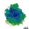
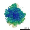
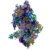
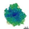
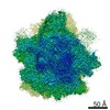
| タイトル | Cryo-EM structure of the Plasmodium falciparum 80S ribosome | |||||||||
 マップデータ マップデータ | Cryo-EM map of the Plasmodium falciparum 80S ribosome | |||||||||
 試料 試料 |
| |||||||||
 キーワード キーワード | Plasmodium falciparum 80S ribosome / Cryo-EM | |||||||||
| 生物種 |  | |||||||||
| 手法 | 単粒子再構成法 / クライオ電子顕微鏡法 / ネガティブ染色法 / 解像度: 3.4 Å | |||||||||
 データ登録者 データ登録者 | Wong W / Bai XC / Brown A / Fernandez IS / Hanssen E / Condron M / Tan YH / Baum J / Scheres SHW | |||||||||
 引用 引用 | ジャーナル: Elife / 年: 2014 タイトル: Cryo-EM structure of the Plasmodium falciparum 80S ribosome bound to the anti-protozoan drug emetine. 著者: Wilson Wong / Xiao-chen Bai / Alan Brown / Israel S Fernandez / Eric Hanssen / Melanie Condron / Yan Hong Tan / Jake Baum / Sjors H W Scheres /   要旨: Malaria inflicts an enormous burden on global human health. The emergence of parasite resistance to front-line drugs has prompted a renewed focus on the repositioning of clinically approved drugs as ...Malaria inflicts an enormous burden on global human health. The emergence of parasite resistance to front-line drugs has prompted a renewed focus on the repositioning of clinically approved drugs as potential anti-malarial therapies. Antibiotics that inhibit protein translation are promising candidates for repositioning. We have solved the cryo-EM structure of the cytoplasmic ribosome from the human malaria parasite, Plasmodium falciparum, in complex with emetine at 3.2 Å resolution. Emetine is an anti-protozoan drug used in the treatment of ameobiasis that also displays potent anti-malarial activity. Emetine interacts with the E-site of the ribosomal small subunit and shares a similar binding site with the antibiotic pactamycin, thereby delivering its therapeutic effect by blocking mRNA/tRNA translocation. As the first cryo-EM structure that visualizes an antibiotic bound to any ribosome at atomic resolution, this establishes cryo-EM as a powerful tool for screening and guiding the design of drugs that target parasite translation machinery. | |||||||||
| 履歴 |
|
- 構造の表示
構造の表示
| ムービー |
 ムービービューア ムービービューア |
|---|---|
| 構造ビューア | EMマップ: SurfViewMolmilJmol/JSmol |
| 添付画像 |

- ダウンロードとリンク
-EMDBアーカイブ
| マップデータ | emd_2661.map.gz | 325.7 MB | EMDBマップデータ形式 | |
|---|---|---|---|---|
| ヘッダ (付随情報) | emd-2661-v30.xmlemd-2661.xml | 9.5 KB 9.5 KB | 表示 表示 | EMDBヘッダ |
| 画像 | 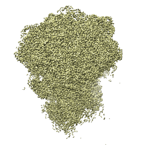 cover_image_EMD2661.png cover_image_EMD2661.png | 322.4 KB | ||
| その他 | emd_2661_half_map_1.map.gzemd_2661_half_map_2.map.gz | 277.2 MB 277.1 MB | ||
| アーカイブディレクトリ |  http://ftp.pdbj.org/pub/emdb/structures/EMD-2661ftp://ftp.pdbj.org/pub/emdb/structures/EMD-2661 http://ftp.pdbj.org/pub/emdb/structures/EMD-2661ftp://ftp.pdbj.org/pub/emdb/structures/EMD-2661 | HTTPS FTP |
-関連構造データ


-リンク
| EMDBのページ | EMDB (EBI/PDBe) / EMDataResource |
|---|---|
| 「今月の分子」の関連する項目 |

-マップ
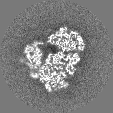
| ファイル | ダウンロード / ファイル: emd_2661.map.gz / 形式: CCP4 / 大きさ: 339.5 MB / タイプ: IMAGE STORED AS FLOATING POINT NUMBER (4 BYTES) | ||||||||||||||||||||||||||||||||||||||||||||||||||||||||||||
|---|---|---|---|---|---|---|---|---|---|---|---|---|---|---|---|---|---|---|---|---|---|---|---|---|---|---|---|---|---|---|---|---|---|---|---|---|---|---|---|---|---|---|---|---|---|---|---|---|---|---|---|---|---|---|---|---|---|---|---|---|---|
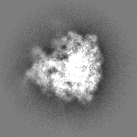
| 注釈 | Cryo-EM map of the Plasmodium falciparum 80S ribosome | ||||||||||||||||||||||||||||||||||||||||||||||||||||||||||||
| 投影像・断面図 | 画像のコントロール
画像は Spider により作成 | ||||||||||||||||||||||||||||||||||||||||||||||||||||||||||||
| ボクセルのサイズ | X=Y=Z: 1.03 Å | ||||||||||||||||||||||||||||||||||||||||||||||||||||||||||||
| 密度 |
| ||||||||||||||||||||||||||||||||||||||||||||||||||||||||||||
| 対称性 | 空間群: 1 | ||||||||||||||||||||||||||||||||||||||||||||||||||||||||||||
| 詳細 | EMDB XML:
CCP4マップ ヘッダ情報:
| ||||||||||||||||||||||||||||||||||||||||||||||||||||||||||||
 Z (Sec.)
 Y (Row.)
 X (Col.)


-添付データ
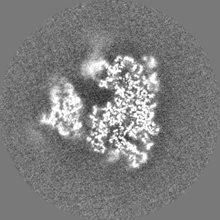
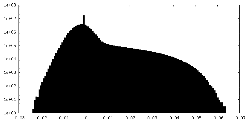
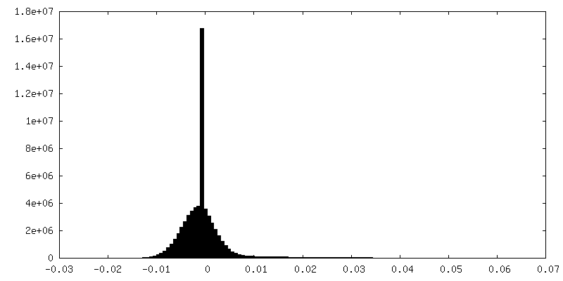
-添付マップデータ: emd 2661 half map 1.map
| ファイル | emd_2661_half_map_1.map | ||||||||||||
|---|---|---|---|---|---|---|---|---|---|---|---|---|---|
| 投影像・断面図 |
| ||||||||||||
| 密度ヒストグラム |


-添付マップデータ: emd 2661 half map 2.map
| ファイル | emd_2661_half_map_2.map | ||||||||||||
|---|---|---|---|---|---|---|---|---|---|---|---|---|---|
| 投影像・断面図 |
| ||||||||||||
| 密度ヒストグラム |


- 試料の構成要素
-全体 : Plasmodium falciparum 80S ribosome
| 全体 | 名称: Plasmodium falciparum 80S ribosome |
|---|---|
| 要素 |
|
-超分子 #1000: Plasmodium falciparum 80S ribosome
| 超分子 | 名称: Plasmodium falciparum 80S ribosome / タイプ: sample / ID: 1000 / Number unique components: 1 |
|---|---|
| 分子量 | 実験値: 4.2 MDa / 理論値: 4.2 MDa |
-超分子 #1: Plasmodium falciparum 80S ribosome
| 超分子 | 名称: Plasmodium falciparum 80S ribosome / タイプ: complex / ID: 1 / 組換発現: No / Ribosome-details: ribosome-eukaryote: ALL |
|---|---|
| 由来(天然) | 生物種: |
| 分子量 | 実験値: 4.2 MDa / 理論値: 4.2 MDa |
-実験情報
-構造解析
| 手法 | ネガティブ染色法, クライオ電子顕微鏡法 |
|---|---|
 解析 解析 | 単粒子再構成法 |
| 試料の集合状態 | particle |
-試料調製
| 濃度 | 0.6 mg/mL |
|---|---|
| 緩衝液 | pH: 7.4 詳細: 20 mM Hepes pH7.4, 40 mM KCH3COO, 10 mM NH4CH3COO, 10 mM Mg(CH3COO)2 and 5 mM 2-mecaptoethanol |
| 染色 | タイプ: NEGATIVE / 詳細: Cryo-EM |
| グリッド | 詳細: 30 s on glow-discharged holey carbon grids (Quantifoil R2/2), onto which a home-made continuous carbon film |
| 凍結 | 凍結剤: ETHANE / チャンバー内湿度: 100 % / チャンバー内温度: 90 K / 装置: FEI VITROBOT MARK IV / 手法: Blot 2.5 seconds before plunging |
- 電子顕微鏡法
| 顕微鏡 | FEI TITAN KRIOS |
|---|---|
| 温度 | 最低: 80 K / 最高: 90 K / 平均: 85 K |
| アライメント法 | Legacy - 非点収差: Objective lens astigmatism was corrected at 78,000 times magnification |
| 日付 | 2014年1月28日 |
| 撮影 | カテゴリ: CCD フィルム・検出器のモデル: FEI FALCON II (4k x 4k) デジタル化 - サンプリング間隔: 14 µm / 実像数: 1307 / 平均電子線量: 20 e/Å2 詳細: An in-house built system was used to intercept the videos from the detector at a rate of 17 frames for the 1 s exposures. |
| 電子線 | 加速電圧: 300 kV / 電子線源:  FIELD EMISSION GUN FIELD EMISSION GUN |
| 電子光学系 | 倍率(補正後): 135922 / 照射モード: FLOOD BEAM / 撮影モード: BRIGHT FIELD / Cs: 2.7 mm / 最大 デフォーカス(公称値): 3.9 µm / 最小 デフォーカス(公称値): 0.7 µm / 倍率(公称値): 78000 |
| 試料ステージ | 試料ホルダーモデル: FEI TITAN KRIOS AUTOGRID HOLDER |
| 実験機器 |  モデル: Titan Krios / 画像提供: FEI Company |
-画像解析
| CTF補正 | 詳細: Each particle |
|---|---|
| 最終 再構成 | 想定した対称性 - 点群: C1 (非対称) / 解像度のタイプ: BY AUTHOR / 解像度: 3.4 Å / 解像度の算出法: OTHER / ソフトウェア - 名称: CTFFIND3, RELION 詳細: Use a newly developed statistical movie processing approach to compensate for beam-induced movement. 使用した粒子像数: 72293 |
