 ムービー
ムービー コントローラー


+ データを開く

- 基本情報
基本情報
| 登録情報 | データベース: EMDB / ID: EMD-2636 | |||||||||
|---|---|---|---|---|---|---|---|---|---|---|
| タイトル | 3D EM map of the sodium proton antiporter MjNhaP1 from Methanocaldococcus jannaschii | |||||||||
 マップデータ マップデータ | A B-factor of -200 was applied. | |||||||||
 試料 試料 |
| |||||||||
 キーワード キーワード | membrane protein / antiporter / transporter / exchanger / CPA | |||||||||
| 機能・相同性 |  機能・相同性情報 機能・相同性情報: / transport / potassium:proton antiporter activity / intracellular potassium ion homeostasis / antiporter activity / sodium ion transport / monoatomic cation transport / membrane => GO:0016020 / monoatomic ion transport / proton transmembrane transport ...: / transport / potassium:proton antiporter activity / intracellular potassium ion homeostasis / antiporter activity / sodium ion transport / monoatomic cation transport / membrane => GO:0016020 / monoatomic ion transport / proton transmembrane transport / transmembrane transport / identical protein binding / membrane / plasma membrane 類似検索 - 分子機能 | |||||||||
| 生物種 |   Methanocaldococcus jannaschii (メタン生成菌) Methanocaldococcus jannaschii (メタン生成菌) | |||||||||
| 手法 | 電子線結晶学 / クライオ電子顕微鏡法 / ネガティブ染色法 / 解像度: 6.0 Å | |||||||||
 データ登録者 データ登録者 | Paulino C / Woehlert D / Yildiz O / Kuhlbrandt W | |||||||||
 引用 引用 | ジャーナル: Elife / 年: 2014 タイトル: Structure and transport mechanism of the sodium/proton antiporter MjNhaP1. 著者: Cristina Paulino / David Wöhlert / Ekaterina Kapotova / Özkan Yildiz / Werner Kühlbrandt /  要旨: Sodium/proton antiporters are essential for sodium and pH homeostasis and play a major role in human health and disease. We determined the structures of the archaeal sodium/proton antiporter MjNhaP1 ...Sodium/proton antiporters are essential for sodium and pH homeostasis and play a major role in human health and disease. We determined the structures of the archaeal sodium/proton antiporter MjNhaP1 in two complementary states. The inward-open state was obtained by x-ray crystallography in the presence of sodium at pH 8, where the transporter is highly active. The outward-open state was obtained by electron crystallography without sodium at pH 4, where MjNhaP1 is inactive. Comparison of both structures reveals a 7° tilt of the 6 helix bundle. (22)Na(+) uptake measurements indicate non-cooperative transport with an activity maximum at pH 7.5. We conclude that binding of a Na(+) ion from the outside induces helix movements that close the extracellular cavity, open the cytoplasmic funnel, and result in a ∼5 Å vertical relocation of the ion binding site to release the substrate ion into the cytoplasm. | |||||||||
| 履歴 |
|
- 構造の表示
構造の表示
| ムービー |
ムービービューア |
|---|---|
| 構造ビューア | EMマップ: SurfViewMolmilJmol/JSmol |
| 添付画像 |

- ダウンロードとリンク
-EMDBアーカイブ
| マップデータ | emd_2636.map.gz | 6 MB | EMDBマップデータ形式 | |
|---|---|---|---|---|
| ヘッダ (付随情報) | emd-2636-v30.xmlemd-2636.xml | 14.6 KB 14.6 KB | 表示 表示 | EMDBヘッダ |
| 画像 | 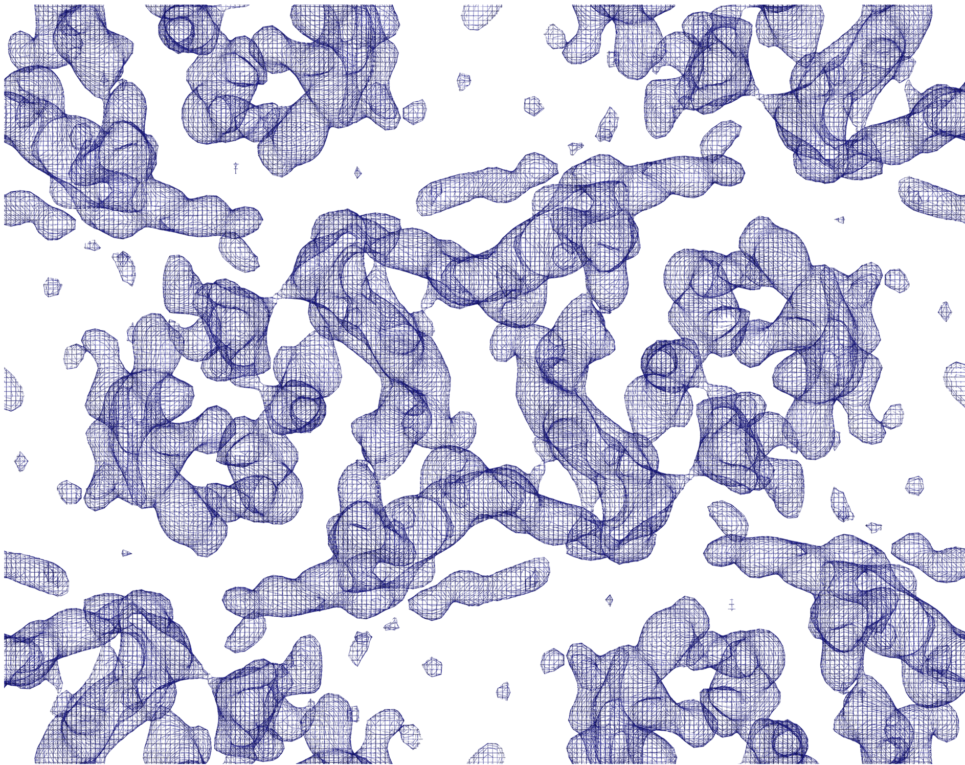 emd_2636.png emd_2636.png | 4 MB | ||
| アーカイブディレクトリ |  http://ftp.pdbj.org/pub/emdb/structures/EMD-2636ftp://ftp.pdbj.org/pub/emdb/structures/EMD-2636 http://ftp.pdbj.org/pub/emdb/structures/EMD-2636ftp://ftp.pdbj.org/pub/emdb/structures/EMD-2636 | HTTPS FTP |
-検証レポート
| 文書・要旨 | emd_2636_validation.pdf.gz | 235.1 KB | 表示 | EMDB検証レポート |
|---|---|---|---|---|
| 文書・詳細版 | emd_2636_full_validation.pdf.gz | 234.2 KB | 表示 | |
| XML形式データ | emd_2636_validation.xml.gz | 4.8 KB | 表示 | |
| アーカイブディレクトリ | https://ftp.pdbj.org/pub/emdb/validation_reports/EMD-2636ftp://ftp.pdbj.org/pub/emdb/validation_reports/EMD-2636 | HTTPS FTP |
-関連構造データ
-リンク
| EMDBのページ | EMDB (EBI/PDBe) / EMDataResource |
|---|
-マップ
| ファイル | ダウンロード / ファイル: emd_2636.map.gz / 形式: CCP4 / 大きさ: 6.3 MB / タイプ: IMAGE STORED AS FLOATING POINT NUMBER (4 BYTES) | ||||||||||||||||||||||||||||||||||||||||||||||||||||||||||||||||||||
|---|---|---|---|---|---|---|---|---|---|---|---|---|---|---|---|---|---|---|---|---|---|---|---|---|---|---|---|---|---|---|---|---|---|---|---|---|---|---|---|---|---|---|---|---|---|---|---|---|---|---|---|---|---|---|---|---|---|---|---|---|---|---|---|---|---|---|---|---|---|
| 注釈 | A B-factor of -200 was applied. | ||||||||||||||||||||||||||||||||||||||||||||||||||||||||||||||||||||
| 投影像・断面図 | 画像のコントロール
画像は Spider により作成 これらの図は立方格子座標系で作成されたものです | ||||||||||||||||||||||||||||||||||||||||||||||||||||||||||||||||||||
| ボクセルのサイズ | X: 1.01875 Å / Y: 0.99327 Å / Z: 1 Å | ||||||||||||||||||||||||||||||||||||||||||||||||||||||||||||||||||||
| 密度 |
| ||||||||||||||||||||||||||||||||||||||||||||||||||||||||||||||||||||
| 対称性 | 空間群: 18 | ||||||||||||||||||||||||||||||||||||||||||||||||||||||||||||||||||||
| 詳細 | EMDB XML:
CCP4マップ ヘッダ情報:
| ||||||||||||||||||||||||||||||||||||||||||||||||||||||||||||||||||||
 Y (Sec.)
 X (Row.)
 Z (Col.)


-添付データ
- 試料の構成要素
-全体 : 3D EM map of the sodium/proton antiporter MjNhaP1
| 全体 | 名称: 3D EM map of the sodium/proton antiporter MjNhaP1 |
|---|---|
| 要素 |
|
-超分子 #1000: 3D EM map of the sodium/proton antiporter MjNhaP1
| 超分子 | 名称: 3D EM map of the sodium/proton antiporter MjNhaP1 / タイプ: sample / ID: 1000 / 詳細: protein was purified in absence of sodium. / 集合状態: Dimer / Number unique components: 1 |
|---|---|
| 分子量 | 実験値: 46 KDa / 理論値: 46 KDa / 手法: SDS-PAGE, MS |
-分子 #1: MjNhaP1
| 分子 | 名称: MjNhaP1 / タイプ: protein_or_peptide / ID: 1 / Name.synonym: NhaP1 / コピー数: 2 / 集合状態: Dimer / 組換発現: Yes |
|---|---|
| 由来(天然) | 生物種: Methanocaldococcus jannaschii (メタン生成菌) 別称: Methanocaldococcus jannaschii / 細胞中の位置: Plasma membrane |
| 分子量 | 実験値: 46 KDa / 理論値: 46 KDa |
| 組換発現 | 生物種:  |
| 配列 | UniProtKB: Na(+)/H(+) antiporter 1 GO: transport, monoatomic ion transport, monoatomic cation transport, sodium ion transport, transmembrane transport, proton transmembrane transport, antiporter activity, GO: 0015299, identical ...GO: transport, monoatomic ion transport, monoatomic cation transport, sodium ion transport, transmembrane transport, proton transmembrane transport, antiporter activity, GO: 0015299, identical protein binding, plasma membrane, membrane, membrane => GO:0016020 InterPro: Cation/H+ exchanger |
-実験情報
-構造解析
| 手法 | ネガティブ染色法, クライオ電子顕微鏡法 |
|---|---|
 解析 解析 | 電子線結晶学 |
| 試料の集合状態 | 2D array |
-試料調製
| 濃度 | 1 mg/mL |
|---|---|
| 緩衝液 | pH: 4 / 詳細: 25mM KAc pH4, 200mM KCl, 5mM glycerol, 5mM MPD |
| 染色 | タイプ: NEGATIVE / 詳細: back injection method with 4% trehalose |
| グリッド | 詳細: 400 mesh copper grid |
| 凍結 | 凍結剤: NITROGEN / チャンバー内湿度: 20 % / チャンバー内温度: 77 K / 装置: OTHER / 詳細: all buffers used were sodium-free Timed resolved state: sample was plunge-frozen in liquid nitrogen 手法: back injection method (Wang & Kuhlbrandt, 1991) with 4% trehalose |
| 詳細 | E.coli polar lipids with a lipid-to-protein ration (LPR) of 0.4-0.5 were used. 2D crystals were grown by slow removal of detergent (0.15% DM) by dialysis. |
| 結晶化 | 詳細: E.coli polar lipids with a lipid-to-protein ration (LPR) of 0.4-0.5 were used. 2D crystals were grown by slow removal of detergent (0.15% DM) by dialysis. |
- 電子顕微鏡法
| 顕微鏡 | JEOL 3000SFF |
|---|---|
| 温度 | 最低: 4 K / 最高: 10 K / 平均: 4 K |
| アライメント法 | Legacy - 非点収差: objective lens was corrected at 60kx and/or 300kx magnification Legacy - Electron beam tilt params: - |
| 特殊光学系 | エネルギーフィルター - 名称: - |
| 日付 | 2012年12月1日 |
| 撮影 | カテゴリ: FILM / フィルム・検出器のモデル: KODAK SO-163 FILM / デジタル化 - スキャナー: ZEISS SCAI / デジタル化 - サンプリング間隔: 7 µm / 実像数: 128 / 平均電子線量: 25 e/Å2 |
| Tilt angle min | 0 |
| 電子線 | 加速電圧: 300 kV / 電子線源:  FIELD EMISSION GUN FIELD EMISSION GUN |
| 電子光学系 | 倍率(補正後): 53000 / 照射モード: SPOT SCAN / 撮影モード: OTHER / Cs: 1.6 mm / 最大 デフォーカス(公称値): 1.8 µm / 最小 デフォーカス(公称値): 0.12 µm / 倍率(公称値): 60000 |
| 試料ステージ | 試料ホルダー: helium-cooled top entry stage with fixed specimen holder. 試料ホルダーモデル: JEOL / Tilt angle max: 45 / Tilt series - Axis1 - Min angle: 0 ° / Tilt series - Axis1 - Max angle: 45 ° |
-画像解析
| 詳細 | Images were processed with the 2dx software. |
|---|---|
| 最終 再構成 | 解像度のタイプ: BY AUTHOR / 解像度: 6.0 Å / 解像度の算出法: OTHER / ソフトウェア - 名称: 2dx 詳細: 6A in plane resolution and 14A resolution in the z direction. |
| 結晶パラメータ | 単位格子 - A: 81.5 Å / 単位格子 - B: 103.3 Å / 単位格子 - C: 200 Å / 単位格子 - γ: 90.0 ° / 単位格子 - α: 90.0 ° / 単位格子 - β: 90.0 ° / 面群: P 2 21 21 |
| CTF補正 | 詳細: 2dx |
-原子モデル構築 1
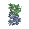
| 初期モデル | PDB ID: Chain - Chain ID: B |
|---|---|
| ソフトウェア | 名称: Coot |
| 詳細 | The X-ray structure of the same protein (4czb) obtained at different conditions was manually fitted to the 3D EM density map. |
| 精密化 | 空間: REAL / プロトコル: RIGID BODY FIT |
| 得られたモデル | 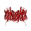 PDB-4d0a: |
