ムービー
ムービー コントローラー


+ データを開く

- 基本情報
基本情報
| 登録情報 | データベース: EMDB / ID: EMD-2633 | |||||||||
|---|---|---|---|---|---|---|---|---|---|---|
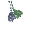
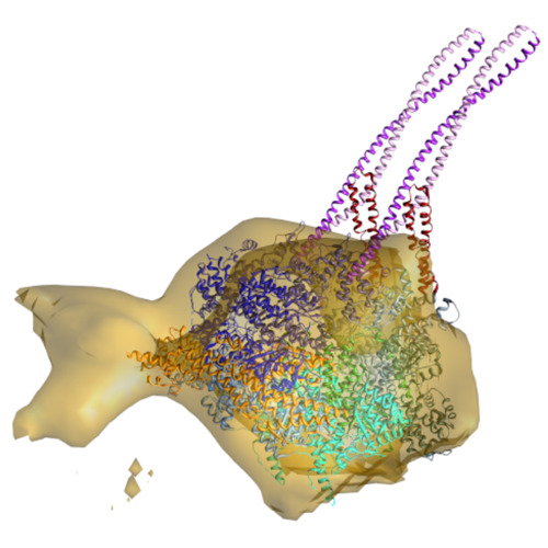
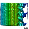
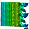
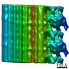
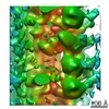
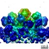
| タイトル | Outer arm dynein dimer from mouse respiratory cilia in pre-power stroke (SH-P) | |||||||||
 マップデータ マップデータ | subtomogram averaging of dynein dimers from mouse respiratory cilia in the SH-P form (pre-power stroke) | |||||||||
 試料 試料 |
| |||||||||
 キーワード キーワード | dynein / ATP / cilia / cryo-electron tomography | |||||||||
| 生物種 |  | |||||||||
| 手法 | サブトモグラム平均法 / クライオ電子顕微鏡法 / 解像度: 40.0 Å | |||||||||
 データ登録者 データ登録者 | Ueno H / Bui KH / Ishikawa T / Imai Y / Yamaguchi T | |||||||||
 引用 引用 | ジャーナル: Cytoskeleton (Hoboken) / 年: 2014 タイトル: Structure of dimeric axonemal dynein in cilia suggests an alternative mechanism of force generation. 著者: Hironori Ueno / Khanh Huy Bui / Takuji Ishikawa / Yohsuke Imai / Takami Yamaguchi / Takashi Ishikawa /  要旨: The mechanism by which the two different heads of the ciliary outer dynein arm produce force to translocate the microtubule during beating is still unknown. In this report we use cryo-electron ...The mechanism by which the two different heads of the ciliary outer dynein arm produce force to translocate the microtubule during beating is still unknown. In this report we use cryo-electron tomography and image processing to analyze the conformational changes and the relative abundance of each conformation of the two dynein heads from mouse respiratory cilia. In the absence of nucleotides the majority of dynein dimers are in the apo form and both heads are tightly packed, whereas they are dissociated and move independently in the presence of nucleotides. The head of the external outer arm dynein heavy chain has a diagonal shift toward both the neighboring B-tubule and the proximal end of the axoneme, while the head of the internal heavy chain shifts only longitudinally toward the proximal end. In the presence of nucleotides a significant number of the dynein dimers have two heads overlapped in the proximal shifting form or overlapped in the apo form. During ciliary bending axonemal dynein translocates microtubules by moving with short steps and two heads stay at the same position longer than cytoplasmic dynein. This demonstrates that the step of the outer arm dynein dimer is not dominated by the hand-over-hand motion, but also indicates the difference between axonemal dynein and cytoplasmic dynein. | |||||||||
| 履歴 |
|
- 構造の表示
構造の表示
| ムービー |
 ムービービューア ムービービューア |
|---|---|
| 構造ビューア | EMマップ: SurfViewMolmilJmol/JSmol |
| 添付画像 |
- ダウンロードとリンク
-EMDBアーカイブ
| マップデータ | emd_2633.map.gz | 9.8 MB | EMDBマップデータ形式 | |
|---|---|---|---|---|
| ヘッダ (付随情報) | emd-2633-v30.xmlemd-2633.xml | 9.3 KB 9.3 KB | 表示 表示 | EMDBヘッダ |
| 画像 | emd_2633.tif | 759.5 KB | ||
| アーカイブディレクトリ |  http://ftp.pdbj.org/pub/emdb/structures/EMD-2633ftp://ftp.pdbj.org/pub/emdb/structures/EMD-2633 http://ftp.pdbj.org/pub/emdb/structures/EMD-2633ftp://ftp.pdbj.org/pub/emdb/structures/EMD-2633 | HTTPS FTP |
-検証レポート
| 文書・要旨 | emd_2633_validation.pdf.gz | 172.1 KB | 表示 | EMDB検証レポート |
|---|---|---|---|---|
| 文書・詳細版 | emd_2633_full_validation.pdf.gz | 171.2 KB | 表示 | |
| XML形式データ | emd_2633_validation.xml.gz | 4.6 KB | 表示 | |
| アーカイブディレクトリ | https://ftp.pdbj.org/pub/emdb/validation_reports/EMD-2633ftp://ftp.pdbj.org/pub/emdb/validation_reports/EMD-2633 | HTTPS FTP |
-関連構造データ
-リンク
| EMDBのページ | EMDB (EBI/PDBe) / EMDataResource |
|---|
-マップ
| ファイル | ダウンロード / ファイル: emd_2633.map.gz / 形式: CCP4 / 大きさ: 11.9 MB / タイプ: IMAGE STORED AS FLOATING POINT NUMBER (4 BYTES) | ||||||||||||||||||||||||||||||||||||||||||||||||||||||||||||||||||||
|---|---|---|---|---|---|---|---|---|---|---|---|---|---|---|---|---|---|---|---|---|---|---|---|---|---|---|---|---|---|---|---|---|---|---|---|---|---|---|---|---|---|---|---|---|---|---|---|---|---|---|---|---|---|---|---|---|---|---|---|---|---|---|---|---|---|---|---|---|---|
| 注釈 | subtomogram averaging of dynein dimers from mouse respiratory cilia in the SH-P form (pre-power stroke) | ||||||||||||||||||||||||||||||||||||||||||||||||||||||||||||||||||||
| 投影像・断面図 | 画像のコントロール
画像は Spider により作成 これらの図は立方格子座標系で作成されたものです | ||||||||||||||||||||||||||||||||||||||||||||||||||||||||||||||||||||
| ボクセルのサイズ | X=Y=Z: 7.25 Å | ||||||||||||||||||||||||||||||||||||||||||||||||||||||||||||||||||||
| 密度 |
| ||||||||||||||||||||||||||||||||||||||||||||||||||||||||||||||||||||
| 対称性 | 空間群: 1 | ||||||||||||||||||||||||||||||||||||||||||||||||||||||||||||||||||||
| 詳細 | EMDB XML:
CCP4マップ ヘッダ情報:
| ||||||||||||||||||||||||||||||||||||||||||||||||||||||||||||||||||||
 Z (Sec.)
 Y (Row.)
 X (Col.)


-添付データ
- 試料の構成要素
-全体 : dynein dimer in the SH-P form (pre-power stroke)
| 全体 | 名称: dynein dimer in the SH-P form (pre-power stroke) |
|---|---|
| 要素 |
|
-超分子 #1000: dynein dimer in the SH-P form (pre-power stroke)
| 超分子 | 名称: dynein dimer in the SH-P form (pre-power stroke) / タイプ: sample / ID: 1000 / Number unique components: 1 |
|---|
-超分子 #1: Cilia
| 超分子 | 名称: Cilia / タイプ: organelle_or_cellular_component / ID: 1 / 集合状態: dimer / 組換発現: No |
|---|---|
| 由来(天然) | 生物種: |
-実験情報
-構造解析
| 手法 | クライオ電子顕微鏡法 |
|---|---|
 解析 解析 | サブトモグラム平均法 |
| 試料の集合状態 | cell |
-試料調製
| グリッド | 詳細: Quantifoil holey grids |
|---|---|
| 凍結 | 凍結剤: ETHANE / チャンバー内湿度: 100 % / チャンバー内温度: 100 K / 装置: FEI VITROBOT MARK IV / 手法: Blot for 2 seconds before plunging |
- 電子顕微鏡法
| 顕微鏡 | FEI TECNAI F20 |
|---|---|
| 温度 | 最低: 90 K / 最高: 100 K / 平均: 95 K |
| 特殊光学系 | エネルギーフィルター - 名称: GIF Tridiem エネルギーフィルター - エネルギー下限: 20.0 eV エネルギーフィルター - エネルギー上限: 25.0 eV |
| 日付 | 2010年12月14日 |
| 撮影 | カテゴリ: CCD フィルム・検出器のモデル: GATAN ULTRASCAN 1000 (2k x 2k) 平均電子線量: 40 e/Å2 |
| 電子線 | 加速電圧: 200 kV / 電子線源:  FIELD EMISSION GUN FIELD EMISSION GUN |
| 電子光学系 | 倍率(補正後): 19303 / 照射モード: FLOOD BEAM / 撮影モード: BRIGHT FIELD / Cs: 1.5 mm / 最大 デフォーカス(公称値): 5.0 µm / 最小 デフォーカス(公称値): 3.0 µm / 倍率(公称値): 30000 |
| 試料ステージ | 試料ホルダー: 626 / 試料ホルダーモデル: GATAN LIQUID NITROGEN / Tilt series - Axis1 - Min angle: -60 ° / Tilt series - Axis1 - Max angle: 60 ° |
| 実験機器 |  モデル: Tecnai F20 / 画像提供: FEI Company |
-画像解析
| 最終 再構成 | 想定した対称性 - 点群: C1 (非対称) / アルゴリズム: OTHER / 解像度のタイプ: BY AUTHOR / 解像度: 40.0 Å / 解像度の算出法: OTHER / ソフトウェア - 名称: IMOD / 使用したサブトモグラム数: 61 |
|---|
-原子モデル構築 1
| 初期モデル | PDB ID: |
|---|---|
| ソフトウェア | 名称: Chimera |
| 詳細 | AAA domains, stalk, and buttress are considered as a rigid body. Only the linker was flexibly fitted. |
| 精密化 | 空間: REAL / プロトコル: FLEXIBLE FIT |