Movie
Movie Controller

[English] 日本語
 Yorodumi
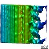
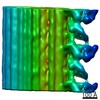
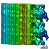
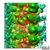
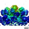
- EMDB-2633: Outer arm dynein dimer from mouse respiratory cilia in pre-power ... -
+ Open data

- Basic information
| Entry | Database: EMDB / ID: EMD-2633 | |||||||||
|---|---|---|---|---|---|---|---|---|---|---|
| Title | Outer arm dynein dimer from mouse respiratory cilia in pre-power stroke (SH-P) | |||||||||
 Map data Map data | subtomogram averaging of dynein dimers from mouse respiratory cilia in the SH-P form (pre-power stroke) | |||||||||
 Sample Sample |
| |||||||||
 Keywords Keywords | dynein / ATP / cilia / cryo-electron tomography | |||||||||
| Biological species |  | |||||||||
| Method | subtomogram averaging / cryo EM / Resolution: 40.0 Å | |||||||||
 Authors Authors | Ueno H / Bui KH / Ishikawa T / Imai Y / Yamaguchi T | |||||||||
 Citation Citation | Journal: Cytoskeleton (Hoboken) / Year: 2014 Title: Structure of dimeric axonemal dynein in cilia suggests an alternative mechanism of force generation. Authors: Hironori Ueno / Khanh Huy Bui / Takuji Ishikawa / Yohsuke Imai / Takami Yamaguchi / Takashi Ishikawa /  Abstract: The mechanism by which the two different heads of the ciliary outer dynein arm produce force to translocate the microtubule during beating is still unknown. In this report we use cryo-electron ...The mechanism by which the two different heads of the ciliary outer dynein arm produce force to translocate the microtubule during beating is still unknown. In this report we use cryo-electron tomography and image processing to analyze the conformational changes and the relative abundance of each conformation of the two dynein heads from mouse respiratory cilia. In the absence of nucleotides the majority of dynein dimers are in the apo form and both heads are tightly packed, whereas they are dissociated and move independently in the presence of nucleotides. The head of the external outer arm dynein heavy chain has a diagonal shift toward both the neighboring B-tubule and the proximal end of the axoneme, while the head of the internal heavy chain shifts only longitudinally toward the proximal end. In the presence of nucleotides a significant number of the dynein dimers have two heads overlapped in the proximal shifting form or overlapped in the apo form. During ciliary bending axonemal dynein translocates microtubules by moving with short steps and two heads stay at the same position longer than cytoplasmic dynein. This demonstrates that the step of the outer arm dynein dimer is not dominated by the hand-over-hand motion, but also indicates the difference between axonemal dynein and cytoplasmic dynein. | |||||||||
| History |
|
- Structure visualization
| Movie |
 Movie viewer Movie viewer |
|---|---|
| Structure viewer | EM map: SurfViewMolmilJmol/JSmol |
| Supplemental images |
- Downloads & links
-EMDB archive
| Map data | emd_2633.map.gz | 9.8 MB | EMDB map data format | |
|---|---|---|---|---|
| Header (meta data) | emd-2633-v30.xmlemd-2633.xml | 9.3 KB 9.3 KB | Display Display | EMDB header |
| Images | emd_2633.tif | 759.5 KB | ||
| Archive directory |  http://ftp.pdbj.org/pub/emdb/structures/EMD-2633ftp://ftp.pdbj.org/pub/emdb/structures/EMD-2633 http://ftp.pdbj.org/pub/emdb/structures/EMD-2633ftp://ftp.pdbj.org/pub/emdb/structures/EMD-2633 | HTTPS FTP |
-Validation report
| Summary document | emd_2633_validation.pdf.gz | 172.1 KB | Display | EMDB validaton report |
|---|---|---|---|---|
| Full document | emd_2633_full_validation.pdf.gz | 171.2 KB | Display | |
| Data in XML | emd_2633_validation.xml.gz | 4.6 KB | Display | |
| Arichive directory | https://ftp.pdbj.org/pub/emdb/validation_reports/EMD-2633ftp://ftp.pdbj.org/pub/emdb/validation_reports/EMD-2633 | HTTPS FTP |
-Related structure data
-Links
| EMDB pages | EMDB (EBI/PDBe) / EMDataResource |
|---|
-Map
| File | Download / File: emd_2633.map.gz / Format: CCP4 / Size: 11.9 MB / Type: IMAGE STORED AS FLOATING POINT NUMBER (4 BYTES) | ||||||||||||||||||||||||||||||||||||||||||||||||||||||||||||||||||||
|---|---|---|---|---|---|---|---|---|---|---|---|---|---|---|---|---|---|---|---|---|---|---|---|---|---|---|---|---|---|---|---|---|---|---|---|---|---|---|---|---|---|---|---|---|---|---|---|---|---|---|---|---|---|---|---|---|---|---|---|---|---|---|---|---|---|---|---|---|---|
| Annotation | subtomogram averaging of dynein dimers from mouse respiratory cilia in the SH-P form (pre-power stroke) | ||||||||||||||||||||||||||||||||||||||||||||||||||||||||||||||||||||
| Projections & slices | Image control
Images are generated by Spider. generated in cubic-lattice coordinate | ||||||||||||||||||||||||||||||||||||||||||||||||||||||||||||||||||||
| Voxel size | X=Y=Z: 7.25 Å | ||||||||||||||||||||||||||||||||||||||||||||||||||||||||||||||||||||
| Density |
| ||||||||||||||||||||||||||||||||||||||||||||||||||||||||||||||||||||
| Symmetry | Space group: 1 | ||||||||||||||||||||||||||||||||||||||||||||||||||||||||||||||||||||
| Details | EMDB XML:
CCP4 map header:
| ||||||||||||||||||||||||||||||||||||||||||||||||||||||||||||||||||||
 Z (Sec.)
 Y (Row.)
 X (Col.)


-Supplemental data
- Sample components
-Entire : dynein dimer in the SH-P form (pre-power stroke)
| Entire | Name: dynein dimer in the SH-P form (pre-power stroke) |
|---|---|
| Components |
|
-Supramolecule #1000: dynein dimer in the SH-P form (pre-power stroke)
| Supramolecule | Name: dynein dimer in the SH-P form (pre-power stroke) / type: sample / ID: 1000 / Number unique components: 1 |
|---|
-Supramolecule #1: Cilia
| Supramolecule | Name: Cilia / type: organelle_or_cellular_component / ID: 1 / Oligomeric state: dimer / Recombinant expression: No |
|---|---|
| Source (natural) | Organism: |
-Experimental details
-Structure determination
| Method | cryo EM |
|---|---|
 Processing Processing | subtomogram averaging |
| Aggregation state | cell |
-Sample preparation
| Grid | Details: Quantifoil holey grids |
|---|---|
| Vitrification | Cryogen name: ETHANE / Chamber humidity: 100 % / Chamber temperature: 100 K / Instrument: FEI VITROBOT MARK IV / Method: Blot for 2 seconds before plunging |
- Electron microscopy
| Microscope | FEI TECNAI F20 |
|---|---|
| Temperature | Min: 90 K / Max: 100 K / Average: 95 K |
| Specialist optics | Energy filter - Name: GIF Tridiem / Energy filter - Lower energy threshold: 20.0 eV / Energy filter - Upper energy threshold: 25.0 eV |
| Date | Dec 14, 2010 |
| Image recording | Category: CCD / Film or detector model: GATAN ULTRASCAN 1000 (2k x 2k) / Average electron dose: 40 e/Å2 |
| Electron beam | Acceleration voltage: 200 kV / Electron source:  FIELD EMISSION GUN FIELD EMISSION GUN |
| Electron optics | Calibrated magnification: 19303 / Illumination mode: FLOOD BEAM / Imaging mode: BRIGHT FIELD / Cs: 1.5 mm / Nominal defocus max: 5.0 µm / Nominal defocus min: 3.0 µm / Nominal magnification: 30000 |
| Sample stage | Specimen holder: 626 / Specimen holder model: GATAN LIQUID NITROGEN / Tilt series - Axis1 - Min angle: -60 ° / Tilt series - Axis1 - Max angle: 60 ° |
| Experimental equipment |  Model: Tecnai F20 / Image courtesy: FEI Company |
-Image processing
| Final reconstruction | Applied symmetry - Point group: C1 (asymmetric) / Algorithm: OTHER / Resolution.type: BY AUTHOR / Resolution: 40.0 Å / Resolution method: OTHER / Software - Name: IMOD / Number subtomograms used: 61 |
|---|
-Atomic model buiding 1
| Initial model | PDB ID: |
|---|---|
| Software | Name: Chimera |
| Details | AAA domains, stalk, and buttress are considered as a rigid body. Only the linker was flexibly fitted. |
| Refinement | Space: REAL / Protocol: FLEXIBLE FIT |