 Movie
Movie Controller

[English] 日本語
 Yorodumi
- EMDB-26305: EBNA1 DNA binding domain (401-641) binds to half Dyad Symmetry element -
+ Open data

- Basic information
| Entry | 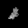 | |||||||||
|---|---|---|---|---|---|---|---|---|---|---|
| Title | EBNA1 DNA binding domain (401-641) binds to half Dyad Symmetry element | |||||||||
 Map data Map data | ||||||||||
 Sample Sample |
| |||||||||
 Keywords Keywords | EBV latency protein EBNA1 Dyad symmetry / VIRAL PROTEIN-DNA complex | |||||||||
| Function / homology |  Function and homology information Function and homology informationhost cell PML body / viral latency / Hydrolases; Acting on ester bonds; Endodeoxyribonucleases producing 5'-phosphomonoesters / symbiont-mediated disruption of host cell PML body / regulation of DNA replication / enzyme-substrate adaptor activity / endonuclease activity / symbiont-mediated suppression of host NF-kappaB cascade / DNA-binding transcription factor activity / hydrolase activity ...host cell PML body / viral latency / Hydrolases; Acting on ester bonds; Endodeoxyribonucleases producing 5'-phosphomonoesters / symbiont-mediated disruption of host cell PML body / regulation of DNA replication / enzyme-substrate adaptor activity / endonuclease activity / symbiont-mediated suppression of host NF-kappaB cascade / DNA-binding transcription factor activity / hydrolase activity / positive regulation of DNA-templated transcription / DNA binding Similarity search - Function | |||||||||
| Biological species |  Human herpesvirus 4 strain B95-8 (Epstein-Barr virus (strain B95-8)) Human herpesvirus 4 strain B95-8 (Epstein-Barr virus (strain B95-8)) | |||||||||
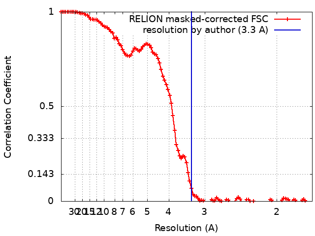
| Method | single particle reconstruction / cryo EM / Resolution: 3.3 Å | |||||||||
 Authors Authors | Mei Y / Lieberman P | |||||||||
| Funding support |  United States, 2 items United States, 2 items
| |||||||||
 Citation Citation | Journal: To Be Published Title: EBNA1 DNA binding domain (401-641) binds to half Dyad Symmetry element Authors: Mei Y / Lieberman P | |||||||||
| History |
|
- Structure visualization
| Supplemental images |
|---|

- Downloads & links
-EMDB archive
| Map data | emd_26305.map.gz | 80.8 MB | EMDB map data format | |
|---|---|---|---|---|
| Header (meta data) | emd-26305-v30.xmlemd-26305.xml | 17.2 KB 17.2 KB | Display Display | EMDB header |
| FSC (resolution estimation) | emd_26305_fsc.xml | 10.7 KB | Display | FSC data file |
| Images |  emd_26305.png emd_26305.png | 28.5 KB | ||
| Filedesc metadata | emd-26305.cif.gz | 6.1 KB | ||
| Archive directory |  http://ftp.pdbj.org/pub/emdb/structures/EMD-26305ftp://ftp.pdbj.org/pub/emdb/structures/EMD-26305 http://ftp.pdbj.org/pub/emdb/structures/EMD-26305ftp://ftp.pdbj.org/pub/emdb/structures/EMD-26305 | HTTPS FTP |
-Related structure data
| Related structure data |  7u1tMC M: atomic model generated by this map C: citing same article ( |
|---|---|
| Similar structure data |
-Links
| EMDB pages | EMDB (EBI/PDBe) / EMDataResource |
|---|
-Map
| File | Download / File: emd_26305.map.gz / Format: CCP4 / Size: 103 MB / Type: IMAGE STORED AS FLOATING POINT NUMBER (4 BYTES) | ||||||||||||||||||||||||||||||||||||
|---|---|---|---|---|---|---|---|---|---|---|---|---|---|---|---|---|---|---|---|---|---|---|---|---|---|---|---|---|---|---|---|---|---|---|---|---|---|
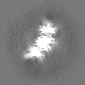
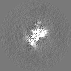
| Projections & slices | Image control
Images are generated by Spider. | ||||||||||||||||||||||||||||||||||||
| Voxel size | X=Y=Z: 0.86 Å | ||||||||||||||||||||||||||||||||||||
| Density |
| ||||||||||||||||||||||||||||||||||||
| Symmetry | Space group: 1 | ||||||||||||||||||||||||||||||||||||
| Details | EMDB XML:
|
 Z (Sec.)
 Y (Row.)
 X (Col.)


-Supplemental data
- Sample components
-Entire : EBNA1 DNA binding domain binds to half Dyad Symmetry region
| Entire | Name: EBNA1 DNA binding domain binds to half Dyad Symmetry region |
|---|---|
| Components |
|
-Supramolecule #1: EBNA1 DNA binding domain binds to half Dyad Symmetry region
| Supramolecule | Name: EBNA1 DNA binding domain binds to half Dyad Symmetry region type: complex / ID: 1 / Parent: 0 / Macromolecule list: all |
|---|---|
| Source (natural) | Organism: Human herpesvirus 4 strain B95-8 (Epstein-Barr virus (strain B95-8)) |
| Molecular weight | Theoretical: 160 KDa |
-Macromolecule #1: Epstein-Barr nuclear antigen 1
| Macromolecule | Name: Epstein-Barr nuclear antigen 1 / type: protein_or_peptide / ID: 1 / Number of copies: 4 / Enantiomer: LEVO |
|---|---|
| Source (natural) | Organism: Human herpesvirus 4 strain B95-8 (Epstein-Barr virus (strain B95-8)) Strain: B95-8 |
| Molecular weight | Theoretical: 19.243031 KDa |
| Recombinant expression | Organism:  |
| Sequence | String: PADDPGEGPS TGPRGQGDGG RRKKGGWFGK HRGQGGSNPK FENIAEGLRA LLARSHVERT TDEGTWVAGV FVYGGSKTSL YNLRRGTAL AIPQCRLTPL SRLPFGMAPG PGPQPGPLRE SIVCYFMVFL QTHIFAEVLK DAIKDLVMTK PAPTCNIRVT V CSFDDGVD LPPWFPPMVE UniProtKB: Epstein-Barr nuclear antigen 1 |
-Macromolecule #2: DNA (59-MER)
| Macromolecule | Name: DNA (59-MER) / type: dna / ID: 2 / Number of copies: 1 / Classification: DNA |
|---|---|
| Source (natural) | Organism: Human herpesvirus 4 strain B95-8 (Epstein-Barr virus (strain B95-8)) |
| Molecular weight | Theoretical: 18.181674 KDa |
| Sequence | String: (DT)(DA)(DA)(DC)(DC)(DC)(DT)(DA)(DA)(DT) (DT)(DC)(DG)(DA)(DT)(DA)(DG)(DC)(DA)(DT) (DA)(DT)(DG)(DC)(DT)(DT)(DC)(DC)(DC) (DG)(DT)(DT)(DG)(DG)(DG)(DT)(DA)(DA)(DC) (DA) (DT)(DA)(DT)(DG)(DC)(DT) ...String: (DT)(DA)(DA)(DC)(DC)(DC)(DT)(DA)(DA)(DT) (DT)(DC)(DG)(DA)(DT)(DA)(DG)(DC)(DA)(DT) (DA)(DT)(DG)(DC)(DT)(DT)(DC)(DC)(DC) (DG)(DT)(DT)(DG)(DG)(DG)(DT)(DA)(DA)(DC) (DA) (DT)(DA)(DT)(DG)(DC)(DT)(DA)(DT) (DT)(DG)(DA)(DA)(DT)(DT)(DA)(DG)(DG)(DG) (DT) GENBANK: GENBANK: M80517.1 |
-Macromolecule #3: DNA (59-MER)
| Macromolecule | Name: DNA (59-MER) / type: dna / ID: 3 / Number of copies: 1 / Classification: DNA |
|---|---|
| Source (natural) | Organism: Human herpesvirus 4 strain B95-8 (Epstein-Barr virus (strain B95-8)) |
| Molecular weight | Theoretical: 18.177703 KDa |
| Sequence | String: (DA)(DC)(DC)(DC)(DT)(DA)(DA)(DT)(DT)(DC) (DA)(DA)(DT)(DA)(DG)(DC)(DA)(DT)(DA)(DT) (DG)(DT)(DT)(DA)(DC)(DC)(DC)(DA)(DA) (DC)(DG)(DG)(DG)(DA)(DA)(DG)(DC)(DA)(DT) (DA) (DT)(DG)(DC)(DT)(DA)(DT) ...String: (DA)(DC)(DC)(DC)(DT)(DA)(DA)(DT)(DT)(DC) (DA)(DA)(DT)(DA)(DG)(DC)(DA)(DT)(DA)(DT) (DG)(DT)(DT)(DA)(DC)(DC)(DC)(DA)(DA) (DC)(DG)(DG)(DG)(DA)(DA)(DG)(DC)(DA)(DT) (DA) (DT)(DG)(DC)(DT)(DA)(DT)(DC)(DG) (DA)(DA)(DT)(DT)(DA)(DG)(DG)(DG)(DT)(DT) (DA) GENBANK: GENBANK: M80517.1 |
-Experimental details
-Structure determination
| Method | cryo EM |
|---|---|
 Processing Processing | single particle reconstruction |
| Aggregation state | particle |
-Sample preparation
| Concentration | 1 mg/mL | ||||||||||||
|---|---|---|---|---|---|---|---|---|---|---|---|---|---|
| Buffer | pH: 7.9 Component:
| ||||||||||||
| Grid | Model: C-flat-2/1 / Material: COPPER / Mesh: 300 / Pretreatment - Type: GLOW DISCHARGE / Pretreatment - Time: 40 sec. / Pretreatment - Atmosphere: AIR / Pretreatment - Pressure: 30.0 kPa | ||||||||||||
| Vitrification | Cryogen name: ETHANE / Chamber humidity: 21 % / Chamber temperature: 293 K / Instrument: FEI VITROBOT MARK IV |
- Electron microscopy
| Microscope | FEI TITAN KRIOS |
|---|---|
| Image recording | Film or detector model: GATAN K3 (6k x 4k) / Number grids imaged: 1 / Number real images: 4938 / Average exposure time: 1.68 sec. / Average electron dose: 1.03 e/Å2 |
| Electron beam | Acceleration voltage: 300 kV / Electron source:  FIELD EMISSION GUN FIELD EMISSION GUN |
| Electron optics | C2 aperture diameter: 100.0 µm / Calibrated defocus max: 30.0 µm / Calibrated defocus min: 10.0 µm / Calibrated magnification: 105000 / Illumination mode: SPOT SCAN / Imaging mode: BRIGHT FIELD / Cs: 2.7 mm / Nominal defocus max: 30.0 µm / Nominal defocus min: 10.0 µm / Nominal magnification: 105000 |
| Sample stage | Specimen holder model: FEI TITAN KRIOS AUTOGRID HOLDER / Cooling holder cryogen: NITROGEN |
| Experimental equipment |  Model: Titan Krios / Image courtesy: FEI Company |
+Image processing

-Atomic model buiding 1
| Initial model |
| ||||||||||||||
|---|---|---|---|---|---|---|---|---|---|---|---|---|---|---|---|
| Refinement | Space: REAL / Protocol: RIGID BODY FIT / Overall B value: 135 | ||||||||||||||
| Output model | PDB-7u1t: |
