negative regulation of endoplasmic reticulum unfolded protein response / oxidized pyrimidine DNA binding / response to TNF agonist / positive regulation of base-excision repair / protein-synthesizing GTPase / positive regulation of respiratory burst involved in inflammatory response / regulation of translational initiation / positive regulation of gastrulation / protein tyrosine kinase inhibitor activity / positive regulation of intrinsic apoptotic signaling pathway in response to DNA damage ...negative regulation of endoplasmic reticulum unfolded protein response / oxidized pyrimidine DNA binding / response to TNF agonist / positive regulation of base-excision repair / protein-synthesizing GTPase / positive regulation of respiratory burst involved in inflammatory response / regulation of translational initiation / positive regulation of gastrulation / protein tyrosine kinase inhibitor activity / positive regulation of intrinsic apoptotic signaling pathway in response to DNA damage / positive regulation of ubiquitin-protein transferase activity / IRE1-RACK1-PP2A complex / positive regulation of DNA-templated transcription initiation / positive regulation of Golgi to plasma membrane protein transport / nucleolus organization / TNFR1-mediated ceramide production / negative regulation of RNA splicing / neural crest cell differentiation / supercoiled DNA binding / NF-kappaB complex / negative regulation of DNA repair / cysteine-type endopeptidase activator activity involved in apoptotic process / oxidized purine DNA binding / cytoplasmic translational initiation / rRNA modification in the nucleus and cytosol / negative regulation of intrinsic apoptotic signaling pathway in response to hydrogen peroxide / regulation of establishment of cell polarity / negative regulation of phagocytosis / negative regulation of bicellular tight junction assembly / ubiquitin-like protein conjugating enzyme binding / erythrocyte homeostasis / cytoplasmic side of rough endoplasmic reticulum membrane / Formation of the ternary complex, and subsequently, the 43S complex / ion channel inhibitor activity / protein kinase A binding / pigmentation / Ribosomal scanning and start codon recognition / positive regulation of mitochondrial depolarization / Translation initiation complex formation / negative regulation of Wnt signaling pathway / fibroblast growth factor binding / Protein hydroxylation / BH3 domain binding / negative regulation of translational frameshifting / regulation of adenylate cyclase-activating G protein-coupled receptor signaling pathway / monocyte chemotaxis / TOR signaling / mTORC1-mediated signalling / SARS-CoV-1 modulates host translation machinery / iron-sulfur cluster binding / positive regulation of GTPase activity / regulation of cell division / cellular response to ethanol / Peptide chain elongation / Selenocysteine synthesis / Formation of a pool of free 40S subunits / negative regulation of protein binding / Eukaryotic Translation Termination / positive regulation of intrinsic apoptotic signaling pathway by p53 class mediator / protein serine/threonine kinase inhibitor activity / SRP-dependent cotranslational protein targeting to membrane / Response of EIF2AK4 (GCN2) to amino acid deficiency / negative regulation of respiratory burst involved in inflammatory response / ubiquitin ligase inhibitor activity / Viral mRNA Translation / endonucleolytic cleavage to generate mature 3'-end of SSU-rRNA from (SSU-rRNA, 5.8S rRNA, LSU-rRNA) / negative regulation of ubiquitin-dependent protein catabolic process / Nonsense Mediated Decay (NMD) independent of the Exon Junction Complex (EJC) / positive regulation of signal transduction by p53 class mediator / GTP hydrolysis and joining of the 60S ribosomal subunit / L13a-mediated translational silencing of Ceruloplasmin expression / Major pathway of rRNA processing in the nucleolus and cytosol / regulation of translational fidelity / positive regulation of microtubule polymerization / phagocytic cup / Nonsense Mediated Decay (NMD) enhanced by the Exon Junction Complex (EJC) / spindle assembly / positive regulation of intrinsic apoptotic signaling pathway / Protein methylation / endonucleolytic cleavage in ITS1 to separate SSU-rRNA from 5.8S rRNA and LSU-rRNA from tricistronic rRNA transcript (SSU-rRNA, 5.8S rRNA, LSU-rRNA) / translation regulator activity / Nuclear events stimulated by ALK signaling in cancer / rough endoplasmic reticulum / positive regulation of cell cycle / ribosomal small subunit export from nucleus / Amplification of signal from unattached kinetochores via a MAD2 inhibitory signal / DNA-(apurinic or apyrimidinic site) endonuclease activity / translation initiation factor activity / gastrulation / Maturation of protein E / signaling adaptor activity / Maturation of protein E / negative regulation of protein ubiquitination / MDM2/MDM4 family protein binding / ER Quality Control Compartment (ERQC) / Myoclonic epilepsy of Lafora / Mitotic Prometaphase / translation initiation factor binding / FLT3 signaling by CBL mutants / liver regeneration Similarity search - Function
Elongation factor Tu-type domain / Elongation factor Tu domain 4 / Translation initiation factor IF- 2, domain 3 / Translation-initiation factor 2 / Translation initiation factor IF- 2 / Translation initiation factor IF-2, domain 3 superfamily / : / Ribosomal protein S26e signature. / Ribosomal protein S21e, conserved site / Ribosomal protein S21e signature. ...Elongation factor Tu-type domain / Elongation factor Tu domain 4 / Translation initiation factor IF- 2, domain 3 / Translation-initiation factor 2 / Translation initiation factor IF- 2 / Translation initiation factor IF-2, domain 3 superfamily / : / Ribosomal protein S26e signature. / Ribosomal protein S21e, conserved site / Ribosomal protein S21e signature. / Ribosomal protein S26e / Ribosomal protein S26e superfamily / Ribosomal protein S26e / Small (40S) ribosomal subunit Asc1/RACK1 / Ribosomal protein S5, eukaryotic/archaeal / Ribosomal protein S19e, conserved site / Ribosomal protein S19e signature. / Ribosomal protein S21e / Ribosomal protein S21e superfamily / Ribosomal protein S21e / 40S Ribosomal protein S10 / S27a-like superfamily / Plectin/S10, N-terminal / Plectin/S10 domain / Ribosomal protein S8e subdomain, eukaryotes / : / Ribosomal protein S7e signature. / Ribosomal protein S27a / Ribosomal protein S27a / Ribosomal protein S27a / Ribosomal protein S3, eukaryotic/archaeal / Ribosomal protein S8e, conserved site / Ribosomal protein S8e signature. / Ribosomal protein S27e signature. / 40S ribosomal protein S4, C-terminal domain / 40S ribosomal protein S4 C-terminus / Ribosomal protein S19A/S15e / Ribosomal protein S19e / Ribosomal protein S19e / Ribosomal_S19e / Ribosomal protein S4e, N-terminal, conserved site / Ribosomal protein S4e signature. / Ribosomal protein S6, eukaryotic / Translational (tr)-type GTP-binding domain / Elongation factor Tu GTP binding domain / Translational (tr)-type guanine nucleotide-binding (G) domain profile. / 40S ribosomal protein S11, N-terminal / Ribosomal_S17 N-terminal / Ribosomal protein S7e / Ribosomal protein S7e / : / Ribosomal S24e conserved site / Ribosomal protein S24e signature. / Ribosomal protein S4e, N-terminal / RS4NT (NUC023) domain / Ribosomal protein S4, KOW domain / Ribosomal protein S4e / Ribosomal protein S4e, central region / Ribosomal protein S4e, central domain superfamily / Ribosomal family S4e / Ribosomal protein S6/S6e/A/B/2, conserved site / Ribosomal protein S17, archaeal/eukaryotic / Ribosomal protein S6e signature. / Ribosomal protein S23, eukaryotic/archaeal / Ribosomal protein S8e / Ribosomal protein S27 / Ribosomal protein S24e / Ribosomal protein S27, zinc-binding domain superfamily / Ribosomal protein S24e / Ribosomal protein S27 / Ribosomal protein S5/S7, eukaryotic/archaeal / Ribosomal protein S6e / Ribosomal protein S6e / Ribosomal protein S6e / Ribosomal protein S4/S9, eukaryotic/archaeal / Ribosomal protein S8e/ribosomal biogenesis NSA2 / Ribosomal protein S8e / : / Ubiquitin domain signature. / Ubiquitin conserved site / Ubiquitin domain / Ribosomal protein S3, conserved site / Ribosomal protein S3 signature. / : / KH domain / Type-2 KH domain profile. / K Homology domain, type 2 / Small GTP-binding protein domain / Ribosomal protein S3, C-terminal / Ribosomal protein S3, C-terminal domain / Ribosomal protein S3, C-terminal domain superfamily / Ribosomal protein S15/S19, conserved site / Ribosomal protein S19 signature. / Ribosomal protein S19/S15 / Ribosomal protein S19/S15, superfamily / Ribosomal protein S19 / Ribosomal protein S5, N-terminal, conserved site / Ribosomal protein S5 signature. / Ribosomal protein S7, conserved site / Ribosomal protein S7 signature. Similarity search - Domain/homology
Small ribosomal subunit protein eS10 / Eukaryotic translation initiation factor 5B / Small ribosomal subunit protein uS5 / Small ribosomal subunit protein uS3 / Small ribosomal subunit protein eS19 / Small ribosomal subunit protein eS27 / Small ribosomal subunit protein uS4 / Small ribosomal subunit protein uS7 / Small ribosomal subunit protein eS7 / Small ribosomal subunit protein eS8 ...Small ribosomal subunit protein eS10 / Eukaryotic translation initiation factor 5B / Small ribosomal subunit protein uS5 / Small ribosomal subunit protein uS3 / Small ribosomal subunit protein eS19 / Small ribosomal subunit protein eS27 / Small ribosomal subunit protein uS4 / Small ribosomal subunit protein uS7 / Small ribosomal subunit protein eS7 / Small ribosomal subunit protein eS8 / Small ribosomal subunit protein uS8 / Small ribosomal subunit protein uS9 / Small ribosomal subunit protein uS12 / Small ribosomal subunit protein uS17 / Small ribosomal subunit protein eS4, X isoform / Small ribosomal subunit protein eS6 / Small ribosomal subunit protein uS19 / Small ribosomal subunit protein eS24 / Small ribosomal subunit protein eS26 / Ubiquitin-ribosomal protein eS31 fusion protein / Small ribosomal subunit protein eS21 / Small ribosomal subunit protein RACK1 Similarity search - Component
Biological species
Homo sapiens (human)
Method
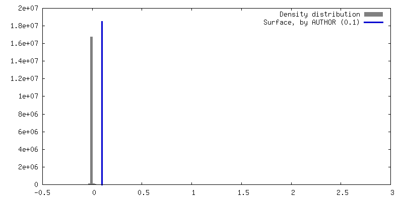
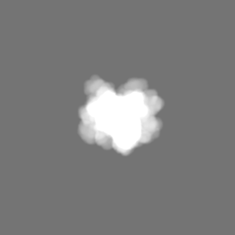
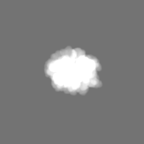
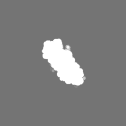
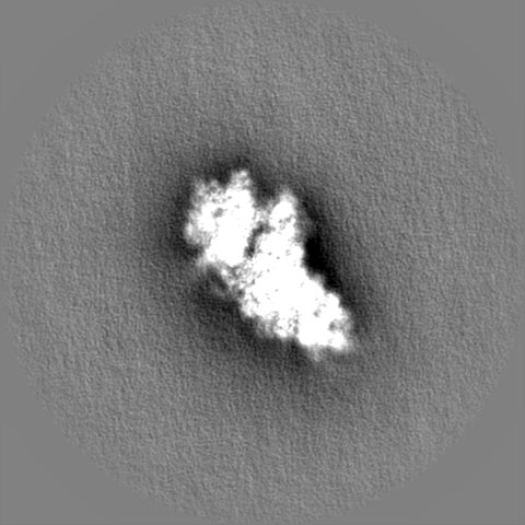
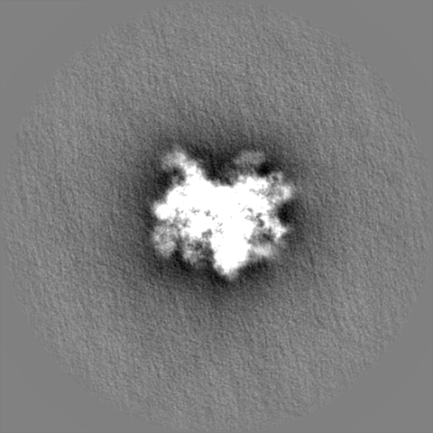
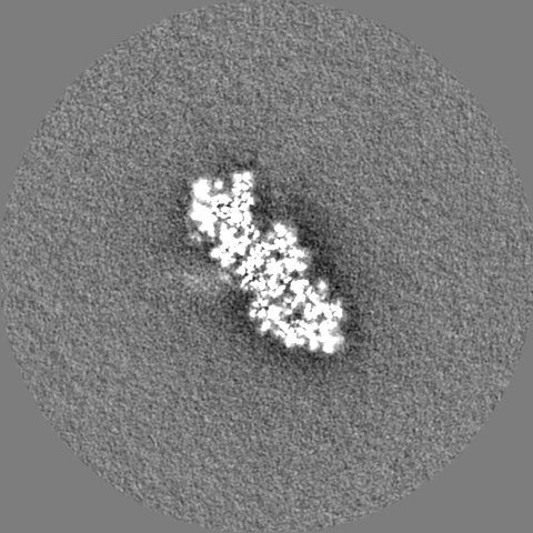
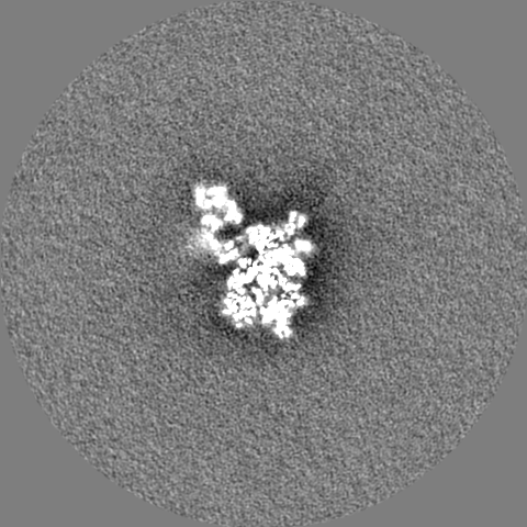
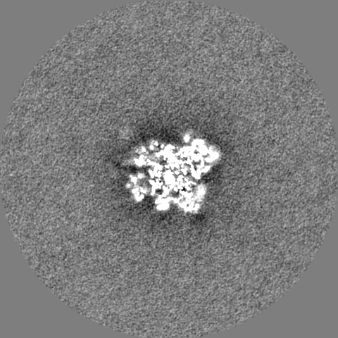
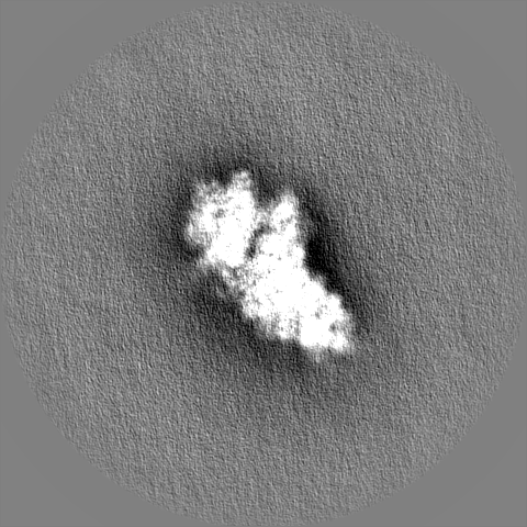
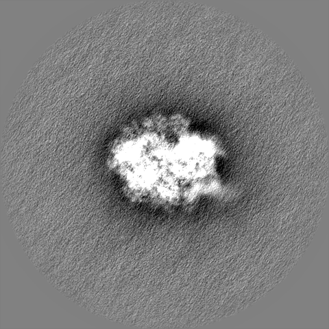
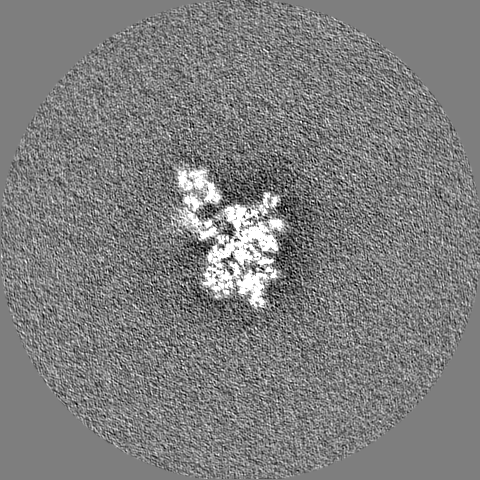
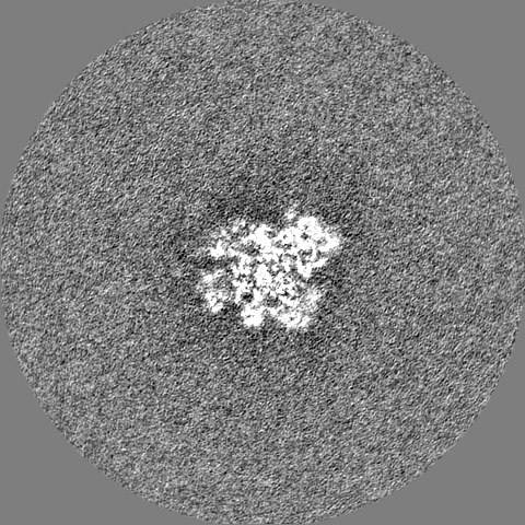
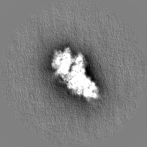
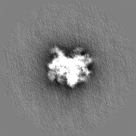
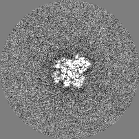
single particle reconstruction / cryo EM / Resolution: 3.2 Å
National Institutes of Health/Eunice Kennedy Shriver National Institute of Child Health & Human Development (NIH/NICHD)
(GM011378
United States
Citation
Journal: Nature / Year: 2022 Title: eIF5B and eIF1A reorient initiator tRNA to allow ribosomal subunit joining. Authors: Christopher P Lapointe / Rosslyn Grosely / Masaaki Sokabe / Carlos Alvarado / Jinfan Wang / Elizabeth Montabana / Nancy Villa / Byung-Sik Shin / Thomas E Dever / Christopher S Fraser / ...Authors: Christopher P Lapointe / Rosslyn Grosely / Masaaki Sokabe / Carlos Alvarado / Jinfan Wang / Elizabeth Montabana / Nancy Villa / Byung-Sik Shin / Thomas E Dever / Christopher S Fraser / Israel S Fernández / Joseph D Puglisi / Abstract: Translation initiation defines the identity and quantity of a synthesized protein. The process is dysregulated in many human diseases. A key commitment step is when the ribosomal subunits join at a ...Translation initiation defines the identity and quantity of a synthesized protein. The process is dysregulated in many human diseases. A key commitment step is when the ribosomal subunits join at a translation start site on a messenger RNA to form a functional ribosome. Here, we combined single-molecule spectroscopy and structural methods using an in vitro reconstituted system to examine how the human ribosomal subunits join. Single-molecule fluorescence revealed when the universally conserved eukaryotic initiation factors eIF1A and eIF5B associate with and depart from initiation complexes. Guided by single-molecule dynamics, we visualized initiation complexes that contained both eIF1A and eIF5B using single-particle cryo-electron microscopy. The resulting structure revealed how eukaryote-specific contacts between the two proteins remodel the initiation complex to orient the initiator aminoacyl-tRNA in a conformation compatible with ribosomal subunit joining. Collectively, our findings provide a quantitative and architectural framework for the molecular choreography orchestrated by eIF1A and eIF5B during translation initiation in humans.
In the structure databanks used in Yorodumi, some data are registered as the other names, "COVID-19 virus" and "2019-nCoV". Here are the details of the virus and the list of structure data.
Jan 31, 2019. EMDB accession codes are about to change! (news from PDBe EMDB page)
EMDB accession codes are about to change! (news from PDBe EMDB page)
The allocation of 4 digits for EMDB accession codes will soon come to an end. Whilst these codes will remain in use, new EMDB accession codes will include an additional digit and will expand incrementally as the available range of codes is exhausted. The current 4-digit format prefixed with “EMD-” (i.e. EMD-XXXX) will advance to a 5-digit format (i.e. EMD-XXXXX), and so on. It is currently estimated that the 4-digit codes will be depleted around Spring 2019, at which point the 5-digit format will come into force.
The EM Navigator/Yorodumi systems omit the EMD- prefix.
Related info.:Q: What is EMD? / ID/Accession-code notation in Yorodumi/EM Navigator
Yorodumi is a browser for structure data from EMDB, PDB, SASBDB, etc.
This page is also the successor to EM Navigator detail page, and also detail information page/front-end page for Omokage search.
The word "yorodu" (or yorozu) is an old Japanese word meaning "ten thousand". "mi" (miru) is to see.
Related info.:EMDB / PDB / SASBDB / Comparison of 3 databanks / Yorodumi Search / Aug 31, 2016. New EM Navigator & Yorodumi / Yorodumi Papers / Jmol/JSmol / Function and homology information / Changes in new EM Navigator and Yorodumi
 Movie
Movie Controller

 Yorodumi
 Open data
 Basic information
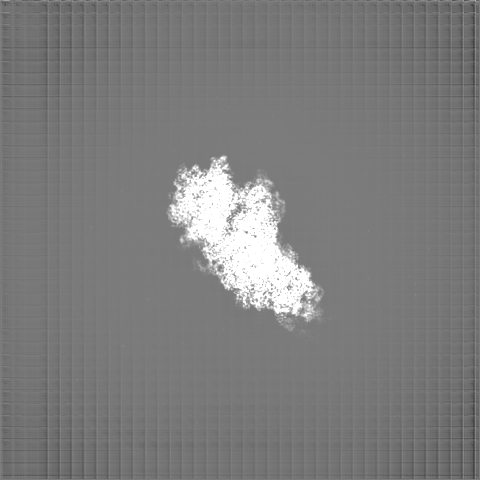
 Map data
 Sample
 Keywords
 Function and homology information
 Homo sapiens (human)
 Authors
 United States,
  Sweden, 3 items
  Citation
 Structure visualization
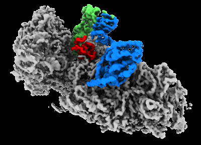
 Downloads & links
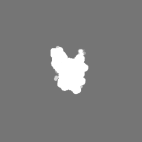
 emd_26067.png
 http://ftp.pdbj.org/pub/emdb/structures/EMD-26067


 Z (Sec.)
 Y (Row.)
 X (Col.)


 Sample components


 Processing
 Electron microscopy
 FIELD EMISSION GUN
