 Movie
Movie Controller

[English] 日本語
 Yorodumi
- EMDB-26003: Cryo-EM structure of the relaxin receptor RXFP1 in complex with h... -
+ Open data

- Basic information
| Entry |  | |||||||||
|---|---|---|---|---|---|---|---|---|---|---|
| Title | Cryo-EM structure of the relaxin receptor RXFP1 in complex with heterotrimeric Gs | |||||||||
 Map data Map data | Primary map used for model building, generated from a combine focused maps job in Phenix. | |||||||||
 Sample Sample |
| |||||||||
 Keywords Keywords | GPCR / Complex / Signaling / MEMBRANE PROTEIN | |||||||||
| Function / homology |  Function and homology information Function and homology informationlung connective tissue development / nipple morphogenesis / Relaxin receptors / parturition / myofibroblast differentiation / G protein-coupled peptide receptor activity / hormone binding / adenylate cyclase-activating G protein-coupled bile acid receptor signaling pathway / adenylate cyclase-activating serotonin receptor signaling pathway / regulation of skeletal muscle contraction ...lung connective tissue development / nipple morphogenesis / Relaxin receptors / parturition / myofibroblast differentiation / G protein-coupled peptide receptor activity / hormone binding / adenylate cyclase-activating G protein-coupled bile acid receptor signaling pathway / adenylate cyclase-activating serotonin receptor signaling pathway / regulation of skeletal muscle contraction / PKA activation in glucagon signalling / hair follicle placode formation / developmental growth / intracellular transport / D1 dopamine receptor binding / vascular endothelial cell response to laminar fluid shear stress / renal water homeostasis / Hedgehog 'off' state / activation of adenylate cyclase activity / adenylate cyclase-activating adrenergic receptor signaling pathway / cellular response to acidic pH / extracellular matrix organization / hormone-mediated signaling pathway / cellular response to glucagon stimulus / intracellular glucose homeostasis / positive regulation of insulin secretion involved in cellular response to glucose stimulus / adenylate cyclase activator activity / trans-Golgi network membrane / negative regulation of inflammatory response to antigenic stimulus / response to prostaglandin E / bone development / platelet aggregation / cognition / G protein-coupled receptor activity / G-protein beta/gamma-subunit complex binding / positive regulation of insulin secretion / Olfactory Signaling Pathway / Activation of the phototransduction cascade / G protein-coupled acetylcholine receptor signaling pathway / G beta:gamma signalling through PLC beta / Presynaptic function of Kainate receptors / Thromboxane signalling through TP receptor / sensory perception of smell / Activation of G protein gated Potassium channels / Inhibition of voltage gated Ca2+ channels via Gbeta/gamma subunits / G-protein activation / Glucagon signaling in metabolic regulation / Prostacyclin signalling through prostacyclin receptor / G beta:gamma signalling through CDC42 / Synthesis, secretion, and inactivation of Glucagon-like Peptide-1 (GLP-1) / G beta:gamma signalling through BTK / photoreceptor disc membrane / ADP signalling through P2Y purinoceptor 12 / Glucagon-type ligand receptors / Sensory perception of sweet, bitter, and umami (glutamate) taste / Adrenaline,noradrenaline inhibits insulin secretion / Vasopressin regulates renal water homeostasis via Aquaporins / Glucagon-like Peptide-1 (GLP1) regulates insulin secretion / G alpha (z) signalling events / cellular response to catecholamine stimulus / ADP signalling through P2Y purinoceptor 1 / ADORA2B mediated anti-inflammatory cytokines production / G beta:gamma signalling through PI3Kgamma / adenylate cyclase-activating dopamine receptor signaling pathway / Cooperation of PDCL (PhLP1) and TRiC/CCT in G-protein beta folding / positive regulation of cold-induced thermogenesis / GPER1 signaling / cellular response to prostaglandin E stimulus / heterotrimeric G-protein complex / G alpha (12/13) signalling events / Inactivation, recovery and regulation of the phototransduction cascade / G-protein beta-subunit binding / extracellular vesicle / sensory perception of taste / Thrombin signalling through proteinase activated receptors (PARs) / adenylate cyclase-activating G protein-coupled receptor signaling pathway / signaling receptor complex adaptor activity / retina development in camera-type eye / GTPase binding / fibroblast proliferation / G protein activity / Ca2+ pathway / High laminar flow shear stress activates signaling by PIEZO1 and PECAM1:CDH5:KDR in endothelial cells / G alpha (i) signalling events / G alpha (s) signalling events / G alpha (q) signalling events / phospholipase C-activating G protein-coupled receptor signaling pathway / Hydrolases; Acting on acid anhydrides; Acting on GTP to facilitate cellular and subcellular movement / Ras protein signal transduction / Extra-nuclear estrogen signaling / cell population proliferation / G protein-coupled receptor signaling pathway / lysosomal membrane / GTPase activity / synapse / GTP binding / protein-containing complex binding / signal transduction / extracellular exosome / membrane Similarity search - Function | |||||||||
| Biological species |  Homo sapiens (human) / Homo sapiens (human) /  | |||||||||
| Method | single particle reconstruction / cryo EM / Resolution: 3.2 Å | |||||||||
 Authors Authors | Erlandson SC / Rawson S | |||||||||
| Funding support |  United States, 2 items United States, 2 items
| |||||||||
 Citation Citation | Journal: Nat Chem Biol / Year: 2023 Title: The relaxin receptor RXFP1 signals through a mechanism of autoinhibition. Authors: Sarah C Erlandson / Shaun Rawson / James Osei-Owusu / Kelly P Brock / Xinyue Liu / Joao A Paulo / Julian Mintseris / Steven P Gygi / Debora S Marks / Xiaojing Cong / Andrew C Kruse /  Abstract: The relaxin family peptide receptor 1 (RXFP1) is the receptor for relaxin-2, an important regulator of reproductive and cardiovascular physiology. RXFP1 is a multi-domain G protein-coupled receptor ...The relaxin family peptide receptor 1 (RXFP1) is the receptor for relaxin-2, an important regulator of reproductive and cardiovascular physiology. RXFP1 is a multi-domain G protein-coupled receptor (GPCR) with an ectodomain consisting of a low-density lipoprotein receptor class A (LDLa) module and leucine-rich repeats. The mechanism of RXFP1 signal transduction is clearly distinct from that of other GPCRs, but remains very poorly understood. In the present study, we determine the cryo-electron microscopy structure of active-state human RXFP1, bound to a single-chain version of the endogenous agonist relaxin-2 and the heterotrimeric G protein. Evolutionary coupling analysis and structure-guided functional experiments reveal that RXFP1 signals through a mechanism of autoinhibition. Our results explain how an unusual GPCR family functions, providing a path to rational drug development targeting the relaxin receptors. #1: Journal: Biorxiv / Year: 2022Title: The relaxin receptor RXFP1 signals through a mechanism of autoinhibition Authors: Erlandson SC / Rawson S / Osei-Owusu J / Brock KP / Liu X / Paulo JA / Mintseris J / Gygi SP / Marks DS / Cong X / Kruse AC | |||||||||
| History |
|
- Structure visualization
| Supplemental images |
|---|

- Downloads & links
-EMDB archive
| Map data | emd_26003.map.gz | 68.1 MB | EMDB map data format | |
|---|---|---|---|---|
| Header (meta data) | emd-26003-v30.xmlemd-26003.xml | 31.9 KB 31.9 KB | Display Display | EMDB header |
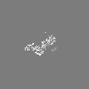
| Images |  emd_26003.png emd_26003.png | 43.5 KB | ||
| Filedesc metadata | emd-26003.cif.gz | 7.3 KB | ||
| Others | emd_26003_additional_1.map.gzemd_26003_additional_2.map.gzemd_26003_additional_3.map.gzemd_26003_additional_4.map.gzemd_26003_half_map_1.map.gzemd_26003_half_map_2.map.gz | 80.8 MB 80.2 MB 81 MB 80.8 MB 84.5 MB 84.5 MB | ||
| Archive directory |  http://ftp.pdbj.org/pub/emdb/structures/EMD-26003ftp://ftp.pdbj.org/pub/emdb/structures/EMD-26003 http://ftp.pdbj.org/pub/emdb/structures/EMD-26003ftp://ftp.pdbj.org/pub/emdb/structures/EMD-26003 | HTTPS FTP |
-Related structure data
| Related structure data |  7tmwMC M: atomic model generated by this map C: citing same article ( |
|---|---|
| Similar structure data |

-Links
| EMDB pages | EMDB (EBI/PDBe) / EMDataResource |
|---|---|
| Related items in Molecule of the Month |


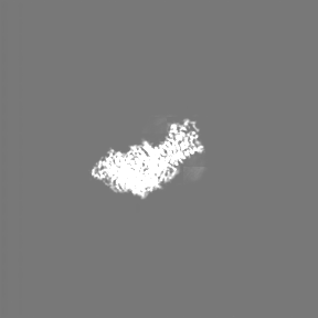
-Map
| File | Download / File: emd_26003.map.gz / Format: CCP4 / Size: 91.1 MB / Type: IMAGE STORED AS FLOATING POINT NUMBER (4 BYTES) | ||||||||||||||||||||||||||||||||||||
|---|---|---|---|---|---|---|---|---|---|---|---|---|---|---|---|---|---|---|---|---|---|---|---|---|---|---|---|---|---|---|---|---|---|---|---|---|---|
| Annotation | Primary map used for model building, generated from a combine focused maps job in Phenix. | ||||||||||||||||||||||||||||||||||||
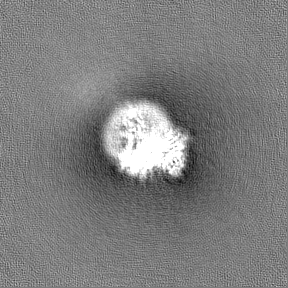
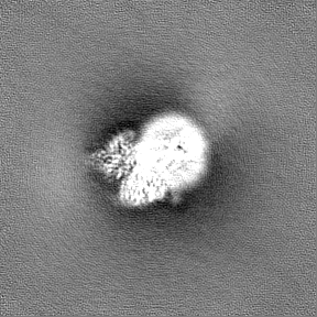
| Projections & slices | Image control
Images are generated by Spider. | ||||||||||||||||||||||||||||||||||||
| Voxel size | X=Y=Z: 1.06 Å | ||||||||||||||||||||||||||||||||||||
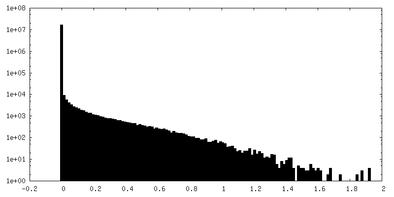
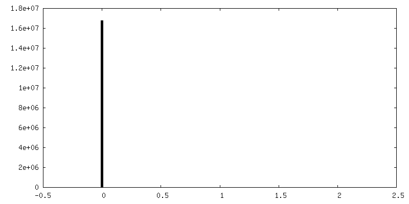
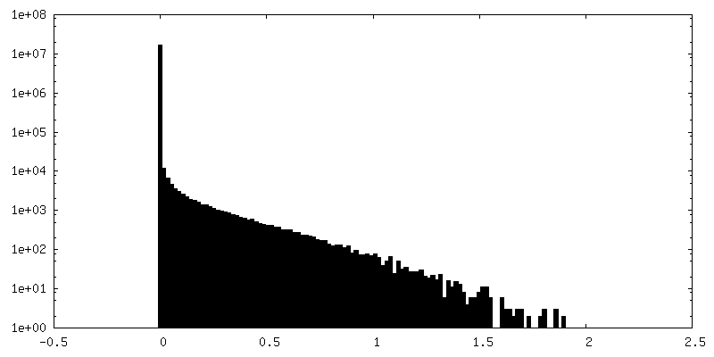
| Density |
| ||||||||||||||||||||||||||||||||||||
| Symmetry | Space group: 1 | ||||||||||||||||||||||||||||||||||||
| Details | EMDB XML:
|
 X (Sec.)
 Y (Row.)
 Z (Col.)


-Supplemental data
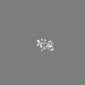
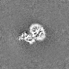
-Additional map: DeepEMhancer post-processed map generated after 3D focused classifications...
| File | emd_26003_additional_1.map | ||||||||||||
|---|---|---|---|---|---|---|---|---|---|---|---|---|---|
| Annotation | DeepEMhancer post-processed map generated after 3D focused classifications of transmembrane helix 1. | ||||||||||||
| Projections & Slices |
| ||||||||||||
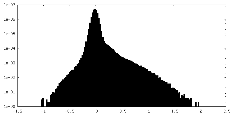
| Density Histograms |


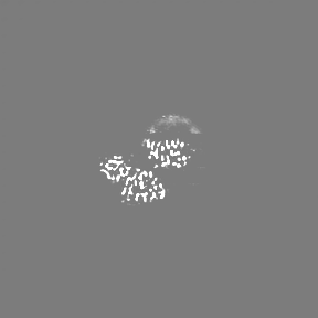
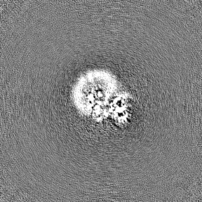
-Additional map: DeepEMhancer post-processed map generated after 3D focused classifications...
| File | emd_26003_additional_2.map | ||||||||||||
|---|---|---|---|---|---|---|---|---|---|---|---|---|---|
| Annotation | DeepEMhancer post-processed map generated after 3D focused classifications of extracellular loops. | ||||||||||||
| Projections & Slices |
| ||||||||||||
| Density Histograms |


-Additional map: DeepEMhancer post-processed map generated after 3D focused classifications...
| File | emd_26003_additional_3.map | ||||||||||||
|---|---|---|---|---|---|---|---|---|---|---|---|---|---|
| Annotation | DeepEMhancer post-processed map generated after 3D focused classifications of extracellular loops. | ||||||||||||
| Projections & Slices |
| ||||||||||||
| Density Histograms |


-Additional map: DeepEMhancer post-processed map generated after 3D focused classifications...
| File | emd_26003_additional_4.map | ||||||||||||
|---|---|---|---|---|---|---|---|---|---|---|---|---|---|
| Annotation | DeepEMhancer post-processed map generated after 3D focused classifications of helix8. | ||||||||||||
| Projections & Slices |
| ||||||||||||
| Density Histograms |


-Half map: Half-map from cryoSPARC refinement for map J8.
| File | emd_26003_half_map_1.map | ||||||||||||
|---|---|---|---|---|---|---|---|---|---|---|---|---|---|
| Annotation | Half-map from cryoSPARC refinement for map J8. | ||||||||||||
| Projections & Slices |
| ||||||||||||
| Density Histograms |


-Half map: Half-map from cryoSPARC refinement for map J8.
| File | emd_26003_half_map_2.map | ||||||||||||
|---|---|---|---|---|---|---|---|---|---|---|---|---|---|
| Annotation | Half-map from cryoSPARC refinement for map J8. | ||||||||||||
| Projections & Slices |
| ||||||||||||
| Density Histograms |


- Sample components
-Entire : Complex of the relaxin receptor RXFP1 and heterotrimeric Gs
| Entire | Name: Complex of the relaxin receptor RXFP1 and heterotrimeric Gs |
|---|---|
| Components |
|
-Supramolecule #1: Complex of the relaxin receptor RXFP1 and heterotrimeric Gs
| Supramolecule | Name: Complex of the relaxin receptor RXFP1 and heterotrimeric Gs type: complex / ID: 1 / Parent: 0 / Macromolecule list: all |
|---|---|
| Molecular weight | Theoretical: 180 KDa |
-Supramolecule #2: Relaxin receptor 1, Guanine nucleotide-binding protein G(s) subun...
| Supramolecule | Name: Relaxin receptor 1, Guanine nucleotide-binding protein G(s) subunit alpha isoforms short fusion type: complex / ID: 2 / Parent: 1 / Macromolecule list: #1 |
|---|---|
| Source (natural) | Organism: Homo sapiens (human) |
-Supramolecule #3: Guanine nucleotide-binding protein G(I)/G(S)/G(T) subunit beta-1,...
| Supramolecule | Name: Guanine nucleotide-binding protein G(I)/G(S)/G(T) subunit beta-1, Guanine nucleotide-binding protein G(I)/G(S)/G(O) subunit gamma-2 type: complex / ID: 3 / Parent: 1 / Macromolecule list: #2-#3 |
|---|---|
| Source (natural) | Organism: Homo sapiens (human) |
-Supramolecule #4: Camelid antibody VHH fragment Nb35
| Supramolecule | Name: Camelid antibody VHH fragment Nb35 / type: complex / ID: 4 / Parent: 1 / Macromolecule list: #4 |
|---|---|
| Source (natural) | Organism: |
-Macromolecule #1: Relaxin receptor 1, Guanine nucleotide-binding protein G(s) subun...
| Macromolecule | Name: Relaxin receptor 1, Guanine nucleotide-binding protein G(s) subunit alpha isoforms short fusion type: protein_or_peptide / ID: 1 / Number of copies: 1 / Enantiomer: LEVO |
|---|---|
| Source (natural) | Organism: Homo sapiens (human) |
| Molecular weight | Theoretical: 113.442891 KDa |
| Recombinant expression | Organism: Homo sapiens (human) |
| Sequence | String: DYKDDDDGGS LEVLFQGPGG SQDVKCSLGY FPCGNITKCL PQLLHCNGVD DCGNQADEDN CGDNNGWSLQ FDKYFASYYK MTSQYPFEA ETPECLVGSV PVQCLCQGLE LDCDETNLRA VPSVSSNVTA MSLQWNLIRK LPPDCFKNYH DLQKLYLQNN K ITSISIYA ...String: DYKDDDDGGS LEVLFQGPGG SQDVKCSLGY FPCGNITKCL PQLLHCNGVD DCGNQADEDN CGDNNGWSLQ FDKYFASYYK MTSQYPFEA ETPECLVGSV PVQCLCQGLE LDCDETNLRA VPSVSSNVTA MSLQWNLIRK LPPDCFKNYH DLQKLYLQNN K ITSISIYA FRGLNSLTKL YLSHNRITFL KPGVFEDLHR LEWLIIEDNH LSRISPPTFY GLNSLILLVL MNNVLTRLPD KP LCQHMPR LHWLDLEGNH IHNLRNLTFI SCSNLTVLVM RKNKINHLNE NTFAPLQKLD ELDLGSNKIE NLPPLIFKDL KEL SQLNLS YNPIQKIQAN QFDYLVKLKS LSLEGIEISN IQQRMFRPLM NLSHIYFKKF QYCGYAPHVR SCKPNTDGIS SLEN LLASI IQRVFVWVVS AVTCFGNIFV ICMRPYIRSE NKLYAMSIIS LCCADCLMGI YLFVIGGFDL KFRGEYNKHA QLWME STHC QLVGSLAILS TEVSVLLLTF LTLEKYICIV YPFRCVRPGK CRTITVLILI WITGFIVAFI PLSNKEFFKN YYGTNG VCF PLHSEDTESI GAQIYSVAIF LGINLAAFII IVFSYGSMFY SVHQSAITAT EIRNQVKKEM ILAKRFFFIV FTDALCW IP IFVVKFLSLL QVEIPGTITS WVVIFILPIN SALNPILYTL TTRPFKEMIH RFWYNYRQRK SMDSKGQKTY APSFIWVE M WPLQEMPPEL MKPDLNSKTE DQRNEEKAQR EANKKIEKQL QKDKQVYRAT HRLLLLGADN SGKSTIVKQM RIYHGGSGG SGGTSGIFET KFQVDKVNFH MFDVGGQRDE RRKWIQCFND VTAIIFVVDS SDYNRLQEAL NLFKSIWNNR WLRTISVILF LNKQDLLAE KVLAGKSKIE DYFPEFARYT TPEDATPEPG EDPRVTRAKY FIRDEFLRIS TASGDGRHYC YPHFTCAVDT E NARRIFND CRDIIQRMHL RQYELL UniProtKB: Relaxin receptor 1, Guanine nucleotide-binding protein G(s) subunit alpha isoforms short |
-Macromolecule #2: Guanine nucleotide-binding protein G(I)/G(S)/G(T) subunit beta-1
| Macromolecule | Name: Guanine nucleotide-binding protein G(I)/G(S)/G(T) subunit beta-1 type: protein_or_peptide / ID: 2 / Number of copies: 1 / Enantiomer: LEVO |
|---|---|
| Source (natural) | Organism: Homo sapiens (human) |
| Molecular weight | Theoretical: 38.534062 KDa |
| Recombinant expression | Organism:   Spodoptera frugiperda (fall armyworm) Spodoptera frugiperda (fall armyworm) |
| Sequence | String: MHHHHHHGSS GSELDQLRQE AEQLKNQIRD ARKACADATL SQITNNIDPV GRIQMRTRRT LRGHLAKIYA MHWGTDSRLL VSASQDGKL IIWDSYTTNK VHAIPLRSSW VMTCAYAPSG NYVACGGLDN ICSIYNLKTR EGNVRVSREL AGHTGYLSCC R FLDDNQIV ...String: MHHHHHHGSS GSELDQLRQE AEQLKNQIRD ARKACADATL SQITNNIDPV GRIQMRTRRT LRGHLAKIYA MHWGTDSRLL VSASQDGKL IIWDSYTTNK VHAIPLRSSW VMTCAYAPSG NYVACGGLDN ICSIYNLKTR EGNVRVSREL AGHTGYLSCC R FLDDNQIV TSSGDTTCAL WDIETGQQTT TFTGHTGDVM SLSLAPDTRL FVSGACDASA KLWDVREGMC RQTFTGHESD IN AICFFPN GNAFATGSDD ATCRLFDLRA DQELMTYSHD NIICGITSVS FSKSGRLLLA GYDDFNCNVW DALKADRAGV LAG HDNRVS CLGVTDDGMA VATGSWDSFL KIWN UniProtKB: Guanine nucleotide-binding protein G(I)/G(S)/G(T) subunit beta-1 |
-Macromolecule #3: Guanine nucleotide-binding protein G(I)/G(S)/G(O) subunit gamma-2
| Macromolecule | Name: Guanine nucleotide-binding protein G(I)/G(S)/G(O) subunit gamma-2 type: protein_or_peptide / ID: 3 / Number of copies: 1 / Enantiomer: LEVO |
|---|---|
| Source (natural) | Organism: Homo sapiens (human) |
| Molecular weight | Theoretical: 7.861143 KDa |
| Recombinant expression | Organism: Spodoptera frugiperda (fall armyworm) |
| Sequence | String: MASNNTASIA QARKLVEQLK MEANIDRIKV SKAAADLMAY CEAHAKEDPL LTPVPASENP FREKKFFCAI L UniProtKB: Guanine nucleotide-binding protein G(I)/G(S)/G(O) subunit gamma-2 |
-Macromolecule #4: Camelid antibody VHH fragment Nb35
| Macromolecule | Name: Camelid antibody VHH fragment Nb35 / type: protein_or_peptide / ID: 4 / Number of copies: 1 / Enantiomer: LEVO |
|---|---|
| Source (natural) | Organism: |
| Molecular weight | Theoretical: 17.444324 KDa |
| Recombinant expression | Organism:  |
| Sequence | String: QVQLQESGGG LVQPGGSLRL SCAASGFTFS NYKMNWVRQA PGKGLEWVSD ISQSGASISY TGSVKGRFTI SRDNAKNTLY LQMNSLKPE DTAVYYCARC PAPFTRDCFD VTSTTYAYRG QGTQVTVSSL EVLFQGPGHH HHHHHHGSED QVDPRLIDGK |
-Experimental details
-Structure determination
| Method | cryo EM |
|---|---|
 Processing Processing | single particle reconstruction |
| Aggregation state | particle |
-Sample preparation
| Concentration | 0.2 mg/mL |
|---|---|
| Buffer | pH: 7.5 |
| Grid | Model: Quantifoil R1.2/1.3 / Material: COPPER / Mesh: 400 / Pretreatment - Type: GLOW DISCHARGE / Pretreatment - Time: 60 sec. |
| Vitrification | Cryogen name: ETHANE / Chamber humidity: 100 % / Chamber temperature: 283.15 K / Instrument: FEI VITROBOT MARK IV |
- Electron microscopy
| Microscope | FEI TITAN KRIOS |
|---|---|
| Image recording | Film or detector model: GATAN K3 BIOQUANTUM (6k x 4k) / Average electron dose: 52.0 e/Å2 |
| Electron beam | Acceleration voltage: 300 kV / Electron source:  FIELD EMISSION GUN FIELD EMISSION GUN |
| Electron optics | Illumination mode: FLOOD BEAM / Imaging mode: BRIGHT FIELD / Nominal defocus max: 2.3000000000000003 µm / Nominal defocus min: 0.8 µm / Nominal magnification: 81000 |
| Sample stage | Specimen holder model: FEI TITAN KRIOS AUTOGRID HOLDER |
| Experimental equipment |  Model: Titan Krios / Image courtesy: FEI Company |
