 Movie
Movie Controller

[English] 日本語
 Yorodumi
- EMDB-25890: Single-molecule 3D density map of HIV cellular entry by liquid-ph... -
+ Open data

- Basic information
| Entry |  | ||||||||||||
|---|---|---|---|---|---|---|---|---|---|---|---|---|---|
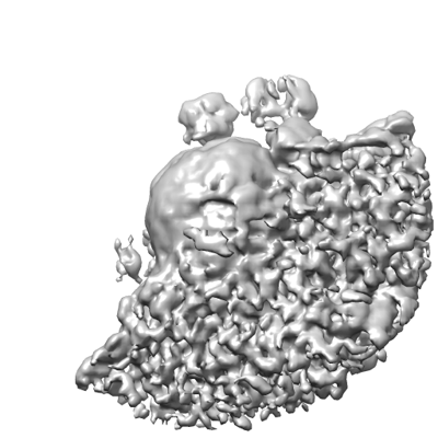
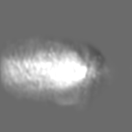
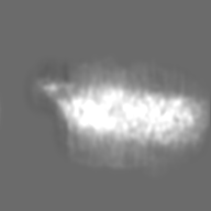
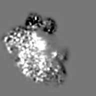
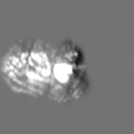
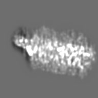
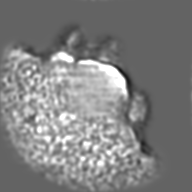
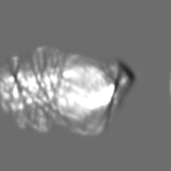
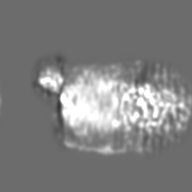
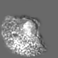
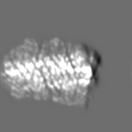
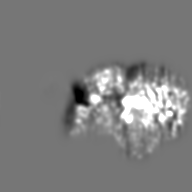
| Title | Single-molecule 3D density map of HIV cellular entry by liquid-phase electron tomography (particle #1) | ||||||||||||
 Map data Map data | |||||||||||||
 Sample Sample |
| ||||||||||||
 Keywords Keywords | HIV cellular entry / CELL INVASION | ||||||||||||
| Biological species |   Human immunodeficiency virus / Human immunodeficiency virus /  Homo sapiens (human) Homo sapiens (human) | ||||||||||||
| Method | electron tomography / Resolution: 74.0 Å | ||||||||||||
 Authors Authors | Kong L / Ren G | ||||||||||||
| Funding support |  United States, 3 items United States, 3 items
| ||||||||||||
 Citation Citation | Journal: Nat Commun / Year: 2023 Title: Facile hermetic TEM grid preparation for molecular imaging of hydrated biological samples at room temperature. Authors: Lingli Kong / Jianfang Liu / Meng Zhang / Zhuoyang Lu / Han Xue / Amy Ren / Jiankang Liu / Jinping Li / Wai Li Ling / Gang Ren /   Abstract: Although structures of vitrified supramolecular complexes have been determined at near-atomic resolution, elucidating in situ molecular structure in living cells remains a challenge. Here, we report ...Although structures of vitrified supramolecular complexes have been determined at near-atomic resolution, elucidating in situ molecular structure in living cells remains a challenge. Here, we report a straightforward liquid cell technique, originally developed for real-time visualization of dynamics at a liquid-gas interface using transmission electron microscopy, to image wet biological samples. Due to the scattering effects from the liquid phase, the micrographs display an amplitude contrast comparable to that observed in negatively stained samples. We succeed in resolving subunits within the protein complex GroEL imaged in a buffer solution at room temperature. Additionally, we capture various stages of virus cell entry, a process for which only sparse structural data exists due to their transient nature. To scrutinize the morphological details further, we used individual particle electron tomography for 3D reconstruction of each virus. These findings showcase this approach potential as an efficient, cost-effective complement to other microscopy technique in addressing biological questions at the molecular level. | ||||||||||||
| History |
|
- Structure visualization
| Supplemental images |
|---|
- Downloads & links
-EMDB archive
| Map data | emd_25890.map.gz | 23.9 MB |  EMDB map data format EMDB map data format | |
|---|---|---|---|---|
| Header (meta data) | emd-25890-v30.xmlemd-25890.xml | 12.6 KB 12.6 KB | Display Display | EMDB header |
| FSC (resolution estimation) | emd_25890_fsc.xml | 7.8 KB | Display | FSC data file |
| Images |  emd_25890.png emd_25890.png | 98.2 KB | ||
| Archive directory |  http://ftp.pdbj.org/pub/emdb/structures/EMD-25890ftp://ftp.pdbj.org/pub/emdb/structures/EMD-25890 http://ftp.pdbj.org/pub/emdb/structures/EMD-25890ftp://ftp.pdbj.org/pub/emdb/structures/EMD-25890 | HTTPS FTP |
-Related structure data
-Links
| EMDB pages | EMDB (EBI/PDBe) / EMDataResource |
|---|
-Map
| File | Download / File: emd_25890.map.gz / Format: CCP4 / Size: 27 MB / Type: IMAGE STORED AS FLOATING POINT NUMBER (4 BYTES) | ||||||||||||||||||||||||||||||||
|---|---|---|---|---|---|---|---|---|---|---|---|---|---|---|---|---|---|---|---|---|---|---|---|---|---|---|---|---|---|---|---|---|---|
| Projections & slices | Image control
Images are generated by Spider. | ||||||||||||||||||||||||||||||||
| Voxel size | X=Y=Z: 21.44 Å | ||||||||||||||||||||||||||||||||
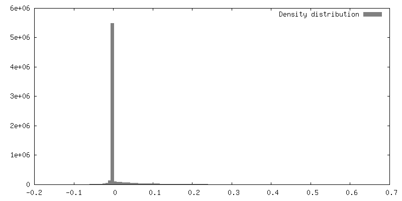
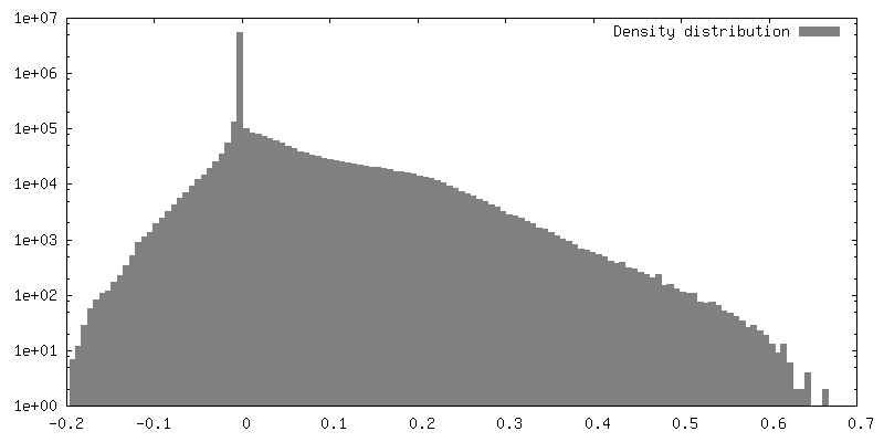
| Density |
| ||||||||||||||||||||||||||||||||
| Symmetry | Space group: 1 | ||||||||||||||||||||||||||||||||
| Details | EMDB XML:
|
 Z (Sec.)
 Y (Row.)
 X (Col.)


-Supplemental data
- Sample components
-Entire : Single molecule 3D map of HIV at its intermediate stage of cellul...
| Entire | Name: Single molecule 3D map of HIV at its intermediate stage of cellular entry (Particle #1, half embedded in cell), determined by liquid-phase electron tomography |
|---|---|
| Components |
|
-Supramolecule #1: Single molecule 3D map of HIV at its intermediate stage of cellul...
| Supramolecule | Name: Single molecule 3D map of HIV at its intermediate stage of cellular entry (Particle #1, half embedded in cell), determined by liquid-phase electron tomography type: organelle_or_cellular_component / ID: 1 / Parent: 0 Details: In the penetrating stage of viral entry, in which a virus half embeds into the cell membrane. |
|---|
-Supramolecule #2: HIV engineered with VSV G glycoprotein
| Supramolecule | Name: HIV engineered with VSV G glycoprotein / type: organelle_or_cellular_component / ID: 2 / Parent: 1 |
|---|---|
| Source (natural) | Organism: Human immunodeficiency virus |
-Supramolecule #3: cell membrane
| Supramolecule | Name: cell membrane / type: organelle_or_cellular_component / ID: 3 / Parent: 1 |
|---|---|
| Source (natural) | Organism: Homo sapiens (human) |
-Experimental details
-Structure determination
 Processing Processing | electron tomography |
|---|---|
| Aggregation state | cell |
-Sample preparation
| Buffer | pH: 7.4 Component:
Details: HeLa cells were grown in Gibco minimum essential media (MEM, containing 10 percent FBS) at 37 degree with 5 percent CO2, and virus-infected cells were prepared by incubating HeLa cells with ...Details: HeLa cells were grown in Gibco minimum essential media (MEM, containing 10 percent FBS) at 37 degree with 5 percent CO2, and virus-infected cells were prepared by incubating HeLa cells with virus at a ratio of 50 to 1 for 12 hours. | ||||||
|---|---|---|---|---|---|---|---|
| Sugar embedding | Material: sanwiched by two formvar films Details: sample solution (native liquid state, label-free, without any staining) was deposited and then sandwiched between two layers of 300 mesh TEM Formvar-coated copper grids at room temperature. | ||||||
| Grid | Model: Homemade / Material: COPPER / Mesh: 300 / Support film - Material: FORMVAR / Support film - topology: CONTINUOUS | ||||||
| Details | HeLa cells were grown in Gibco minimum essential media and virus-infected cells were prepared by incubating HeLa cells with virus at a ratio of 50:1 for 12 hours. | ||||||
| Cryo protectant | Liquid-phase | ||||||
| Sectioning | Other: NO SECTIONING |
- Electron microscopy
| Microscope | ZEISS LIBRA120PLUS |
|---|---|
| Specialist optics | Energy filter - Name: In-column Omega Filter / Energy filter - Slit width: 20 eV |
| Image recording | Film or detector model: GATAN ULTRASCAN 4000 (4k x 4k) / Number real images: 97 / Average electron dose: 1.0 e/Å2 Details: The total illumination electron doses were ~30 electron per angstrom square. |
| Electron beam | Acceleration voltage: 120 kV / Electron source: LAB6 |
| Electron optics | Calibrated defocus max: 11.0 µm / Illumination mode: FLOOD BEAM / Imaging mode: BRIGHT FIELD / Cs: 2.4 mm / Nominal defocus max: 11.0 µm / Nominal defocus min: 3.0 µm |
-Image processing
| Details | The tilt series of the targeted particles, including HeLa cells, viruses and nanoparticles, was reconstructed by IPET software (PLoS ONE, 2012, 7(1): e30249). In brief, a circular mask with a Gaussian edge was applied to each image, followed by 3D reconstruction via an iteration refinement process with a series of soft-boundary masks and filters. To display the objects with positive or negative contrast, a superimposed 3D density map was generated by combining the positive contour map of the final IPET 3D density maps with its negative contour map by using Chimera software. The resolution of the final 3D map was estimated based on the intra-Fourier shell correlation (FSC). Briefly, the aligned images were then split into two groups based on having an odd- or even-numbered tilting index so that two 3D reconstructions were generated to compute the FSC curves, and the frequency at which the FSC curve fell to a value of 0.5 was used to represent the resolution. |
|---|---|
| Final reconstruction | Algorithm: FOURIER SPACE / Resolution.type: BY AUTHOR / Resolution: 74.0 Å / Resolution method: FSC 0.5 CUT-OFF / Software - Name: IPET (ver. 1.0) / Software - details: PLoS ONE, 2012, 7(1): e30249 / Number images used: 97 |
| FSC plot (resolution estimation) | 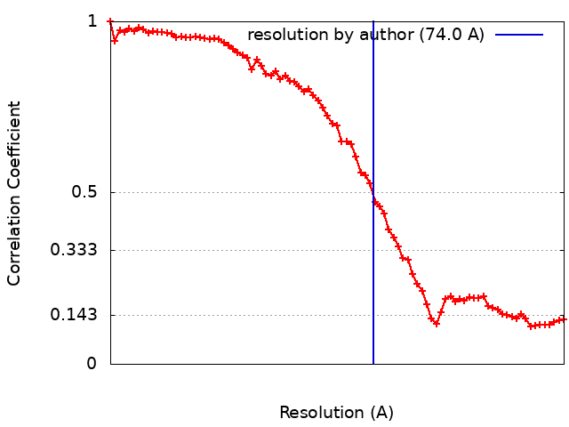 |
