National Institutes of Health/National Institute of General Medical Sciences (NIH/NIGMS)
R35GM133498
United States
National Institutes of Health/National Institute of General Medical Sciences (NIH/NIGMS)
T32GM119999
United States
National Institutes of Health/National Cancer Institute (NIH/NCI)
F99CA253730
United States
Citation
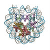
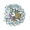
Journal: Nucleic Acids Res / Year: 2022 Title: Multivalent DNA and nucleosome acidic patch interactions specify VRK1 mitotic localization and activity. Authors: Gabrielle R Budziszewski / Yani Zhao / Cathy J Spangler / Katarzyna M Kedziora / Michael R Williams / Dalal N Azzam / Aleksandra Skrajna / Yuka Koyama / Andrew P Cesmat / Holly C Simmons / ...Authors: Gabrielle R Budziszewski / Yani Zhao / Cathy J Spangler / Katarzyna M Kedziora / Michael R Williams / Dalal N Azzam / Aleksandra Skrajna / Yuka Koyama / Andrew P Cesmat / Holly C Simmons / Eyla C Arteaga / Joshua D Strauss / Dmitri Kireev / Robert K McGinty / Abstract: A key role of chromatin kinases is to phosphorylate histone tails during mitosis to spatiotemporally regulate cell division. Vaccinia-related kinase 1 (VRK1) is a serine-threonine kinase that ...A key role of chromatin kinases is to phosphorylate histone tails during mitosis to spatiotemporally regulate cell division. Vaccinia-related kinase 1 (VRK1) is a serine-threonine kinase that phosphorylates histone H3 threonine 3 (H3T3) along with other chromatin-based targets. While structural studies have defined how several classes of histone-modifying enzymes bind to and function on nucleosomes, the mechanism of chromatin engagement by kinases is largely unclear. Here, we paired cryo-electron microscopy with biochemical and cellular assays to demonstrate that VRK1 interacts with both linker DNA and the nucleosome acidic patch to phosphorylate H3T3. Acidic patch binding by VRK1 is mediated by an arginine-rich flexible C-terminal tail. Homozygous missense and nonsense mutations of this acidic patch recognition motif in VRK1 are causative in rare adult-onset distal spinal muscular atrophy. We show that these VRK1 mutations interfere with nucleosome acidic patch binding, leading to mislocalization of VRK1 during mitosis, thus providing a potential new molecular mechanism for pathogenesis.
In the structure databanks used in Yorodumi, some data are registered as the other names, "COVID-19 virus" and "2019-nCoV". Here are the details of the virus and the list of structure data.
Jan 31, 2019. EMDB accession codes are about to change! (news from PDBe EMDB page)
EMDB accession codes are about to change! (news from PDBe EMDB page)
The allocation of 4 digits for EMDB accession codes will soon come to an end. Whilst these codes will remain in use, new EMDB accession codes will include an additional digit and will expand incrementally as the available range of codes is exhausted. The current 4-digit format prefixed with “EMD-” (i.e. EMD-XXXX) will advance to a 5-digit format (i.e. EMD-XXXXX), and so on. It is currently estimated that the 4-digit codes will be depleted around Spring 2019, at which point the 5-digit format will come into force.
The EM Navigator/Yorodumi systems omit the EMD- prefix.
Related info.:Q: What is EMD? / ID/Accession-code notation in Yorodumi/EM Navigator
Yorodumi is a browser for structure data from EMDB, PDB, SASBDB, etc.
This page is also the successor to EM Navigator detail page, and also detail information page/front-end page for Omokage search.
The word "yorodu" (or yorozu) is an old Japanese word meaning "ten thousand". "mi" (miru) is to see.
Related info.:EMDB / PDB / SASBDB / Comparison of 3 databanks / Yorodumi Search / Aug 31, 2016. New EM Navigator & Yorodumi / Yorodumi Papers / Jmol/JSmol / Function and homology information / Changes in new EM Navigator and Yorodumi
 Movie
Movie Controller


 Open data
 Basic information
 Map data
 Sample
 Homo sapiens (human) / synthetic construct (others)
 Authors
 United States, 3 items
  Citation
 Structure visualization
 Downloads & links
 EMDB map data format
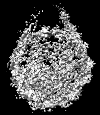 emd_25778.png
 http://ftp.pdbj.org/pub/emdb/structures/EMD-25778


 Z (Sec.)
 Y (Row.)
 X (Col.)


 Sample components
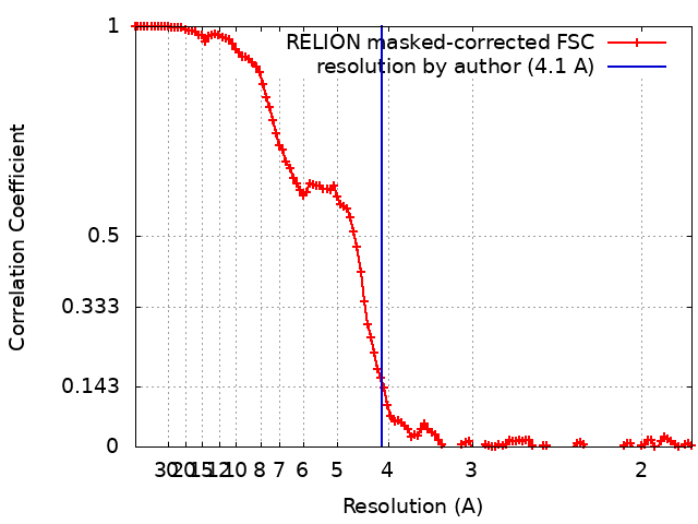
 Processing
 Electron microscopy
 FIELD EMISSION GUN