 Movie
Movie Controller


+ Open data

- Basic information
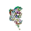
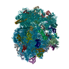
| Entry | Database: EMDB / ID: EMD-2567 | |||||||||
|---|---|---|---|---|---|---|---|---|---|---|
| Title | Structure of a bacterial Type IV secretion system | |||||||||
 Map data Map data | Structure of a bacterial Type IV secretion system | |||||||||
 Sample Sample |
| |||||||||
 Keywords Keywords | Type IV secretion system / R388 conjugative plasmid / Escherichia coli / negative stain electron microscopy | |||||||||
| Biological species |  | |||||||||
| Method | single particle reconstruction / negative staining / Resolution: 20.0 Å | |||||||||
 Authors Authors | Low HH / Gubellini F / Rivera-Calzada A / Braun N / Connery S / Dujeancourt A / Lu F / Redzej A / Fronzes R / Orlova EV / Waksman G | |||||||||
 Citation Citation | Journal: Nature / Year: 2014 Title: Structure of a type IV secretion system. Authors: Harry H Low / Francesca Gubellini / Angel Rivera-Calzada / Nathalie Braun / Sarah Connery / Annick Dujeancourt / Fang Lu / Adam Redzej / Rémi Fronzes / Elena V Orlova / Gabriel Waksman /   Abstract: Bacterial type IV secretion systems translocate virulence factors into eukaryotic cells, distribute genetic material between bacteria and have shown potential as a tool for the genetic modification ...Bacterial type IV secretion systems translocate virulence factors into eukaryotic cells, distribute genetic material between bacteria and have shown potential as a tool for the genetic modification of human cells. Given the complex choreography of the substrate through the secretion apparatus, the molecular mechanism of the type IV secretion system has proved difficult to dissect in the absence of structural data for the entire machinery. Here we use electron microscopy to reconstruct the type IV secretion system encoded by the Escherichia coli R388 conjugative plasmid. We show that eight proteins assemble in an intricate stoichiometric relationship to form an approximately 3 megadalton nanomachine that spans the entire cell envelope. The structure comprises an outer membrane-associated core complex connected by a central stalk to a substantial inner membrane complex that is dominated by a battery of 12 VirB4 ATPase subunits organized as side-by-side hexameric barrels. Our results show a secretion system with markedly different architecture, and consequently mechanism, to other known bacterial secretion systems. | |||||||||
| History |
|
- Structure visualization
| Movie |
 Movie viewer Movie viewer |
|---|---|
| Structure viewer | EM map: SurfViewMolmilJmol/JSmol |
| Supplemental images |

- Downloads & links
-EMDB archive
| Map data | emd_2567.map.gz | 802.3 KB | EMDB map data format | |
|---|---|---|---|---|
| Header (meta data) | emd-2567-v30.xmlemd-2567.xml | 8.9 KB 8.9 KB | Display Display | EMDB header |
| Images | EMD-2567.tif | 636.5 KB | ||
| Archive directory |  http://ftp.pdbj.org/pub/emdb/structures/EMD-2567ftp://ftp.pdbj.org/pub/emdb/structures/EMD-2567 http://ftp.pdbj.org/pub/emdb/structures/EMD-2567ftp://ftp.pdbj.org/pub/emdb/structures/EMD-2567 | HTTPS FTP |
-Related structure data
| Similar structure data |
|---|


-Links
| EMDB pages | EMDB (EBI/PDBe) / EMDataResource |
|---|
-Map
| File | Download / File: emd_2567.map.gz / Format: CCP4 / Size: 26.4 MB / Type: IMAGE STORED AS FLOATING POINT NUMBER (4 BYTES) | ||||||||||||||||||||||||||||||||||||||||||||||||||||||||||||||||||||
|---|---|---|---|---|---|---|---|---|---|---|---|---|---|---|---|---|---|---|---|---|---|---|---|---|---|---|---|---|---|---|---|---|---|---|---|---|---|---|---|---|---|---|---|---|---|---|---|---|---|---|---|---|---|---|---|---|---|---|---|---|---|---|---|---|---|---|---|---|---|
| Annotation | Structure of a bacterial Type IV secretion system | ||||||||||||||||||||||||||||||||||||||||||||||||||||||||||||||||||||
| Projections & slices | Image control
Images are generated by Spider. | ||||||||||||||||||||||||||||||||||||||||||||||||||||||||||||||||||||
| Voxel size | X=Y=Z: 3.3 Å | ||||||||||||||||||||||||||||||||||||||||||||||||||||||||||||||||||||
| Density |
| ||||||||||||||||||||||||||||||||||||||||||||||||||||||||||||||||||||
| Symmetry | Space group: 1 | ||||||||||||||||||||||||||||||||||||||||||||||||||||||||||||||||||||
| Details | EMDB XML:
CCP4 map header:
| ||||||||||||||||||||||||||||||||||||||||||||||||||||||||||||||||||||
 Z (Sec.)
 Y (Row.)
 X (Col.)


-Supplemental data
- Sample components
-Entire : Structure of a bacterial Type IV secretion system from Escherichi...
| Entire | Name: Structure of a bacterial Type IV secretion system from Escherichia coli R388 conjugative plasmid |
|---|---|
| Components |
|
-Supramolecule #1000: Structure of a bacterial Type IV secretion system from Escherichi...
| Supramolecule | Name: Structure of a bacterial Type IV secretion system from Escherichia coli R388 conjugative plasmid type: sample / ID: 1000 / Number unique components: 1 |
|---|---|
| Molecular weight | Experimental: 3 MDa / Theoretical: 3.4 MDa Method: Calculation based on stoichiometry determination of individual components within the complex |
-Macromolecule #1: Type IV secretion system from R388 conjugative plasmid
| Macromolecule | Name: Type IV secretion system from R388 conjugative plasmid type: protein_or_peptide / ID: 1 / Recombinant expression: Yes |
|---|---|
| Source (natural) | Organism: |
| Molecular weight | Experimental: 3 MDa / Theoretical: 3.4 MDa |
| Recombinant expression | Organism: |
-Experimental details
-Structure determination
| Method | negative staining |
|---|---|
 Processing Processing | single particle reconstruction |
| Aggregation state | particle |
-Sample preparation
| Concentration | 0.01 mg/mL |
|---|---|
| Buffer | pH: 8 Details: 50 mM Tris-HCl pH 8.0, 200 mM NaCl, 0.06 % w/v DM-NPG, 0.1 % w/v digitonin, 1 mM DTT and 1mM EDTA |
| Staining | Type: NEGATIVE Details: Grids with adsorbed protein floated on 2% w/v uranyl acetate for 30 seconds. |
| Grid | Details: Glow-discharged carbon-coated copper grids (Agar Scientific) |
| Vitrification | Cryogen name: NONE / Instrument: OTHER |
- Electron microscopy
| Microscope | FEI TECNAI F20 |
|---|---|
| Date | Jun 1, 2012 |
| Image recording | Category: CCD / Film or detector model: GATAN ULTRASCAN 4000 (4k x 4k) / Number real images: 693 / Average electron dose: 15 e/Å2 |
| Electron beam | Acceleration voltage: 200 kV / Electron source:  FIELD EMISSION GUN FIELD EMISSION GUN |
| Electron optics | Calibrated magnification: 45500 / Illumination mode: FLOOD BEAM / Imaging mode: BRIGHT FIELD / Nominal defocus max: 3.0 µm / Nominal defocus min: 1.0 µm / Nominal magnification: 45500 |
| Sample stage | Specimen holder model: SIDE ENTRY, EUCENTRIC |
| Experimental equipment |  Model: Tecnai F20 / Image courtesy: FEI Company |
-Image processing
| Final reconstruction | Applied symmetry - Point group: C1 (asymmetric) / Resolution.type: BY AUTHOR / Resolution: 20.0 Å / Resolution method: OTHER / Number images used: 3200 |
|---|---|
| Final two d classification | Number classes: 803 |
