 Movie
Movie Controller

[English] 日本語
 Yorodumi
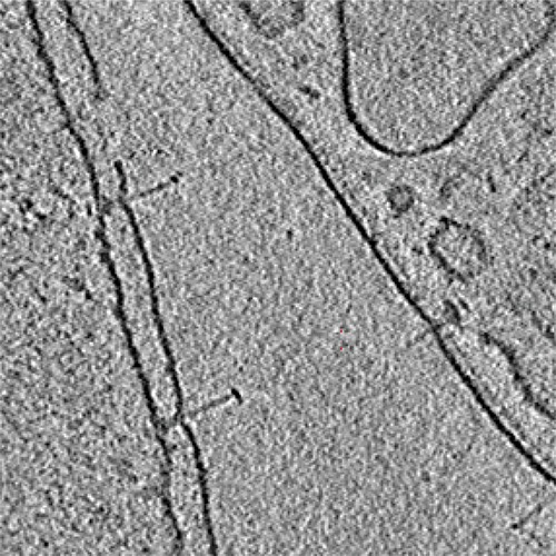
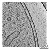
- EMDB-2480: Tomogram of Injectisome (wild type) from Salmonella typhiumurium ... -
+ Open data

- Basic information
| Entry | Database: EMDB / ID: EMD-2480 | |||||||||
|---|---|---|---|---|---|---|---|---|---|---|
| Title | Tomogram of Injectisome (wild type) from Salmonella typhiumurium (substrate trapped) in situ | |||||||||
 Map data Map data | Tomogram of substrate trapped type-3 secretion systems from Salmonella typhimurium | |||||||||
 Sample Sample |
| |||||||||
 Keywords Keywords | Injectisome / Pathogenic Type-3 Secretion System / Protein delivery machine / Salmonella typhimurium / cryo electron tomography | |||||||||
| Biological species |  Salmonella enterica subsp. enterica serovar Typhimurium (bacteria) Salmonella enterica subsp. enterica serovar Typhimurium (bacteria) | |||||||||
| Method | electron tomography / cryo EM | |||||||||
 Authors Authors | Radics J / Konigsmaier L / Marlovits TC | |||||||||
 Citation Citation | Journal: Nat Struct Mol Biol / Year: 2014 Title: Structure of a pathogenic type 3 secretion system in action. Authors: Julia Radics / Lisa Königsmaier / Thomas C Marlovits /   Abstract: Type 3 secretion systems use 3.5-megadalton syringe-like, membrane-embedded 'injectisomes', each containing an ~800-Å-long needle complex to connect intracellular compartments of infectious bacteria ...Type 3 secretion systems use 3.5-megadalton syringe-like, membrane-embedded 'injectisomes', each containing an ~800-Å-long needle complex to connect intracellular compartments of infectious bacteria and hosts. Here we identify requirements for substrate association with, transport through and exit from the injectisome of Salmonella enterica serovar Typhimurium. This guided the design of substrates that become trapped within the secretion path and enabled visualization of injectisomes in action in situ. We used cryo-EM to define the secretion path, providing a structural explanation as to why effector proteins must be unfolded during transport. Furthermore, trapping of a heterologous substrate in the needle prevents secretion of natural bacterial effectors. Together, the data reveal the path of protein secretion across multiple membranes and show that mechanisms rejecting unacceptable substrates can be undermined, and transport of bacterial effectors across an already assembled type 3 secretion system can be inhibited. | |||||||||
| History |
|
- Structure visualization
| Movie |
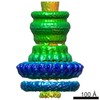
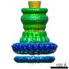
 Movie viewer Movie viewer |
|---|---|
| Structure viewer | EM map: SurfViewMolmilJmol/JSmol |
| Supplemental images |
- Downloads & links
-EMDB archive
| Map data | emd_2480.map.gz | 27.7 MB | EMDB map data format | |
|---|---|---|---|---|
| Header (meta data) | emd-2480-v30.xmlemd-2480.xml | 9.6 KB 9.6 KB | Display Display | EMDB header |
| Images | emd_2480.tif | 1.5 MB | ||
| Archive directory |  http://ftp.pdbj.org/pub/emdb/structures/EMD-2480ftp://ftp.pdbj.org/pub/emdb/structures/EMD-2480 http://ftp.pdbj.org/pub/emdb/structures/EMD-2480ftp://ftp.pdbj.org/pub/emdb/structures/EMD-2480 | HTTPS FTP |
-Related structure data
-Links
| EMDB pages | EMDB (EBI/PDBe) / EMDataResource |
|---|
-Map
| File | Download / File: emd_2480.map.gz / Format: CCP4 / Size: 52.2 MB / Type: IMAGE STORED AS SIGNED BYTE | ||||||||||||||||||||||||||||||||||||||||||||||||||||||||||||||||||||
|---|---|---|---|---|---|---|---|---|---|---|---|---|---|---|---|---|---|---|---|---|---|---|---|---|---|---|---|---|---|---|---|---|---|---|---|---|---|---|---|---|---|---|---|---|---|---|---|---|---|---|---|---|---|---|---|---|---|---|---|---|---|---|---|---|---|---|---|---|---|
| Annotation | Tomogram of substrate trapped type-3 secretion systems from Salmonella typhimurium | ||||||||||||||||||||||||||||||||||||||||||||||||||||||||||||||||||||
| Voxel size | X=Y=Z: 19.88 Å | ||||||||||||||||||||||||||||||||||||||||||||||||||||||||||||||||||||
| Density |
| ||||||||||||||||||||||||||||||||||||||||||||||||||||||||||||||||||||
| Symmetry | Space group: 1 | ||||||||||||||||||||||||||||||||||||||||||||||||||||||||||||||||||||
| Details | EMDB XML:
CCP4 map header:
| ||||||||||||||||||||||||||||||||||||||||||||||||||||||||||||||||||||
-Supplemental data
- Sample components
-Entire : Type-3 secretion system from Salmonella typhimurium in situ (subs...
| Entire | Name: Type-3 secretion system from Salmonella typhimurium in situ (substrate trapped): Visualization of unfolded protein transport across membranes |
|---|---|
| Components |
|
-Supramolecule #1000: Type-3 secretion system from Salmonella typhimurium in situ (subs...
| Supramolecule | Name: Type-3 secretion system from Salmonella typhimurium in situ (substrate trapped): Visualization of unfolded protein transport across membranes type: sample / ID: 1000 / Oligomeric state: 1 / Number unique components: 1 |
|---|---|
| Molecular weight | Theoretical: 3.5 MDa |
-Supramolecule #1: type 3 secretion system
| Supramolecule | Name: type 3 secretion system / type: organelle_or_cellular_component / ID: 1 / Name.synonym: injectisome / Recombinant expression: No / Database: NCBI |
|---|---|
| Source (natural) | Organism: Salmonella enterica subsp. enterica serovar Typhimurium (bacteria) Strain: SB905 / Location in cell: Plasma membrane |
| Molecular weight | Theoretical: 3.5 MDa |
-Experimental details
-Structure determination
| Method | cryo EM |
|---|---|
 Processing Processing | electron tomography |
| Aggregation state | cell |
-Sample preparation
| Buffer | pH: 8 / Details: 10 mM Tris, 5 mM EDTA |
|---|---|
| Grid | Details: 400 mesh Mo-Grid Quantifoil R2/1 |
| Vitrification | Cryogen name: ETHANE / Chamber humidity: 65 % / Chamber temperature: 91 K / Instrument: LEICA EM GP Method: 1. grids glowdischarged (300sec/20mAmp) 2. sample 5ul applied for 5sec (cell settling) 3. blotting for 2sec/distance 189 |
- Electron microscopy
| Microscope | FEI POLARA 300 |
|---|---|
| Temperature | Min: 83 K / Max: 108 K / Average: 93 K |
| Alignment procedure | Legacy - Astigmatism: objective lens astigmatism was corrected at appr 200.000 magnification |
| Date | Mar 2, 2012 |
| Image recording | Category: CCD / Film or detector model: GATAN ULTRASCAN 4000 (4k x 4k) / Digitization - Sampling interval: 15 µm / Number real images: 60 / Average electron dose: 100 e/Å2 / Details: Image acquisition using SerialEM |
| Electron beam | Acceleration voltage: 300 kV / Electron source:  FIELD EMISSION GUN FIELD EMISSION GUN |
| Electron optics | Calibrated magnification: 30120 / Illumination mode: FLOOD BEAM / Imaging mode: BRIGHT FIELD / Cs: 2 mm / Nominal defocus max: 9.0 µm / Nominal magnification: 23000 |
| Sample stage | Specimen holder: liquid nitrogen cooled / Specimen holder model: GATAN HELIUM / Tilt series - Axis1 - Max angle: 60 ° / Tilt series - Axis1 - Angle increment: 2 ° |
| Experimental equipment |  Model: Tecnai Polara / Image courtesy: FEI Company |
-Image processing
| Details | IMOD (weighted back-projection) |
|---|---|
| Final reconstruction | Algorithm: OTHER / Software - Name: IMOD / Number images used: 60 |
