National Institutes of Health/National Institute Of Allergy and Infectious Diseases (NIH/NIAID)
AI094386/DE028583/DE025567
United States
Citation
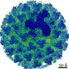
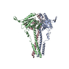
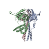
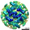
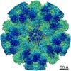
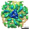
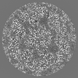
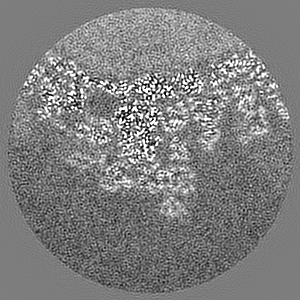
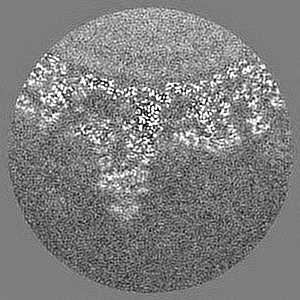
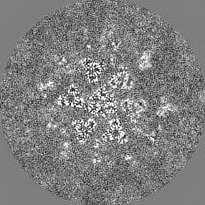
Journal: Nat Microbiol / Year: 2021 Title: Bluetongue virus capsid protein VP5 perforates membranes at low endosomal pH during viral entry. Authors: Xian Xia / Weining Wu / Yanxiang Cui / Polly Roy / Z Hong Zhou / Abstract: Bluetongue virus (BTV) is a non-enveloped virus and causes substantial morbidity and mortality in ruminants such as sheep. Fashioning a receptor-binding protein (VP2) and a membrane penetration ...Bluetongue virus (BTV) is a non-enveloped virus and causes substantial morbidity and mortality in ruminants such as sheep. Fashioning a receptor-binding protein (VP2) and a membrane penetration protein (VP5) on the surface, BTV releases its genome-containing core (VP3 and VP7) into the host cell cytosol after perforation of the endosomal membrane. Unlike enveloped ones, the entry mechanisms of non-enveloped viruses into host cells remain poorly understood. Here we applied single-particle cryo-electron microscopy, cryo-electron tomography and structure-guided functional assays to characterize intermediate states of BTV cell entry in endosomes. Four structures of BTV at the resolution range of 3.4-3.9 Å show the different stages of structural rearrangement of capsid proteins on exposure to low pH, including conformational changes of VP5, stepwise detachment of VP2 and a small shift of VP7. In detail, sensing of the low-pH condition by the VP5 anchor domain triggers three major VP5 actions: projecting the hidden dagger domain, converting a surface loop to a protonated β-hairpin that anchors VP5 to the core and stepwise refolding of the unfurling domains into a six-helix stalk. Cryo-electron tomography structures of BTV interacting with liposomes show a length decrease of the VP5 stalk from 19.5 to 15.5 nm after its insertion into the membrane. Our structures, functional assays and structure-guided mutagenesis experiments combined indicate that this stalk, along with dagger domain and the WHXL motif, creates a single pore through the endosomal membrane that enables the viral core to enter the cytosol. Our study unveils the detailed mechanisms of BTV membrane penetration and showcases general methods to study cell entry of other non-enveloped viruses.
History
Deposition
Aug 13, 2021
-
Header (metadata) release
Nov 3, 2021
-
Map release
Nov 3, 2021
-
Update
Nov 10, 2021
-
Current status
Nov 10, 2021
Processing site: RCSB / Status: Released
-
Structure visualization
Movie
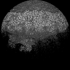
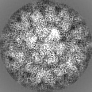
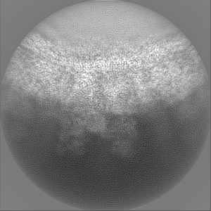
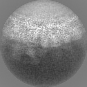
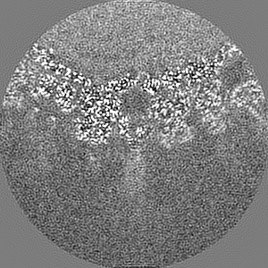
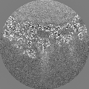
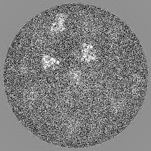
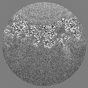
Surface view with section colored by density value
Model: PELCO Ultrathin Carbon with Lacey Carbon / Material: COPPER / Mesh: 400 / Support film - Material: CARBON / Support film - topology: CONTINUOUS / Pretreatment - Type: GLOW DISCHARGE / Pretreatment - Atmosphere: AIR
Vitrification
Cryogen name: ETHANE / Chamber humidity: 100 % / Instrument: FEI VITROBOT MARK IV
-
Electron microscopy
Microscope
FEI TITAN KRIOS
Specialist optics
Energy filter - Name: GIF Quantum LS / Energy filter - Slit width: 20 eV
Image recording
Film or detector model: GATAN K2 SUMMIT (4k x 4k) / Detector mode: SUPER-RESOLUTION / Number grids imaged: 1 / Number real images: 3609 / Average exposure time: 8.0 sec. / Average electron dose: 50.0 e/Å2
Electron beam
Acceleration voltage: 300 kV / Electron source: FIELD EMISSION GUN
In the structure databanks used in Yorodumi, some data are registered as the other names, "COVID-19 virus" and "2019-nCoV". Here are the details of the virus and the list of structure data.
Jan 31, 2019. EMDB accession codes are about to change! (news from PDBe EMDB page)
EMDB accession codes are about to change! (news from PDBe EMDB page)
The allocation of 4 digits for EMDB accession codes will soon come to an end. Whilst these codes will remain in use, new EMDB accession codes will include an additional digit and will expand incrementally as the available range of codes is exhausted. The current 4-digit format prefixed with “EMD-” (i.e. EMD-XXXX) will advance to a 5-digit format (i.e. EMD-XXXXX), and so on. It is currently estimated that the 4-digit codes will be depleted around Spring 2019, at which point the 5-digit format will come into force.
The EM Navigator/Yorodumi systems omit the EMD- prefix.
Related info.:Q: What is EMD? / ID/Accession-code notation in Yorodumi/EM Navigator
Yorodumi is a browser for structure data from EMDB, PDB, SASBDB, etc.
This page is also the successor to EM Navigator detail page, and also detail information page/front-end page for Omokage search.
The word "yorodu" (or yorozu) is an old Japanese word meaning "ten thousand". "mi" (miru) is to see.
Related info.:EMDB / PDB / SASBDB / Comparison of 3 databanks / Yorodumi Search / Aug 31, 2016. New EM Navigator & Yorodumi / Yorodumi Papers / Jmol/JSmol / Function and homology information / Changes in new EM Navigator and Yorodumi
 Movie
Movie Controller

 Yorodumi
 Open data
 Basic information
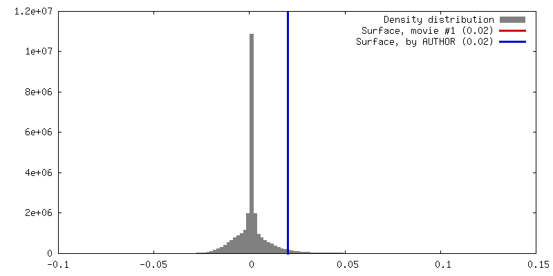
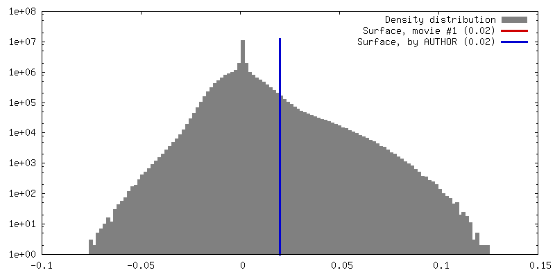
 Map data
 Sample
 Function and homology information
 Bluetongue virus (serotype 1 / isolate South Africa)
 Authors
 United States, 1 items
  Citation
 Structure visualization
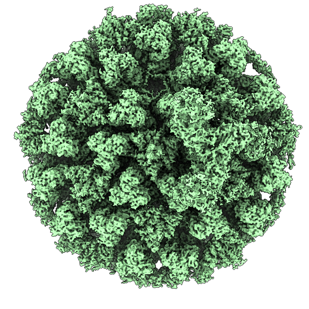
 Downloads & links
 emd_24687.png
 http://ftp.pdbj.org/pub/emdb/structures/EMD-24687
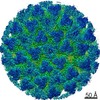


 Z (Sec.)
 Y (Row.)
 X (Col.)


 Sample components
 Processing
 Electron microscopy
 FIELD EMISSION GUN
