 Movie
Movie Controller


+ Open data

- Basic information
| Entry | Database: EMDB / ID: EMD-24496 | |||||||||
|---|---|---|---|---|---|---|---|---|---|---|
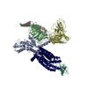
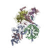
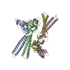
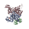
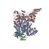
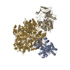
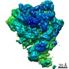
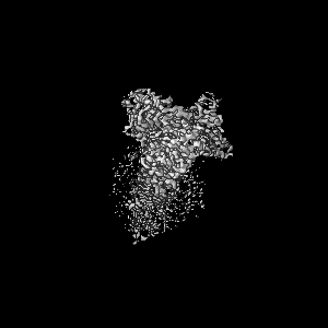
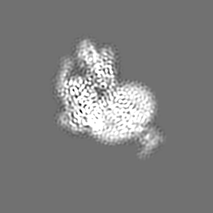
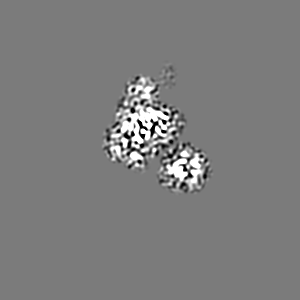
| Title | Structure of CX3CL1-US28-G11iN18-scFv16 in TL-state | |||||||||
 Map data Map data | Full sharpen map. | |||||||||
 Sample Sample |
| |||||||||
 Keywords Keywords | Viral GPCR / HCMV / cytomegalovirus / G protein complex / GDP-bound state / MEMBRANE PROTEIN | |||||||||
| Function / homology |  Function and homology information Function and homology informationCXCR1 chemokine receptor binding / positive regulation of calcium-independent cell-cell adhesion / negative regulation of interleukin-1 alpha production / leukocyte adhesive activation / CX3C chemokine receptor binding / negative regulation of glutamate receptor signaling pathway / regulation of melanocyte differentiation / autocrine signaling / lymphocyte chemotaxis / synapse pruning ...CXCR1 chemokine receptor binding / positive regulation of calcium-independent cell-cell adhesion / negative regulation of interleukin-1 alpha production / leukocyte adhesive activation / CX3C chemokine receptor binding / negative regulation of glutamate receptor signaling pathway / regulation of melanocyte differentiation / autocrine signaling / lymphocyte chemotaxis / synapse pruning / positive regulation of microglial cell migration / regulation of lipopolysaccharide-mediated signaling pathway / negative regulation of microglial cell activation / negative regulation of hippocampal neuron apoptotic process / negative regulation of neuron migration / positive regulation of transforming growth factor beta1 production / phospholipase C-activating G protein-coupled acetylcholine receptor signaling pathway / endothelin receptor signaling pathway / Fatty Acids bound to GPR40 (FFAR1) regulate insulin secretion / Acetylcholine regulates insulin secretion / developmental pigmentation / CCR chemokine receptor binding / phospholipase C-activating dopamine receptor signaling pathway / leukocyte migration involved in inflammatory response / microglial cell proliferation / positive regulation of actin filament bundle assembly / cellular response to pH / PLC beta mediated events / entrainment of circadian clock / cranial skeletal system development / integrin activation / C-C chemokine receptor activity / eosinophil chemotaxis / C-C chemokine binding / leukocyte chemotaxis / angiogenesis involved in wound healing / chemokine activity / Chemokine receptors bind chemokines / negative regulation of interleukin-1 beta production / positive regulation of cell-matrix adhesion / symbiont-mediated transformation of host cell / positive regulation of neuroblast proliferation / positive chemotaxis / neuron remodeling / ligand-gated ion channel signaling pathway / chemoattractant activity / negative regulation of interleukin-6 production / macrophage chemotaxis / phototransduction, visible light / negative regulation of cell-substrate adhesion / negative regulation of apoptotic signaling pathway / negative regulation of tumor necrosis factor production / action potential / regulation of neurogenesis / negative regulation of extrinsic apoptotic signaling pathway in absence of ligand / photoreceptor outer segment / enzyme regulator activity / extrinsic apoptotic signaling pathway in absence of ligand / cell projection / neutrophil chemotaxis / positive regulation of smooth muscle cell proliferation / Turbulent (oscillatory, disturbed) flow shear stress activates signaling by PIEZO1 and integrins in endothelial cells / response to ischemia / negative regulation of cell migration / positive regulation of release of sequestered calcium ion into cytosol / chemokine-mediated signaling pathway / cell chemotaxis / skeletal system development / calcium-mediated signaling / defense response / microglial cell activation / neuron cellular homeostasis / cell-cell adhesion / positive regulation of neuron projection development / regulation of synaptic plasticity / G protein-coupled receptor binding / integrin binding / regulation of blood pressure / chemotaxis / G-protein beta/gamma-subunit complex binding / cytokine-mediated signaling pathway / adenylate cyclase-modulating G protein-coupled receptor signaling pathway / positive regulation of angiogenesis / positive regulation of insulin secretion / Olfactory Signaling Pathway / Activation of the phototransduction cascade / positive regulation of inflammatory response / G protein-coupled acetylcholine receptor signaling pathway / G beta:gamma signalling through PLC beta / Presynaptic function of Kainate receptors / Thromboxane signalling through TP receptor / Activation of G protein gated Potassium channels / Inhibition of voltage gated Ca2+ channels via Gbeta/gamma subunits / G-protein activation / Glucagon signaling in metabolic regulation / Prostacyclin signalling through prostacyclin receptor / G beta:gamma signalling through CDC42 / Synthesis, secretion, and inactivation of Glucagon-like Peptide-1 (GLP-1) / G beta:gamma signalling through BTK / photoreceptor disc membrane Similarity search - Function | |||||||||
| Biological species |  Homo sapiens (human) / Homo sapiens (human) /    Human betaherpesvirus 5 / Human cytomegalovirus Human betaherpesvirus 5 / Human cytomegalovirus | |||||||||
| Method | single particle reconstruction / cryo EM / Resolution: 4.0 Å | |||||||||
 Authors Authors | Tsutsumi N / Maeda S | |||||||||
| Funding support |  United States, 2 items United States, 2 items
| |||||||||
 Citation Citation | Journal: Sci Adv / Year: 2022 Title: Atypical structural snapshots of human cytomegalovirus GPCR interactions with host G proteins Authors: Tsutsumi N / Maeda S / Qu Q / Voegele M / Jude KM / Suomivuori CM / Panova O / Waghray D / Kato HE / Velasco A / Dror RO / Skiniotis G / Kobilka BK / Garcia KC | |||||||||
| History |
|
- Structure visualization
| Movie |
Movie viewer |
|---|---|
| Structure viewer | EM map: SurfViewMolmilJmol/JSmol |
| Supplemental images |

- Downloads & links
-EMDB archive
| Map data | emd_24496.map.gz | 8.8 MB | EMDB map data format | |
|---|---|---|---|---|
| Header (meta data) | emd-24496-v30.xmlemd-24496.xml | 22.7 KB 22.7 KB | Display Display | EMDB header |
| Images |  emd_24496.png emd_24496.png | 87.9 KB | ||
| Filedesc metadata | emd-24496.cif.gz | 7.5 KB | ||
| Archive directory |  http://ftp.pdbj.org/pub/emdb/structures/EMD-24496ftp://ftp.pdbj.org/pub/emdb/structures/EMD-24496 http://ftp.pdbj.org/pub/emdb/structures/EMD-24496ftp://ftp.pdbj.org/pub/emdb/structures/EMD-24496 | HTTPS FTP |
-Related structure data
| Related structure data | 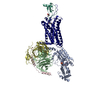 7rkfMC  7rkmC  7rknC  7rkxC  7rkyC M: atomic model generated by this map C: citing same article ( |
|---|---|
| Similar structure data |


-Links
| EMDB pages | EMDB (EBI/PDBe) / EMDataResource |
|---|---|
| Related items in Molecule of the Month |


-Map
| File | Download / File: emd_24496.map.gz / Format: CCP4 / Size: 103 MB / Type: IMAGE STORED AS FLOATING POINT NUMBER (4 BYTES) | ||||||||||||||||||||||||||||||||||||||||||||||||||||||||||||
|---|---|---|---|---|---|---|---|---|---|---|---|---|---|---|---|---|---|---|---|---|---|---|---|---|---|---|---|---|---|---|---|---|---|---|---|---|---|---|---|---|---|---|---|---|---|---|---|---|---|---|---|---|---|---|---|---|---|---|---|---|---|
| Annotation | Full sharpen map. | ||||||||||||||||||||||||||||||||||||||||||||||||||||||||||||
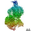
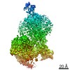
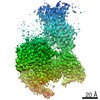
| Projections & slices | Image control
Images are generated by Spider. | ||||||||||||||||||||||||||||||||||||||||||||||||||||||||||||
| Voxel size | X=Y=Z: 0.85 Å | ||||||||||||||||||||||||||||||||||||||||||||||||||||||||||||
| Density |
| ||||||||||||||||||||||||||||||||||||||||||||||||||||||||||||
| Symmetry | Space group: 1 | ||||||||||||||||||||||||||||||||||||||||||||||||||||||||||||
| Details | EMDB XML:
CCP4 map header:
| ||||||||||||||||||||||||||||||||||||||||||||||||||||||||||||
 Z (Sec.)
 Y (Row.)
 X (Col.)


-Supplemental data
- Sample components
+Entire : CX3CL1-US28-G11iN18-scFv16 complex
+Supramolecule #1: CX3CL1-US28-G11iN18-scFv16 complex
+Supramolecule #2: G11iN18 heterotrimer
+Supramolecule #3: scFv16
+Supramolecule #4: CX3CL1-US28
+Macromolecule #1: Guanine nucleotide-binding protein subunit alpha-11
 Trichoplusia ni (cabbage looper)
+Macromolecule #2: Guanine nucleotide-binding protein G(I)/G(S)/G(T) subunit beta-1
+Macromolecule #3: Guanine nucleotide-binding protein G(I)/G(S)/G(O) subunit gamma-2
+Macromolecule #4: Antibody fragment scFv16
+Macromolecule #5: Fractalkine
+Macromolecule #6: G-protein coupled receptor homolog US28
+Macromolecule #7: GUANOSINE-5'-DIPHOSPHATE

+Macromolecule #8: 2-acetamido-2-deoxy-beta-D-glucopyranose

-Experimental details
-Structure determination
| Method | cryo EM |
|---|---|
 Processing Processing | single particle reconstruction |
| Aggregation state | particle |
-Sample preparation
| Concentration | 25 mg/mL | ||||||||||||
|---|---|---|---|---|---|---|---|---|---|---|---|---|---|
| Buffer | pH: 7.2 Component:
| ||||||||||||
| Grid | Model: Quantifoil R1.2/1.3 / Material: GOLD / Mesh: 200 / Pretreatment - Type: GLOW DISCHARGE | ||||||||||||
| Vitrification | Cryogen name: ETHANE / Chamber humidity: 100 % / Chamber temperature: 295 K / Instrument: FEI VITROBOT MARK IV / Details: 1 s blotting before plunging. |
- Electron microscopy
| Microscope | FEI TITAN KRIOS |
|---|---|
| Image recording | Film or detector model: GATAN K3 (6k x 4k) / Detector mode: SUPER-RESOLUTION / Number grids imaged: 1 / Number real images: 1330 / Average electron dose: 67.0 e/Å2 |
| Electron beam | Acceleration voltage: 300 kV / Electron source:  FIELD EMISSION GUN FIELD EMISSION GUN |
| Electron optics | Calibrated magnification: 58679 / Illumination mode: FLOOD BEAM / Imaging mode: BRIGHT FIELD / Cs: 2.7 mm / Nominal defocus max: -2.0 µm / Nominal defocus min: -1.0 µm / Nominal magnification: 29000 |
| Sample stage | Specimen holder model: FEI TITAN KRIOS AUTOGRID HOLDER / Cooling holder cryogen: NITROGEN |
| Experimental equipment |  Model: Titan Krios / Image courtesy: FEI Company |
+Image processing
-Atomic model buiding 1
| Refinement | Space: REAL / Protocol: FLEXIBLE FIT |
|---|---|
| Output model | PDB-7rkf: |
