Movie
Movie Controller

[English] 日本語
 Yorodumi
- EMDB-24407: High-resolution structure of photosystem II from the mesophilic c... -
+ Open data

- Basic information
| Entry | Database: EMDB / ID: EMD-24407 | |||||||||
|---|---|---|---|---|---|---|---|---|---|---|
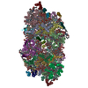
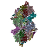
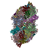
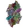
| Title | High-resolution structure of photosystem II from the mesophilic cyanobacterium, Synechocystis sp. PCC 6803 | |||||||||
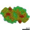
 Map data Map data | Photosystem II from Synechocystis sp. PCC 6803 at low dose sharpened | |||||||||
 Sample Sample |
| |||||||||
 Keywords Keywords | Photosystem II / Cyanobacteria / Synechocystis 6803 / Mesophile / CyanoQ / PHOTOSYNTHESIS | |||||||||
| Function / homology |  Function and homology information Function and homology informationplasma membrane-derived thylakoid photosystem II / photosystem II oxygen evolving complex / photosystem II assembly / thylakoid / oxygen evolving activity / photosystem II stabilization / photosystem II reaction center / photosystem II / photosynthetic electron transport chain / oxidoreductase activity, acting on diphenols and related substances as donors, oxygen as acceptor ...plasma membrane-derived thylakoid photosystem II / photosystem II oxygen evolving complex / photosystem II assembly / thylakoid / oxygen evolving activity / photosystem II stabilization / photosystem II reaction center / photosystem II / photosynthetic electron transport chain / oxidoreductase activity, acting on diphenols and related substances as donors, oxygen as acceptor / response to herbicide / photosystem II / extrinsic component of membrane / plasma membrane-derived thylakoid membrane / photosynthetic electron transport in photosystem II / chlorophyll binding / photosynthesis, light reaction / phosphate ion binding / photosynthesis / respiratory electron transport chain / manganese ion binding / electron transfer activity / protein stabilization / iron ion binding / heme binding / calcium ion binding / metal ion binding / identical protein binding Similarity search - Function | |||||||||
| Biological species |  | |||||||||
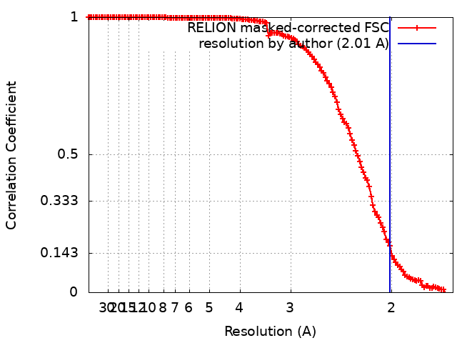
| Method | single particle reconstruction / cryo EM / Resolution: 2.01 Å | |||||||||
 Authors Authors | Gisriel CJ / Brudvig GW | |||||||||
| Funding support |  United States, 1 items United States, 1 items
| |||||||||
 Citation Citation | Journal: Proc Natl Acad Sci U S A / Year: 2022 Title: High-resolution cryo-electron microscopy structure of photosystem II from the mesophilic cyanobacterium, sp. PCC 6803. Authors: Christopher J Gisriel / Jimin Wang / Jinchan Liu / David A Flesher / Krystle M Reiss / Hao-Li Huang / Ke R Yang / William H Armstrong / M R Gunner / Victor S Batista / Richard J Debus / Gary W Brudvig / Abstract: Photosystem II (PSII) enables global-scale, light-driven water oxidation. Genetic manipulation of PSII from the mesophilic cyanobacterium sp. PCC 6803 has provided insights into the mechanism of ...Photosystem II (PSII) enables global-scale, light-driven water oxidation. Genetic manipulation of PSII from the mesophilic cyanobacterium sp. PCC 6803 has provided insights into the mechanism of water oxidation; however, the lack of a high-resolution structure of oxygen-evolving PSII from this organism has limited the interpretation of biophysical data to models based on structures of thermophilic cyanobacterial PSII. Here, we report the cryo-electron microscopy structure of PSII from sp. PCC 6803 at 1.93-Å resolution. A number of differences are observed relative to thermophilic PSII structures, including the following: the extrinsic subunit PsbQ is maintained, the C terminus of the D1 subunit is flexible, some waters near the active site are partially occupied, and differences in the PsbV subunit block the Large (O1) water channel. These features strongly influence the structural picture of PSII, especially as it pertains to the mechanism of water oxidation. | |||||||||
| History |
|
- Structure visualization
| Movie |
Movie viewer |
|---|---|
| Structure viewer | EM map: SurfViewMolmilJmol/JSmol |
| Supplemental images |

- Downloads & links
-EMDB archive
| Map data | emd_24407.map.gz | 29.4 MB | EMDB map data format | |
|---|---|---|---|---|
| Header (meta data) | emd-24407-v30.xmlemd-24407.xml | 39.1 KB 39.1 KB | Display Display | EMDB header |
| FSC (resolution estimation) | emd_24407_fsc.xml | 13.5 KB | Display | FSC data file |
| Images | 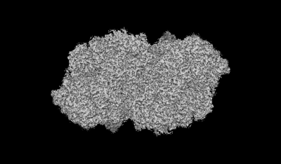 emd_24407.png emd_24407.png | 57.4 KB | ||
| Filedesc metadata | emd-24407.cif.gz | 8.7 KB | ||
| Others | emd_24407_additional_1.map.gz | 169.3 MB | ||
| Archive directory |  http://ftp.pdbj.org/pub/emdb/structures/EMD-24407ftp://ftp.pdbj.org/pub/emdb/structures/EMD-24407 http://ftp.pdbj.org/pub/emdb/structures/EMD-24407ftp://ftp.pdbj.org/pub/emdb/structures/EMD-24407 | HTTPS FTP |
-Related structure data
| Related structure data |  7rcvMC  7n8oC M: atomic model generated by this map C: citing same article ( |
|---|---|
| Similar structure data |


-Links
| EMDB pages | EMDB (EBI/PDBe) / EMDataResource |
|---|---|
| Related items in Molecule of the Month |


-Map
| File | Download / File: emd_24407.map.gz / Format: CCP4 / Size: 216 MB / Type: IMAGE STORED AS FLOATING POINT NUMBER (4 BYTES) | ||||||||||||||||||||||||||||||||||||||||||||||||||||||||||||||||||||
|---|---|---|---|---|---|---|---|---|---|---|---|---|---|---|---|---|---|---|---|---|---|---|---|---|---|---|---|---|---|---|---|---|---|---|---|---|---|---|---|---|---|---|---|---|---|---|---|---|---|---|---|---|---|---|---|---|---|---|---|---|---|---|---|---|---|---|---|---|---|
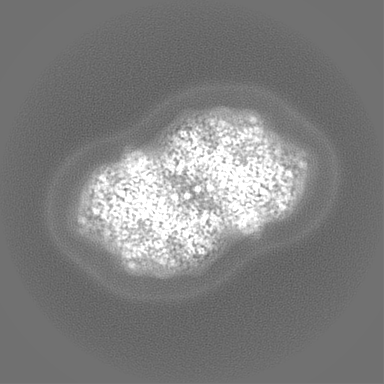
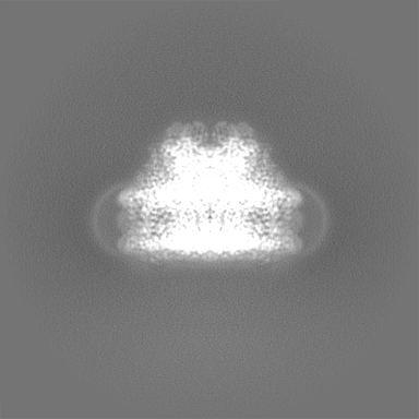
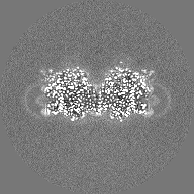
| Annotation | Photosystem II from Synechocystis sp. PCC 6803 at low dose sharpened | ||||||||||||||||||||||||||||||||||||||||||||||||||||||||||||||||||||
| Projections & slices | Image control
Images are generated by Spider. | ||||||||||||||||||||||||||||||||||||||||||||||||||||||||||||||||||||
| Voxel size | X=Y=Z: 0.832 Å | ||||||||||||||||||||||||||||||||||||||||||||||||||||||||||||||||||||
| Density |
| ||||||||||||||||||||||||||||||||||||||||||||||||||||||||||||||||||||
| Symmetry | Space group: 1 | ||||||||||||||||||||||||||||||||||||||||||||||||||||||||||||||||||||
| Details | EMDB XML:
CCP4 map header:
| ||||||||||||||||||||||||||||||||||||||||||||||||||||||||||||||||||||
 Z (Sec.)
 Y (Row.)
 X (Col.)


-Supplemental data
-Additional map: Photosystem II from Synechocystis sp. PCC 6803 at...
| File | emd_24407_additional_1.map | ||||||||||||
|---|---|---|---|---|---|---|---|---|---|---|---|---|---|
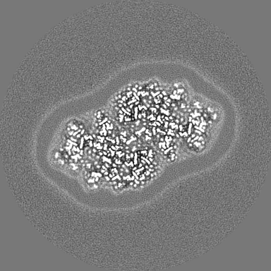
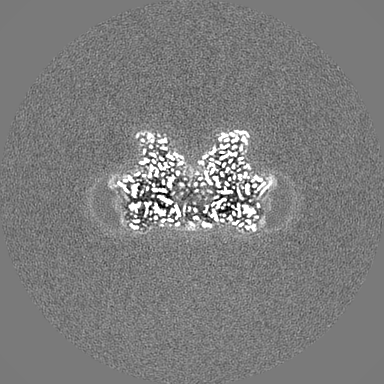
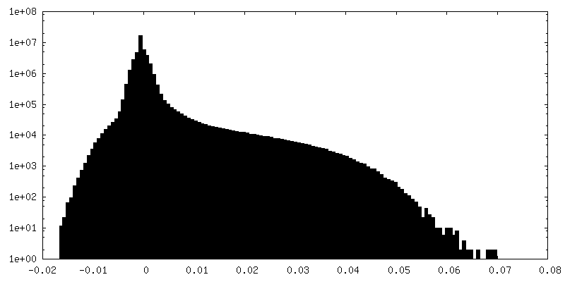
| Annotation | Photosystem II from Synechocystis sp. PCC 6803 at low dose unsharpened | ||||||||||||
| Projections & Slices |
| ||||||||||||
| Density Histograms |


- Sample components
+Entire : Photosystem II
+Supramolecule #1: Photosystem II
+Macromolecule #1: Photosystem II protein D1 2
+Macromolecule #2: Photosystem II CP47 reaction center protein
+Macromolecule #3: Photosystem II CP43 reaction center protein
+Macromolecule #4: Photosystem II D2 protein
+Macromolecule #5: Cytochrome b559 subunit alpha
+Macromolecule #6: Cytochrome b559 subunit beta
+Macromolecule #7: Photosystem II reaction center protein H
+Macromolecule #8: Photosystem II reaction center protein I
+Macromolecule #9: Photosystem II reaction center protein J
+Macromolecule #10: Photosystem II reaction center protein K
+Macromolecule #11: Photosystem II reaction center protein L
+Macromolecule #12: Photosystem II reaction center protein M
+Macromolecule #13: Photosystem II manganese-stabilizing polypeptide
+Macromolecule #14: Sll1638 protein
+Macromolecule #15: Photosystem II protein Y
+Macromolecule #16: Photosystem II reaction center protein T
+Macromolecule #17: Photosystem II 12 kDa extrinsic protein
+Macromolecule #18: Cytochrome c-550
+Macromolecule #19: Photosystem II reaction center X protein
+Macromolecule #20: Photosystem II reaction center protein Ycf12
+Macromolecule #21: Photosystem II reaction center protein Z
+Macromolecule #22: CA-MN4-O5 CLUSTER

+Macromolecule #23: FE (II) ION
+Macromolecule #24: CHLORIDE ION
+Macromolecule #25: CHLOROPHYLL A

+Macromolecule #26: PHEOPHYTIN A

+Macromolecule #27: BETA-CAROTENE

+Macromolecule #28: 1,2-DI-O-ACYL-3-O-[6-DEOXY-6-SULFO-ALPHA-D-GLUCOPYRANOSYL]-SN-GLYCEROL

+Macromolecule #29: 1,2-DISTEAROYL-MONOGALACTOSYL-DIGLYCERIDE

+Macromolecule #30: 2,3-DIMETHYL-5-(3,7,11,15,19,23,27,31,35-NONAMETHYL-2,6,10,14,18,...
+Macromolecule #31: DODECYL-BETA-D-MALTOSIDE

+Macromolecule #32: BICARBONATE ION

+Macromolecule #33: 1,2-DIPALMITOYL-PHOSPHATIDYL-GLYCEROLE

+Macromolecule #34: DIGALACTOSYL DIACYL GLYCEROL (DGDG)

+Macromolecule #35: PROTOPORPHYRIN IX CONTAINING FE

+Macromolecule #36: CALCIUM ION
+Macromolecule #37: (3R)-beta,beta-caroten-3-ol

+Macromolecule #38: water

-Experimental details
-Structure determination
| Method | cryo EM |
|---|---|
 Processing Processing | single particle reconstruction |
| Aggregation state | particle |
-Sample preparation
| Buffer | pH: 6.8 |
|---|---|
| Vitrification | Cryogen name: ETHANE |
- Electron microscopy
| Microscope | FEI TITAN KRIOS |
|---|---|
| Image recording | Film or detector model: GATAN K3 (6k x 4k) / Average electron dose: 40.8 e/Å2 |
| Electron beam | Acceleration voltage: 300 kV / Electron source:  FIELD EMISSION GUN FIELD EMISSION GUN |
| Electron optics | Illumination mode: FLOOD BEAM / Imaging mode: BRIGHT FIELD |
| Experimental equipment |  Model: Titan Krios / Image courtesy: FEI Company |