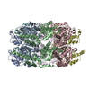
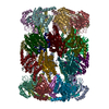
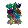
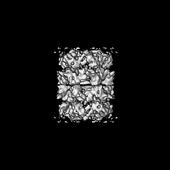
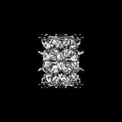
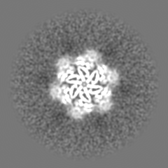
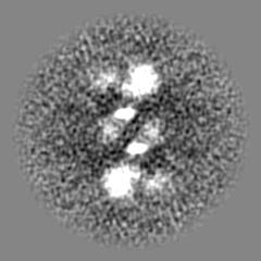
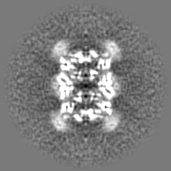
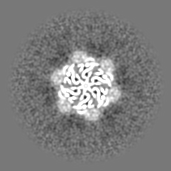
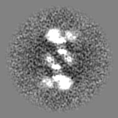
登録情報 データベース : EMDB / ID : EMD-24306タイトル Structure of the p97(R155H) dodecamer II Structure of the p97(R155H) dodecamer II 複合体 : p97 R155H mutantタンパク質・ペプチド : p97 R155H mutant in dodecameric form機能・相同性 分子機能 ドメイン・相同性 構成要素
/ / / / / / / / / / / / / / / / / / / / / / / / / / / / / / / / / / / / / / / / / / / / / / / / / / / / / / / / / / / / / / / / / / / / / / / / / / / / / / / / / / / / / / / / / / / / / / / / / / / / / / / / / / / / / / / / / / / / / / / / / / / / / / / / / / / / / / / 生物種 Homo sapiens (ヒト)手法 / / 解像度 : 6.1 Å Nandi P / Li S / Coulmbres RCA / Wang F / Williams DR / Poh Y-P / Chou T-F / Chiu P-L 資金援助 Organization Grant number 国 National Institutes of Health/National Institute of Neurological Disorders and Stroke (NIH/NINDS) R01NS102279 National Science Foundation (NSF, United States) MRI 1531991
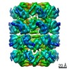
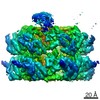
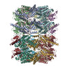
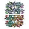
ジャーナル : Int J Mol Sci / 年 : 2021タイトル : Structural and Functional Analysis of Disease-Linked p97 ATPase Mutant Complexes.著者 : Purbasha Nandi / Shan Li / Rod Carlo A Columbres / Feng Wang / Dewight R Williams / Yu-Ping Poh / Tsui-Fen Chou / Po-Lin Chiu / 要旨 : IBMPFD/ALS is a genetic disorder caused by a single amino acid mutation on the p97 ATPase, promoting ATPase activity and cofactor dysregulation. The disease mechanism underlying p97 ATPase ... IBMPFD/ALS is a genetic disorder caused by a single amino acid mutation on the p97 ATPase, promoting ATPase activity and cofactor dysregulation. The disease mechanism underlying p97 ATPase malfunction remains unclear. To understand how the mutation alters the ATPase regulation, we assembled a full-length p97 with its p47 cofactor and first visualized their structures using single-particle cryo-EM. More than one-third of the population was the dodecameric form. Nucleotide presence dissociates the dodecamer into two hexamers for its highly elevated function. The N-domains of the p97 mutant all show up configurations in ADP- or ATPS-bound states. Our functional and structural analyses showed that the p47 binding is likely to impact the p97 ATPase activities via changing the conformations of arginine fingers. These functional and structural analyses underline the ATPase dysregulation with the miscommunication between the functional modules of the p97. 履歴 登録 2021年6月25日 - ヘッダ(付随情報) 公開 2021年8月25日 - マップ公開 2021年8月25日 - 更新 2021年8月25日 - 現状 2021年8月25日 処理サイト : RCSB / 状態 : 公開
すべて表示 表示を減らす
 ムービー
ムービー コントローラー


 データを開く
 基本情報
基本情報 マップデータ
 試料
試料 機能・相同性情報
 Homo sapiens (ヒト)
 データ登録者
 米国, 2件
  引用
引用 構造の表示
構造の表示
 ダウンロードとリンク
 emd_24306.png
 http://ftp.pdbj.org/pub/emdb/structures/EMD-24306


 Z (Sec.)
 Y (Row.)
 X (Col.)


 試料の構成要素
 解析
解析 電子顕微鏡法
 FIELD EMISSION GUN
