 Movie
Movie Controller


+ Open data

- Basic information
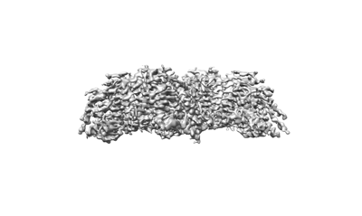
| Entry | Database: EMDB / ID: EMD-24208 | |||||||||
|---|---|---|---|---|---|---|---|---|---|---|
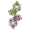
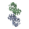
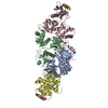
| Title | Structure of PPPA bound human ACAT2 | |||||||||
 Map data Map data | Structure of PPPA bound human ACAT2 | |||||||||
 Sample Sample |
| |||||||||
 Keywords Keywords | Inhibitor / MEMBRANE PROTEIN / Transferase-Inhibitor complex | |||||||||
| Function / homology |  Function and homology information Function and homology informationpositive regulation of intestinal cholesterol absorption / sterol O-acyltransferase / sterol O-acyltransferase activity / cholesterol O-acyltransferase activity / cholesterol storage / macrophage derived foam cell differentiation / very-low-density lipoprotein particle assembly / fatty-acyl-CoA binding / low-density lipoprotein particle clearance / intestinal cholesterol absorption ...positive regulation of intestinal cholesterol absorption / sterol O-acyltransferase / sterol O-acyltransferase activity / cholesterol O-acyltransferase activity / cholesterol storage / macrophage derived foam cell differentiation / very-low-density lipoprotein particle assembly / fatty-acyl-CoA binding / low-density lipoprotein particle clearance / intestinal cholesterol absorption / LDL clearance / cholesterol efflux / cholesterol binding / acyltransferase activity / brush border / cholesterol metabolic process / cholesterol homeostasis / endoplasmic reticulum membrane / endoplasmic reticulum Similarity search - Function | |||||||||
| Biological species |  Homo sapiens (human) Homo sapiens (human) | |||||||||
| Method | single particle reconstruction / cryo EM / Resolution: 3.87 Å | |||||||||
 Authors Authors | Li X / Long T | |||||||||
| Funding support | 1 items
| |||||||||
 Citation Citation | Journal: Structure / Year: 2021 Title: Molecular structures of human ACAT2 disclose mechanism for selective inhibition. Authors: Tao Long / Yang Liu / Xiaochun Li /  Abstract: Endoplasmic reticulum-localized acyl-CoA:cholesterol acyltransferases (ACAT), including ACAT1 and ACAT2, convert cholesterol to cholesteryl esters that become incorporated into lipoproteins or stored ...Endoplasmic reticulum-localized acyl-CoA:cholesterol acyltransferases (ACAT), including ACAT1 and ACAT2, convert cholesterol to cholesteryl esters that become incorporated into lipoproteins or stored in cytosolic lipid droplets. Selective inhibition of ACAT2 has been shown to considerably attenuate hypercholesterolemia and atherosclerosis in mice. Here, we report cryogenic electron microscopy structures of human ACAT2 bound to its specific inhibitor pyripyropene A or the general ACAT inhibitor nevanimibe. Structural analysis reveals that ACAT2 has a topology in membranes similar to that of ACAT1. A catalytic core with an entry site occupied by a cholesterol molecule and another site for allosteric activation of ACAT2 is observed in these structures. Enzymatic assays show that mutations within sites of cholesterol entry or allosteric activation attenuate ACAT2 activity in vitro. Together, these results reveal mechanisms for ACAT2-mediated esterification of cholesterol, providing a blueprint to design new ACAT2 inhibitors for use in the prevention of cardiovascular disease. | |||||||||
| History |
|
- Structure visualization
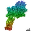
| Movie |
Movie viewer |
|---|---|
| Structure viewer | EM map: SurfViewMolmilJmol/JSmol |
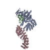
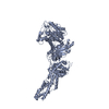
| Supplemental images |
- Downloads & links
-EMDB archive
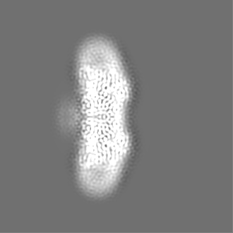
| Map data | emd_24208.map.gz | 26.7 MB | EMDB map data format | |
|---|---|---|---|---|
| Header (meta data) | emd-24208-v30.xmlemd-24208.xml | 9.5 KB 9.5 KB | Display Display | EMDB header |
| Images |  emd_24208.png emd_24208.png | 35.5 KB | ||
| Filedesc metadata | emd-24208.cif.gz | 5.1 KB | ||
| Archive directory |  http://ftp.pdbj.org/pub/emdb/structures/EMD-24208ftp://ftp.pdbj.org/pub/emdb/structures/EMD-24208 http://ftp.pdbj.org/pub/emdb/structures/EMD-24208ftp://ftp.pdbj.org/pub/emdb/structures/EMD-24208 | HTTPS FTP |
-Related structure data
| Related structure data |  7n6qMC  7n6rC M: atomic model generated by this map C: citing same article ( |
|---|---|
| Similar structure data |


-Links
| EMDB pages | EMDB (EBI/PDBe) / EMDataResource |
|---|---|
| Related items in Molecule of the Month |

-Map
| File | Download / File: emd_24208.map.gz / Format: CCP4 / Size: 137.1 MB / Type: IMAGE STORED AS FLOATING POINT NUMBER (4 BYTES) | ||||||||||||||||||||||||||||||||||||||||||||||||||||||||||||||||||||
|---|---|---|---|---|---|---|---|---|---|---|---|---|---|---|---|---|---|---|---|---|---|---|---|---|---|---|---|---|---|---|---|---|---|---|---|---|---|---|---|---|---|---|---|---|---|---|---|---|---|---|---|---|---|---|---|---|---|---|---|---|---|---|---|---|---|---|---|---|---|
| Annotation | Structure of PPPA bound human ACAT2 | ||||||||||||||||||||||||||||||||||||||||||||||||||||||||||||||||||||
| Projections & slices | Image control
Images are generated by Spider. | ||||||||||||||||||||||||||||||||||||||||||||||||||||||||||||||||||||
| Voxel size | X=Y=Z: 0.833 Å | ||||||||||||||||||||||||||||||||||||||||||||||||||||||||||||||||||||
| Density |
| ||||||||||||||||||||||||||||||||||||||||||||||||||||||||||||||||||||
| Symmetry | Space group: 1 | ||||||||||||||||||||||||||||||||||||||||||||||||||||||||||||||||||||
| Details | EMDB XML:
CCP4 map header:
| ||||||||||||||||||||||||||||||||||||||||||||||||||||||||||||||||||||
 X (Sec.)
 Y (Row.)
 Z (Col.)


-Supplemental data
- Sample components
-Entire : human ACAT2 with PPPA
| Entire | Name: human ACAT2 with PPPA |
|---|---|
| Components |
|
-Supramolecule #1: human ACAT2 with PPPA
| Supramolecule | Name: human ACAT2 with PPPA / type: organelle_or_cellular_component / ID: 1 / Parent: 0 / Macromolecule list: #1 |
|---|---|
| Source (natural) | Organism: Homo sapiens (human) |
-Macromolecule #1: Sterol O-acyltransferase 2
| Macromolecule | Name: Sterol O-acyltransferase 2 / type: protein_or_peptide / ID: 1 / Number of copies: 4 / Enantiomer: LEVO / EC number: sterol O-acyltransferase |
|---|---|
| Source (natural) | Organism: Homo sapiens (human) |
| Molecular weight | Theoretical: 60.893 KDa |
| Recombinant expression | Organism: Homo sapiens (human) |
| Sequence | String: MDYKDDDDKE PGGARLRLQR TGGLGGERER QPCGDGNTET HRAPDLVQWT RHMEAVKAQL LEQAQGQLRE LLDRAMREAI QSYPSQDKP LPPPPPGSLS RTQEPSLGKQ KVFIIRKSLL DELMEVQHFR TIYHMFIAGL CVFIISTLAI DFIDEGRLLL E FDLLIFSF ...String: MDYKDDDDKE PGGARLRLQR TGGLGGERER QPCGDGNTET HRAPDLVQWT RHMEAVKAQL LEQAQGQLRE LLDRAMREAI QSYPSQDKP LPPPPPGSLS RTQEPSLGKQ KVFIIRKSLL DELMEVQHFR TIYHMFIAGL CVFIISTLAI DFIDEGRLLL E FDLLIFSF GQLPLALVTW VPMFLSTLLA PYQALRLWAR GTWTQATGLG CALLAAHAVV LCALPVHVAV EHQLPPASRC VL VFEQVRF LMKSYSFLRE AVPGILRARR GEGIQAPSFS SYLYFLFCPT LIYRETYPRT PYVRWNYVAK NFAQALGCVL YAC FILGRL CVPVFANMSR EPFSTRALVL SILHATLPGI FMLLLIFFAF LHCWLNAFAE MLRFGDRMFY RDWWNSTSFS NYYR TWNVV VHDWLYSYVY QDGLRLLGAR ARGVAMLGVF LVSAVAHEYI FCFVLGFFYP VMLILFLVIG GMLNFMMHDQ RTGPA WNVL MWTMLFLGQG IQVSLYCQEW YARRHCPLPQ ATFWGLVTPR SWSCHT UniProtKB: Sterol O-acyltransferase 2 |
-Macromolecule #2: (3S,4R,4aR,6S,6aS,12R,12aS,12bS)-4-[(acetyloxy)methyl]-12-hydroxy...
| Macromolecule | Name: (3S,4R,4aR,6S,6aS,12R,12aS,12bS)-4-[(acetyloxy)methyl]-12-hydroxy-4,6a,12b-trimethyl-11-oxo-9-(pyridin-3-yl)-1,3,4,4a,5,6,6a,12,12a,12b-decahydro-2H,11H-naphtho[2,1-b]pyrano[3,4-e]pyran-3,6-diyl diacetate type: ligand / ID: 2 / Number of copies: 4 / Formula: 7T8 |
|---|---|
| Molecular weight | Theoretical: 583.626 Da |
| Chemical component information | 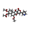 ChemComp-7T8: |
-Macromolecule #3: CHOLESTEROL
| Macromolecule | Name: CHOLESTEROL / type: ligand / ID: 3 / Number of copies: 8 / Formula: CLR |
|---|---|
| Molecular weight | Theoretical: 386.654 Da |
| Chemical component information |  ChemComp-CLR: |
-Macromolecule #4: OLEIC ACID
| Macromolecule | Name: OLEIC ACID / type: ligand / ID: 4 / Number of copies: 4 / Formula: OLA |
|---|---|
| Molecular weight | Theoretical: 282.461 Da |
| Chemical component information |  ChemComp-OLA: |
-Experimental details
-Structure determination
| Method | cryo EM |
|---|---|
 Processing Processing | single particle reconstruction |
| Aggregation state | particle |
-Sample preparation
| Buffer | pH: 7.5 |
|---|---|
| Vitrification | Cryogen name: ETHANE |
- Electron microscopy
| Microscope | FEI TITAN KRIOS |
|---|---|
| Image recording | Film or detector model: GATAN K3 (6k x 4k) / Average electron dose: 60.0 e/Å2 |
| Electron beam | Acceleration voltage: 300 kV / Electron source:  FIELD EMISSION GUN FIELD EMISSION GUN |
| Electron optics | Illumination mode: FLOOD BEAM / Imaging mode: DARK FIELD |
| Experimental equipment |  Model: Titan Krios / Image courtesy: FEI Company |
-Image processing
| Startup model | Type of model: PDB ENTRY |
|---|---|
| Final reconstruction | Resolution.type: BY AUTHOR / Resolution: 3.87 Å / Resolution method: FSC 0.143 CUT-OFF / Number images used: 153208 |
| Initial angle assignment | Type: ANGULAR RECONSTITUTION |
| Final angle assignment | Type: ANGULAR RECONSTITUTION |
